Fatemeh Abyar and
Hossein Farrokhpour*
Chemistry Department, Isfahan University of Technology, Isfahan, 84156-83111, Iran. E-mail: farrokhphossein@gmail.com; h-farrokh@cc.iut.ac.ir; Fax: +98 311 3912350; Tel: +98 311 3913243
First published on 25th July 2014
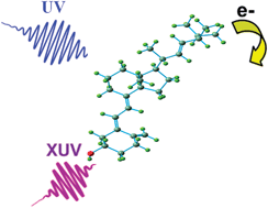
The electronic structures and photoelectron spectra of several fat-soluble vitamins including A (all-trans-retinol and its two derivatives, 13-cis-retinoic acid and all-trans-retinoic acid), D2, D3, E (consisting of α-tocopherol, β-tocopherol, γ-tocopherol and δ-tocopherol) and K were studied theoretically in this work. The vertical ionization energies of these compounds, including considerations of their electron correlations were calculated in the gas phase. The direct symmetry-adapted cluster/configuration interaction method, which employs single and double excitation operators (direct-SAC-CI SD-R), and the D95(df,pd) basis set were used for the calculations. The results indicate that more than one conformer contributes to the photoelectron spectrum of vitamin A, all-trans-retinoic acid, vitamin D2 and D3, suggesting that there is more than one biologically active form for each of these vitamins. The photoelectron spectrum of each vitamin was simulated and assigned, and the previously reported assignments from the literature were revisited. The ionization of vitamin D from highest occupied molecular orbit − 2 (HOMO − 2) and the lone electron pairs of oxygen was found to not take place below 10 eV. The first ionization band of vitamin E was assigned to the ionization from πCC and π*C
C of its aromatic ring, unlike the previous assignment relating this ionization band to the lone electron pair of oxygen. In addition, the ionizations of vitamin A and its derivatives from the lone electron pairs of oxygen were found to not occur below 11 eV in the gas phase.
The intrinsic properties of biomolecules, which are hidden in the complex media of natural biological systems, can be revealed by theoretical and experimental studies conducted in the gas phase, in an isolated environment.12 In this case, a detailed understanding of the structures and dynamics of biomolecules together with the distinction between the intrinsic properties and those due to environmental interactions can be obtained. For example, the effects of bulk hydration and the interaction of other ions in the biological environment cause significant changes in the electronic structures of biological molecules. This in turn affects the chemical and physical properties of the molecules. Changes in the electronic structure of molecules in biological media are accompanied by changes in their orbital energies and possible reordering of the orbital energies. This has an effect on the vertical ionization energies and vertical excitation energies of the molecules. Comparing the valence ionization and excitation spectrum of a biomolecule in the gas phase with those in solution provides an understanding of how the biomolecule bonds and interacts with its surroundings. For example, Ghosh et al. theoretically studied the effect of hydration on the vertical ionization energy of thymine.13 They found that microsolvation reduces the ionization energy by about 0.1 eV per water molecule, while the first solvation shell increases the ionization energy by 0.1 eV compared to the ionization energy in the gas phase. As an another example, Slavicek et al. used both theoretical and experimental techniques to study the effect of solvation with water on the vertical ionization energies of cytidine and deoxythymidine in comparison to the gas phase.14 They reported that upon bulk hydration, the ionization potential of the base becomes insensitive to the presence of the sugar and phosphate, unlike that which is observed in the gas phase. Based on these studies, the ionization energies of biomolecules in the gas phase are very important.
The gas phase ionization energy is one of the most important and fundamental properties of vitamins, and these energies could be used to better understand some of the biological phenomena contributed by these molecules. For example, the vertical ionization energies of different forms of vitamin E can be used as diagnostic indicators for the variation in activation energy associated with the antioxidant reaction of this vitamin.15 In addition, measuring the ionization energies of vitamins provides information about the oxidative potential of these molecules and can be used as a scale for comparing their oxidation potential in electrochemical reactions. The assignment of the photoelectron spectra of vitamins is also important for elucidating the mechanism of vitamin damage caused by photoionization because it determines where ionization occurs in the vitamin molecule. Knowing the location of ionization in the different ionic states of a vitamin allows us to obtain information about how the vitamin is fragmented in its excited ionic states after ionization.
One of the best methods for measuring the ionization energies of molecules in the gas phase is photoelectron spectroscopy (PES).16–18 The most challenging part of gas phase PES, especially for large biomolecules, is the decomposition and degradation of the sample before it reaches the gas phase. In this case, calculating the gas phase ionization energies and photoelectron spectra of biomolecules with a very accurate computational method is useful and provides information about gas phase ionization. Although there have been many experimental studies on the interaction of UV light with vitamins in solution, there are a limited number of studies in the literature related to the ionization of vitamins in the gas phase using UV light and X-ray radiation. Jericevic et al.19 recorded the He–I photoelectron spectra for some vitamin A derivatives including trans-retinoic acid, trans-retinal and β-carotene in the gas phase. Katsumata and Ikehata recorded the first He–I photoelectron spectrum of vitamin A in the gas phase.20 Novak and Potts studied the electronic structures of vitamins D2 and D3 using a PES technique5 and found that, based on their similar photoelectron spectra, D vitamins have steroid structures. There are no high level theoretical calculations on the ionization energies and photoelectron spectra of vitamin D and its derivatives in the literature. Nagaoka et al.15 recorded the He–I photoelectron spectra of vitamin E and its derivatives, but there is no experimental photoelectron spectrum for vitamin K in the literature. The aim of this current work is: (i) to calculate the vertical ionization energies of fat-soluble vitamins including A (all-trans-retinol and its two derivatives, 13-cis-retinoic acid and all-trans-retinoic acid), D2, D3, E (consisting of α-tocopherol, β-tocopherol, γ-tocopherol and δ-tocopherol) and K, taking into consideration their electron correlations to study their electronic structures; and (ii) to simulate and assign their photoelectron spectra.
![]() | ||
Fig. 2 Comparison of the calculated photoelectron spectra and the BWP spectrum of the vitamin D2 conformers with the experimental photoelectron spectrum of this vitamin.5 Vertical lines show the calculated positions and intensities of ionization bands. The letters A, B and C indicate the dominant features in the experimental spectrum. |
![]() | ||
Fig. 3 Comparison of the calculated photoelectron spectra and the BWP spectrum of the vitamin D3 conformers with the experimental photoelectron spectrum of this vitamin.5 Vertical lines show the calculated positions and intensities of ionization bands. The letters A, B and C indicate the dominant features in the experimental spectrum. |
Novak and Potts reported the first separate He–I photoelectron spectra of vitamins D2 and D3.5 They also performed semi-empirical Austin Model 1 (AM1) calculations to obtain the ionization and molecular orbital energies of vitamins D2 and D3. It should be mentioned that they did not perform conformational analysis on these vitamins to determine their ionization energies and conformer populations in the gas phase. Therefore, the ionization energies of these vitamins were calculated considering only one geometrical structure for each vitamin in the work of Novak and Potts. In addition, the molecular structures used for calculating the ionization energies in their work were optimized using molecular mechanics, and the observed features in the recorded photoelectron spectra were assigned based on Koopmans' approximation. Considering the above deficiencies, we decided in this work to calculate and assign the photoelectron spectra of vitamins D2 and D3 using a theoretical method that considers the electron correlations.
Fig. 2 indicates five ionization bands for the two conformers of vitamin D2 below 10 eV, while Novak and Potts predicted four ionization bands in this region based on Koopmans' approximation. In addition, the observed relative energies and intensities of the calculated ionization bands of conformers 1 and 2 are different. To obtain the calculated photoelectron spectrum of vitamin D2, the sum of the Boltzmann weighted individual calculated spectra of conformers 1 and 2 was considered (indicated by BWP in Fig. 2). As can be seen, there is an excellent agreement between the BWP spectrum and the experimental spectrum of vitamin D2 in Fig. 2. It is important to note that the experimental photoelectron spectrum of vitamin D2 is very similar to the calculated spectrum of conformer 2. This observation is in agreement with the population ratios of the vitamin D2 conformers obtained in this work (25.6% conformer 1; 74.3% conformer 2). Therefore, the assignment of the experimental photoelectron spectrum of D2 is explained based on the SAC-CI and NBO results for conformer 2.
Feature A of the experimental spectrum is related mainly to the first ionization band of conformers 1 and 2. The wave function of the first ionic state of D2 is a single HF-ionized determinant related to highest occupied molecular orbital (HOMO), which is a π molecular orbital due to π(C9–C15), π(C17–C19) and π(C23–C29) of the triene system (Tables S1† and 1). The calculated first ionization energy of conformer 2 obtained using the direct SAC-CI and HF methods are 5.945 and 7.75 eV, respectively. The position of feature A in the experimental spectrum of vitamin D2 is approximately 7.528 eV, while the first ionization energy of vitamin D2 calculated by Novak and Potts is approximately 8.72 eV. Thus, considering the electron correlations for vitamin D2 decreases the ionization energy. Therefore, the BWP spectrum of vitamin D2 (Fig. 2) was shifted by 1.58 eV towards the higher binding energy to match the position of the first peak of the BWP spectrum with the position of feature A in the experimental spectrum. The difference between the calculated and experimental first ionization energy of vitamin D2 can be attributed primarily to the intrinsic error present in the direct SAC-CI SD-R method. The average error of the direct SAC-CI method is approximately 0.5 eV in small molecules because of the cut-off approximation in the unlinked terms; this error increases with the size of the molecule.32 However, comparison of the BWP spectrum with the experimental spectrum shows that the direct SAC-CI SD-R method with the D95(df,pd) basis set can predict the relative energies and intensities of the photoelectron lines quite accurately. The numbers in parenthesis in Table 1 indicate the calculated ionization energies of the vitamin D conformers considering the value of the energy shift.
State (2A) | Main electronic configuration | Ionization energy (eV) | Intensity | ||
---|---|---|---|---|---|
Vitamin D2 | Conformer 2 | 1 | 0.94(HOMO) | 5.945 (7.528) | 0.914 |
2 | −0.89(HOMO − 1) + 0.24(HOMO − 3) | 7.274 (8.854) | 0.913 | ||
3 | −0.9(HOMO − 4) + 0.21(HOMO − 3) | 7.957 (9.537) | 0.904 | ||
4 | −0.20(HOMO − 2) − 0.69(HOMO − 3)−0.51(HOMO − 4) − 0.30(HOMO − 1) | 7.990 (9.57) | 0.904 | ||
5 | −0.49(HOMO − 3) + 0.72(HOMO − 4) | 8.373 (9.953) | 0.900 | ||
Vitamin D3 | Conformer 2 | 1 | 0.94(HOMO) | 5.858 (7.458) | 0.914 |
2 | 0.92(HOMO − 1) | 7.498 (9.098) | 0.902 | ||
3 | −0.87(HOMO − 2) + 0.30(HOMO − 3) | 7.868 (9.468) | 0.903 | ||
4 | 0.53(HOMO − 4) − 0.64(HOMO − 3) | 8.196 (9.796) | 8.196 | ||
5 | 0.52(HOMO − 3) + 0.72(HOMO − 4) | 8.332 (9.932) | 0.898 | ||
Conformer 3 | 1 | 0.94(HOMO) | 5.987 (7.587) | 0.914 | |
2 | 0.39(HOMO − 1) − 0.84(HOMO − 2) | 7.518 (9.118) | 0.902 | ||
3 | 0.38(HOMO − 2) + 0.84(HOMO − 1) | 7.906 (9.506) | 0.905 | ||
4 | 0.21(HOMO − 2) + 0.70(HOMO − 3) − 0.23(HOMO − 4) − 0.48(HOMO − 5) | 8.153 (9.753) | 0.901 | ||
5 | −0.88(HOMO − 3) − 0.21(HOMO − 4) | 8.250 (9.85) | 0.903 | ||
Vitamin K (MK-1) | 1 | −0.67(HOMO) + 0.57(HOMO − 4) + 0.24(HOMO − 3) | 7.386 | 0.899 | |
2 | −0.87(HOMO − 1)-0.26(HOMO − 3) −0.24(HOMO − 2) | 7.819 | 0.907 | ||
3 | 0.75(HOMO − 2) + 0.47(HOMO − 3) − 0.25(HOMO − 4) | 8.142 | 0.909 | ||
4 | 0.67(HOMO − 3) + 0.41(HOMO − 2) + 0.35(HOMO − 1) + 0.33(HOMO − 5) | 8.432 | 0.897 | ||
5 | 0.67(HOMO − 4) − 0.65(HOMO) | 8.472 | 0.891 |
The second ionization band of conformers 1 and 2 corresponds to feature B of the experimental spectrum. The second ionization band of vitamin D2 should occur from HOMO − 1 based on Koopmans' approximation, however, the SAC-CI result shows that this ionization can also originate from HOMO − 3 as well as HOMO − 1 because of the presence of electron correlations. HOMO − 1 is a π molecular orbital primarily localized on the C14C16 bond of the side chain of the molecule. The assignment of the first and second ionization bands of vitamin D2 are in agreement with the assignment performed by Novak and Potts.5 Notably, the third ionization band of vitamin D2 originates mostly from HOMO − 4, unlike the prediction by Koopmans' approximation (Table 1). HOMO − 4 is a π molecular orbital related only to π(C9–C15) and π(C17–C19) of the triene (Table S1†). This assignment is different to that reported by Novak and Potts.5 The wave function of the fourth ionization band is a linear combination of four single HF-ionized determinants including HOMO − 1, HOMO − 2, HOMO − 3 and HOMO − 4, demonstrating that the amount of electron correlations have been increased in this ionic state (see Table 1). One of the most important points related to vitamin D2 is that its ionization from HOMO − 2 and the lone electron pairs of its OH group do not occur below 10 eV.
Three conformers of vitamin D3 are observed in the gas phase (Fig. 1). The difference between vitamin D2 and D3 is mainly related to the side chain (there is no CC bond in the side chain of D3). Fig. 3 shows the calculated photoelectron spectra of the vitamin D3 conformers together with their BWP spectrum. The experimental spectrum of vitamin D3 recorded by Novak and Potts5 is also included in this figure. The calculated spectra needed to be shifted by 1.6 eV towards the higher binding energy in order to match the experimental spectrum. The experimental photoelectron spectrum of D3 is similar to that of D2, however, the first peak in the spectrum of D3 is broader than that in the spectrum of D2.5 This difference has been attributed to the more varied gas phase conformer population for vitamin D3 compared to D2,5 which is in agreement with the thermochemistry results obtained in this work, suggesting that the number of gas phase populated conformers of D3 is higher than for D2. Comparison of theoretical results with experimental results shows that feature A of the vitamin D3 experimental spectrum is composed only of the first ionization band of the conformers. Fig. 3 shows that the ionization energies of conformer 1 are higher than those of the other two conformers. For example, the order of the first ionization energies of the vitamin D3 conformers are: conformer 1 > conformer 3 > conformer 2. The difference between the first ionization energies of the vitamin D3 conformers causes the broadening of feature A in the spectrum of vitamin D3 compared to that in the spectrum of vitamin D2.
In a similar way to vitamin D2, the first ionization band of vitamin D3 is related to the ionization from HOMO, which is a π molecular orbital related to the triene system of the molecule (Tables 1 and S1†). Table S1† shows that the HOMOs of vitamin D3 conformers 2 and 3 are mostly localized on π(C15C9) and π(C18
C20). A small feature observed in the experimental spectrum of vitamin D3 (assigned with an asterisk in Fig. 3) is at first glance assumed to be the second ionization band of vitamin D3, however, the calculated BWP spectrum presented in this work does not show or confirm this feature. Feature B of the experimental spectrum is only related to the second ionization band of conformers 2 and 3. Therefore, it is possible to follow the change in the total population of conformers 2 and 3 relative to conformer 1 with temperature using the change in intensity of this feature detected by photoelectron spectroscopy. The wave function of the second ionization band of conformer 2 is mainly related to HOMO − 1; in contrast, it is related to both HOMO − 1 and HOMO − 2 (most contribution) for conformer 3. It can be seen that Koopmans' approximation is not valid for conformer 3, whereas it is for conformer 2. The HOMO − 1s of conformers 2 and 1 are mostly composed of π(C24
C28) and π(C9
C15), respectively, and HOMO − 2 has σ character. The third ionization band of conformer 2 is related to HOMO − 2, whereas it is related to HOMO − 1 in conformer 3, however, there is a little contribution from HOMO − 3 and HOMO − 2 to the main configuration of this ionic state for conformers 2 and 3, respectively. The wave function of the fourth ionization band of conformer 2 is a linear combination of HOMO − 4 and HOMO − 3, while in conformer 3 it is related to HOMO − 3 and HOMO − 5 with nearly the same contribution. HOMO − 3 is a π molecular orbital related to C18
C20 and C9
C15. The shapes of the molecular orbitals of the two vitamin D3 conformers are illustrated in Fig. S1.†
![]() | ||
Fig. 4 Calculated photoelectron spectrum of vitamin K (MK-1). Vertical lines show the calculated positions and intensities of ionization bands. |
The first ionization band of MK-1 is related to the ionization from HOMO, HOMO − 3 and HOMO − 4, and the contributions of HOMO and HOMO − 4 to the wave function are greater than that of HOMO − 3 (Table 1). This means that Koopmans' approximation is not valid for this vitamin. Based on the NBO calculations, the HOMO is a π molecular orbital that is mostly localized on the side chain C14C10 bond, whereas HOMO − 4 is a non-bonding orbital related to the lone electron pairs of the menaquinone oxygen atoms (Table S1 and Fig. S3†). Therefore, the ionization of MK-1 occurs from two different places in the molecule with the nearly same probability. Interestingly, the first ionization of this vitamin does not occur from the π electrons of the aromatic ring. The wave function of the second ionization band is a linear combination of three ionized HF determinants including HOMO − 1 (greatest contribution), HOMO − 2 and HOMO − 3. HOMO − 1 is a π molecular orbital localized on the aromatic ring (π(C13
C16); π(C12
C15); π*(C6
C7); see Fig. S3†); the ionization probability from π(C13
C16) and π(C12
C15) is higher than that from π*(C6
C7). The third ionization band is related to HOMO − 2 (more contribution), HOMO − 3 and HOMO − 4 with different probabilities. HOMO − 2 is a π molecular orbital related to the π system of the aromatic ring and C3
C4. HOMO − 3 is a π molecular orbital mostly related to the menaquinone C3
C4 and localized primarily on this bond (Fig. S3†). The assignment of the fifth ionization band is similar to that of the first band because the wave function of this ionic state is a linear combination of two ionized determinants related to HOMO and HOMO − 4. Finally, it is important to note that the first ionization of vitamin K does not take place from the aromatic ring, but instead occurs from the unsaturated C14
C10 bond of the side chain and the menaquinone oxygen atoms.
![]() | ||
Fig. 5 The molecular structures of different forms of vitamin E including (a) α-tocopherol, (b) β-tocopherol, (c) γ-tocopherol, (d) δ-tocopherol and (e) tocopherol. |
![]() | ||
Fig. 6 Comparison of the calculated photoelectron spectra of tocopherol, α-tocopherol and β-tocopherol with their experimental spectra.15 Vertical lines show the calculated positions and intensities of ionization bands. |
![]() | ||
Fig. 7 Comparison of the calculated photoelectron spectra of γ-tocopherol and δ-tocopherol with their experimental spectra.15 Vertical lines show the calculated positions and intensities of ionization bands. |
State (2A) | Main electronic configuration | Ionization energy (eV) | Intensity | |
---|---|---|---|---|
Tocopherol | 1 | −0.93(HOMO) | 5.865 (7.465) | 0.912 |
2 | 0.95(HOMO − 1) | 7.494 (9.094) | 0.910 | |
3 | −0.91(HOMO − 2) | 8.035 (9.635) | 0.907 | |
4 | 0.93(HOMO − 3) | 8.256 (9.856) | 0.909 | |
5 | 0.94(HOMO − 4) | 8.416 (10.016) | 0.911 | |
α-Tocopherol | 1 | −0.94(HOMO) | 5.550 (7.15) | 0.910 |
2 | 0.95(HOMO − 1) | 6.636 (8.236) | 0.911 | |
3 | −0.89(HOMO − 2) | 7.978 (9.578) | 0.895 | |
4 | −0.8(HOMO − 3) − 0.20(HOMO − 4) + 0.44(HOMO − 5) | 8.399 (9.999) | 0.901 | |
5 | −0.95(HOMO − 4) | 8.486 (10.086) | 0.911 | |
β-Tocopherol | 1 | 0.93(HOMO) | 5.669 (7.269) | 0.912 |
2 | 0.94(HOMO − 1) | 6.934 (8.534) | 0.911 | |
3 | −0.77(HOMO − 2) + 0.40(HOMO − 4) | 8.154 (9.754) | 0.902 | |
4 | −0.91(HOMO − 2) − 0.20(HOMO − 3) | 8.347 (9.947) | 0.910 | |
5 | −0.81(HOMO − 4) − 0.460(HOMO − 2) | 8.457 (10.057) | 0.912 | |
γ-Tocopherol | 1 | 0.94(HOMO) | 5.696 (7.396) | 0.911 |
2 | −0.95(HOMO − 1) | 6.868 (8.568) | 0.912 | |
3 | 0.94(HOMO − 2) | 8.163 (9.863) | 0.902 | |
4 | −0.72(HOMO − 3) + 0.59(HOMO − 4) | 8.350 (10.05) | 0.910 | |
5 | 0.76(HOMO − 4) + 0.59(HOMO − 3 | 8.459 (10.159) | 0.912 | |
δ-Tocopherol | 1 | −0.94(HOMO) | 5.79 (7.29) | 0.912 |
2 | 0.95(HOMO − 1) | 7.213 (8.713) | 0.911 | |
3 | 0.92(HOMO − 2) | 8.213 (9.713) | 0.912 | |
4 | 0.8(HOMO − 3) − 0.45(HOMO − 4) | 8.348 (9.848) | 0.909 | |
5 | −0.20(HOMO − 6) + 0.77(HOMO − 4) + 0.45(HOMO − 3) | 8.423 (9.923) | 0.904 |
The first ionization bands of all forms of vitamin E are related to the ionization from HOMO. This molecular orbital is mostly localized on the aromatic ring (Fig. S2 and S3†) and related to two πCC and one π*C
C of the ring (Table S2†). Table S2† also shows that there is a small probability for the ionization of vitamin E from the lone electron pairs of oxygen. Based on calculations using the HF/STO-3G method, Nagaoka et al.15 assigned the first ionization band of vitamin E only to the ionization of lone electron pairs of oxygen; in the present study, this ionization band was attributed primarily to the πC
C and π*C
C of the aromatic ring based on the SAC-CI calculation. The second ionization band of all forms of vitamin E is mainly related to the ionization of HOMO − 1 localized on the phenyl ring. HOMO − 1 is a π molecular orbital related to the π C
C of the aromatic ring. The shapes of HOMO and HOMO − 1 for all forms of vitamin E are illustrated in Fig. S2 and S3.† Based on the above assignments, the first and second ionizations of vitamin E originate from the π electrons of the aromatic ring. The calculated spectra in Fig. 6 and 7 show that the energy difference between the first and second ionization band decreases from α-tocopherol, β-tocopherol, γ-tocopherol and δ-tocopherol compared to tocol. The third ionization band in the calculated spectrum of α-tocopherol is a resolved feature that has been identified with an asterisk in Fig. 6. The third ionization band is related to HOMO − 2, which is an σ orbital. The other molecular orbitals of all forms of vitamin E have σ character. Most importantly, the ionization of vitamin E from the lone electron pairs of oxygen does not take place below 10.5 eV; this conclusion is in contrast to the assignments made by Nagaoka et al.15
There are 30, 37 and 22 conformers for vitamin A, 13-cis-retinoic acid and all-trans-retinoic acid, respectively. The thermochemistry calculations performed in this work showed that only two conformers are populated in the gas phase for vitamin A and all-trans-retinoic acid, while only one conformer is populated for 13-cis-retinoic acid. The molecular structures of the conformers are depicted in Fig. 8. Fig. 9 shows the calculated photoelectron and BWP spectra of the vitamin A conformers and compares them with the experimental photoelectron spectrum of vitamin A.20 The calculated and experimental spectra of 13-cis-retinoic acid are also included in this figure. Fig. 10 shows the calculated photoelectron and BMP spectra of the conformers of all-trans-retinoic acid and compares them with its experimental spectrum.20
![]() | ||
Fig. 9 Comparison of the calculated photoelectron spectra of vitamin A conformers, their BWP spectrum and the calculated spectrum of 13-cis-retinoic acid with the experimental photoelectron spectra of vitamin A20 and 13-cis-retinoic acid.20 The numbers above the experimental spectra show the visible features identified by Katsumata et al.20 |
![]() | ||
Fig. 10 Comparison of the calculated photoelectron spectra and the BWP spectrum of the conformers of all-trans-retinoic acid with the experimental photoelectron spectrum of all-trans-retinoic acid.20 |
The calculated photoelectron spectra of the two conformers of vitamin A are very similar (Fig. 9). The calculated BWP spectrum of vitamin A describes its photoelectron spectra20 very well. Unfortunately, the resolution of the experimental spectrum17 is poor, however, the features can still be made out. The calculated photoelectron spectra of the conformers of all-trans-retinoic acid (Fig. 10) and 13-cis-retinoic acid are very similar to those of vitamin A. This means that the calculated ionization bands originate from the same region of the molecule in all three compounds. The calculated BWP spectra of vitamin A (1.5 eV) and all-trans-retinoic acid (1.3 eV) along with the spectrum of 13-cis-retinoic acid (1.25 eV) were shifted to higher binding energies to obtain the best agreement with the experiment. The main configuration of the ionic wave functions of vitamin A, all-trans-retinoic acid and 13-cis-retinoic acid obtained from SAC-CI calculations (Table 3) shows that the electronic correlations are not negligible in the calculation of ionization energies in the energy region considered; this is because the wave function of some ionic states are a linear combination of two or more single HF-ionized determinants. To see the effect of the correlation energy on the relative energy positions of the ionization bands, the calculated ionization energies of the vitamin A conformers relative to the energy of the first ionization band were plotted against the ionization band number (Fig. 11). There is a considerable difference between the plot obtained using HF theory (without electron correlation) and that obtained using SAC-CI theory. The plot obtained using the calculated ionization energies of Katsumata et al.20 has also been included in this figure. The electronic correlations are absent in the calculations of Katsumata et al., as shown by the similarity of their plot to those obtained in this work using the HF/D95(df,pd) method. The comparison of theory with experimental work in Fig. 9 and 10 again confirms that although the calculated photoelectron spectra were shifted to higher binding energies, the direct SAC-CI method can provide accurate predictions of the relative energies of ionization bands.
State (2A) | Main electronic configuration | Ionization energy (eV) | Intensity | ||
---|---|---|---|---|---|
Vitamin A | Conformer I | 1 | −0.94(HOMO) | 5.611 (7.111) | 0.91 |
2 | −0.92(HOMO − 1) | 6.798 (8.298) | 0.904 | ||
3 | −0.87(HOMO − 2) − 0.25(HOMO − 3) | 7.823 (9.323) | 0.889 | ||
4 | −0.72(HOMO − 3) − 0.43(HOMO − 4) | 8.260 (9.76) | 0.893 | ||
5 | 0.22(HOMO − 2) − 0.38(HOMO − 3) − 0.81(HOMO − 4) | 8.734 (10.234) | 0.884 | ||
Conformer II | 1 | −0.94(HOMO) | 5.594 (7.094) | 0.910 | |
2 | 0.92(HOMO − 1) | 6.796 (8.296) | 0.903 | ||
3 | 0.88(HOMO − 2) + 0.24(HOMO − 3) | 7.835 (9.335) | 0.888 | ||
4 | −0.72(HOMO − 3) + 0.44(HOMO − 4) | 8.255 (9.755) | 0.894 | ||
5 | −0.20(HOMO − 2) − 0.39(HOMO − 3) + 0.81(HOMO − 4) | 8.733 (10.233) | 0.883 | ||
All-trans-retinoic acid | Conformer I | 1 | −0.93(HOMO) | 5.800 (7.1) | 0.905 |
2 | −0.92(HOMO − 1) | 6.973 (8.273) | 0.898 | ||
3 | 0.85(HOMO − 2) + 0.31(HOMO − 3) | 8.049 (9.349) | 0.882 | ||
4 | −0.64(HOMO − 3) − 0.45(HOMO − 6) | 8.407 (9.707) | 0.890 | ||
5 | −0.28(HOMO − 2) −0.38(HOMO − 3) − 0.79(HOMO − 4) | 8.909 (10.209) | 0.884 | ||
Conformer II | 1 | 0.94(HOMO) | 5.822 (7.122) | 0.90662 | |
2 | 0.93(HOMO − 1) | 6.917 (8.217) | 0.90185 | ||
3 | −0.90(HOMO − 2) | 7.933 (9.233) | 0.88685 | ||
4 | −0.82(HOMO − 3) + 0.30(HOMO − 6) | 8.428 (9.728) | 0.89329 | ||
5 | −0.89(HOMO − 4) − 0.21(HOMO − 3) | 8.806 (10.106) | 0.88183 | ||
13-cis-Retinoic | 1 | 0.93(HOMO) | 5.726 (6.976) | 0.906 | |
2 | 0.92(HOMO − 1) | 6.908 (8.158) | 0.899 | ||
3 | −0.83(HOMO − 2) − 0.32(HOMO − 3) | 7.943 (9.193) | 0.882 | ||
4 | 0.68(HOMO − 3) − 0.49(HOMO − 6) | 8.316 (9.566) | 0.889 | ||
5 | 0.33(HOMO − 2) − 0.41(HOMO − 3) − 0.76(HOMO − 4) | 8.8139 (10.063) | 0.878 |
![]() | ||
Fig. 11 Variation in the energy positions of the ionization bands of vitamin A conformers relative to their first ionization bands versus the ionization band number. |
The first and second ionization bands of vitamin A, all-trans-retinoic acid and 13-cis-retinoic acid correspond to HOMO and HOMO − 1, respectively (Table 3). HOMO is a π molecular orbital related to five conjugate πCC but primarily localized on three C
C bonds (identified with black asterisks in Fig. 8). Similarly, HOMO − 1 is a π molecular orbital, but it is mostly concentrated on only one C
C bond (identified with a blue asterisk in Fig. 8; see also Table S3†). The shape of the molecular orbitals of vitamin A, all-trans-retinoic acid and 13-cis-retinoic acid are shown in Fig. S4 and S5.† The wave function of the third ionization band of the vitamin A conformers, 13-cis-retinoic acid and conformer 1 of all-trans-retinoic acid is a linear combination of two single HF-ionized determinants including HOMO − 2 (major contribution) and HOMO − 3. The ionic wave function of conformer 2 of all-trans-retinoic acid mainly originates from HOMO − 2. The third feature in the experimental spectra of these compounds corresponds to the third ionization band of each conformer. HOMO − 2 is a π molecular orbital mostly concentrated on one C
C bond (Table S3†). For example, this C
C bond, for conformer 1 of vitamin A is C18
C20, which has a π contribution in HOMO − 2 of approximately 30.3%. Katsumata et al.20 proposed that the fourth feature of the experimental photoelectron spectra of vitamin A and its derivatives are composed of the fourth and fifth ionization bands based on the assignment using Koopmans' approximation. The SAC-CI calculations showed that the fourth feature in the experimental spectrum is composed of only the fourth ionization band. The reason for this discrepancy is related to the absence of electron correlations in the calculations of Katsumata et al.20 The wave function of the fourth ionic state of vitamin A is a linear combination of three single ionized HF determinants including HOMO − 3 and HOMO − 4 (major contribution; Table 3). The main configuration of the fourth ionic states of all-trans-retinoic acid and 13-cis-retinoic acid is a linear combination of HOMO − 3 and HOMO − 6. The assignment of the fourth ionization band of vitamin A is different from all-trans-retinoic acid and 13-cis-retinoic acid. HOMO − 3 is related to the chain of five πC
C for all compounds, while HOMO − 4 is related to the σC–C of the cyclohexene ring. The position of the fifth ionization band of vitamin A identified by Katsumata et al.20 in their experimental spectra (see Fig. 8 and 9) is not confirmed by the calculated spectra in this work. For example, our theoretical calculations show that the fifth ionization band of all-trans-retinoic is around 10.2 eV, while Katsumata et al.20 considered this feature to be around 10.8 eV, containing three ionization bands (the fifth, sixth and seventh). Finally, the theoretical calculations show that the ionization of vitamin A and its derivatives from the lone electron pairs of oxygen does not take place below 11 eV.
Footnote |
† Electronic supplementary information (ESI) available. See DOI: 10.1039/c4ra05271d |
This journal is © The Royal Society of Chemistry 2014 |