Amidoxime-functionalized magnetic mesoporous silica for selective sorption of U(VI)

Yingguo Zhaoabc, Jiaxing Li*a, Shouwei Zhanga and Xiangke Wangad
aKey Laboratory of Novel Thin Film Solar Cells, Institute of Plasma Physics, Chinese Academy of Sciences, P.O. Box 1126, Hefei, 230031, PR China. E-mail: lijx@ipp.ac.cn; Fax: +86-551-65591310; Tel: +86-551-655933088
bSchool of Chemistry and Materials Science, University of Science and Technology of China, Hefei 230026, PR China
cSchool of Chemistry and Chemical Engineering, Anqing Normal College, Anqing 246011, PR China
dFaculty of Engineering, King Abdulaziz University, Jeddah 21589, Saudi Arabia

Received 30th May 2014 , Accepted 14th July 2014

First published on 16th July 2014


Abstract

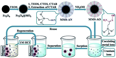
Amidoxime-functionalized magnetic mesoporous silica (MMS-AO) microspheres were synthesized through co-condensation of tetraethyl orthosilicate (TEOS) and 2-cyanoethyltriethoxysilane (CTES) on the surface of silica-coated Fe3O4 followed by chemical modification of nitrile into amidoxime. The synthesized microspheres exhibit a typical sandwich structure with an inner core of Fe3O4, a middle layer of nonporous silica and an outer layer of amidoxime-functionalized mesoporous silica. Owing to the mesoporous structure and amidoxime functionalization, sorption of U(VI) by MMS-AO reaches equilibrium in 2 h of contact time with a maximum sorption capacity of 1.165 mmol g−1 (277.3 mg g−1) at pH = 5.0 ± 0.1 and T = 298 K, which is much higher than the results previously reported for other magnetic materials. The sorption process is strongly dependent on pH but independent of ionic strength, indicating that the predominant sorption mechanism is inner-sphere surface complexation. The selectivity of MMS-AO for U(VI) is remarkably improved in comparison with that of magnetic mesoporous silica without amidoxime functionalization. U(VI)-loaded MMS-AO can be conveniently separated from aqueous solutions with an external magnetic field and efficiently regenerated using 1 mol L−1 HCl with only a small decrease in U(VI) sorption capacity. These results suggest that MMS-AO shows promise as a future candidate for selective separation of U(VI) from aqueous solutions in possible real applications.


Introduction

Uranium has attracted significant interest because of its strategic and ecological significance. Uncertainty in fossil fuel-based energy sources and global warming impel more and more countries to develop nuclear power to guarantee their energy security and cut down on greenhouse gas emissions. Due to the expected shortage of uranium in the near future, there are increasing efforts to acquire uranium from unconventional resources such as sea water and ground water.1 On the other hand, uranium-containing wastewater discharged from uranium mining and nuclear fuel cycle activities is dangerous for human health and other organisms because of its high toxicity and radioactivity.2 Therefore, efficient extraction of uranium from aqueous phase is of considerable realistic significance from the viewpoints of both energy security and environmental protection.

Sorption is one of the most widely used recovery methods because of its low cost, easy operation and wide adaptability.3,4 Convenient separation of sorbents from aqueous phase is of special importance considering the recyclability of sorbents and the recoverability of sorbates that are either toxic or useful and precious. Traditional separation methods such as centrifugation and filtration are usually labor-consumptive, uneconomical and thus impractical for large-scale water treatment. Magnetic sorbents exhibit special superiority due to convenient separation by an external magnetic field.5–7 However, the most commonly used magnetic sorbents are based on Fe3O4 particles which are susceptible to leaching under acidic conditions and may aggregate when dispersed in electrolyte solutions. To compensate for these disadvantages, surface modifications of Fe3O4 particles based on physical coating have been extensively studied.8–11

Of various core–shell structured microspheres, magnetic mesoporous silica (MMS) is of particular interest owing to the typical sandwich structure with an inner core of Fe3O4, a middle layer of nonporous silica and an outer layer of ordered mesoporous silica.12 In addition to the magnet-responsive function, the nonporous silica layer offers protection of the inner Fe3O4 core and the mesoporous silica shell exhibits high surface area, large pore volume, tunable pore size and low cytotoxicity.13 However, most reported materials have pure silica as the mesoporous shell and show poor selectivity toward specific sorbates. Replacement of pure silica by organic–inorganic hybrid materials incorporated with various organic groups would definitely offer advanced properties that are often difficult to achieve either from totally organic or from totally inorganic materials. The emergence of silica-based organic–inorganic hybrids appears as a breakthrough since they combine in a single solid both the properties of a stable three-dimensional inorganic network with the specific chemical reactivity of organic functional groups.14–17 In comparison to the widely studied functionalization of pure mesoporous silica, incorporation of organic functional groups into magnetic mesoporous silica is rare.18

Among various functional groups studied for uranium recovery from aqueous solutions, the amidoxime group has attracted extensive attention due to its special structure.19–22 Amidoxime is amphoteric with both acidic oxime and basic amino groups. Lone pairs of electrons in amino nitrogen and oxime oxygen can be donated to the positive metal center to form a stable five-membered chelate with U(VI). Consequently, the amidoxime group has been used to functionalize various substrates for the sorption of U(VI), and the results indicate that amidoxime functionalization can remarkably enhance sorption capacity and selectivity. In this study, amidoxime-functionalized magnetic mesoporous silica (MMS-AO) was prepared, carefully characterized and used to adsorb U(VI) from aqueous solutions, and the sorption kinetics, sorption thermodynamics, selectivity and reusability were investigated. Based on the experimental results, application possibility of MMS-AO for the sorption of U(VI) from aqueous solutions in real work was evaluated.

Experimental

Preparation of MMS-AO

Chemical materials used in this study are listed in (ESI). Prior to the deposition of mesoporous structure, Fe3O4 microspheres were first coated with a thin nonporous silica layer (Fe3O4@nSiO2, see ESI). The nonporous silica shell present on the surface of Fe3O4 ensures that the subsequent nucleation of mesoporous silica is initiated at the surface of Fe3O4@nSiO2 and thus offers very effective encapsulation of every microsphere by mesoporous silica. Amidoxime-functionalized magnetic mesoporous silica was prepared as follows (schematically described in Fig. 1). 0.1 g Fe3O4@nSiO2 microspheres were dispersed in a mixture containing 0.30 g CTAB, 80 mL Milli-Q water, 1.00 g concentrated ammonia aqueous solution and 60 mL ethanol. The mixed solution was sonicated for 0.5 h to form a uniform dispersion. A mixture containing 0.3 g TEOS and 0.1 g CTES was added dropwise to the dispersion with continuous stirring. After reaction for 6 h, the product was collected with a magnet bar and washed with ethanol and Milli-Q water alternatively to remove nonmagnetic by-products. Finally, the purified microspheres were redispersed in 100 mL of ethanol containing 0.3 g NH4NO3 and refluxed at 80 °C for 6 h to remove the template CTAB. The extraction was repeated for three times and the obtained microspheres were washed with Milli-Q water and ethanol several times and denoted as MMS-AN. The obtained MMS-AN was then treated with 1.0 g K2CO3 and 1.0 g NH2OH·HCl in a 10/90 H2O–C2H5OH solution (100 mL) for 6 h at 80 °C in a closed flask. The final product was separated by a permanent magnet, rinsed with Milli-Q water and ethanol, dried under vacuum at 60 °C and denoted as MMS-AO. For comparison, magnetic mesoporous silica without organic functionalization was also prepared and denoted as MMS.
image file: c4ra05128a-f1.tif
Fig. 1 Schematic illustration of the preparation procedure of MMS-AO.

Characterization

Small angle X-ray diffraction (SAXRD) patterns were recorded by a Philips X'Pert Pro Super X-ray diffractometer with Cu-Kα source (λ = 1.54178 Å). Nitrogen adsorption–desorption isotherms were measured at −196 °C using a TriStar 3000 volumetric adsorption analyzer (Micromeritics Instrument Corp., Norcross, GA). Before measurements, the samples were degassed in a vacuum at 200 °C for at least 6 h. Scanning electron microscopy (SEM) images were recorded with a JEOL JSM-6330F microscope. Transmission electron microscopy (TEM) images were performed on a JEOL-2010 microscope. Fourier transformed infrared (FT-IR) spectra were recorded on a Nicolet Magana-IR 750 spectrometer with KBr pellets at room temperature. The TGA measurement was carried out using a Shimadzu TGA-50 thermogravimetric analyzer. Magnetic measurements were conducted in a MPMS-XL SQUID magnetometer.

Sorption experiments

Sorption experiments were carried out using batch method. Specific amounts of stock solutions of sorbents and U(VI), Milli-Q water and NaClO4 solutions were mixed in polyethylene tubes. Afterwards, pH was adjusted using negligible amounts of HClO4 and/or NaOH. After being shaken for 24 h to achieve sorption equilibrium, the U(VI)-loaded sorbents were collected from the solution using a permanent magnet. Concentrations of U(VI) in the supernatant were determined by inductively coupled plasma atomic emission spectroscopy (ICP-AES, Thermo Elemental, USA). The sorption percentage, the amount of U(VI) adsorbed (qe) and the distribution coefficient (Kd) were calculated as follows:
image file: c4ra05128a-t1.tif

image file: c4ra05128a-t2.tif

image file: c4ra05128a-t3.tif
where V is the volume of the suspension, m is the mass of the dry sorbent, and C0 and Ce are the initial and equilibrium U(VI) concentrations, respectively. All of the experimental data were the averages of triplicate determinations. The relative errors of the data were about 5%.

Results and discussion

Characterization

SAXRD patterns of MMS and MMS-AO are shown in Fig. 2(a). Both samples exhibit an intense diffraction peak centered approximately at 2θ = 2.3°, confirming the presence of mesoporous structure. Compared with MMS, the diffraction peaks of MMS-AO become weaker and broader, indicating that the structural ordering of organic–inorganic hybrids suffers some disturbance due to the incorporation of organic moieties. Mesoporous structure can be further identified through N2 adsorption–desorption isotherms. As shown in Fig. 2(b), adsorption–desorption isotherms show representative type-IV curves according to the IUPAC classification, with a sharp capillary condensation step at intermediate relative pressures (p/p0 < 0.5) corresponding to framework mesopores. Specific surface area was calculated to be 287.1 m2 g−1 through the Brunauer–Emmett–Teller (BET) method using adsorption data in the relative pressure range from 0.05 to 0.35. The total pore volume was estimated to be 0.273 cm3 g−1 from the adsorbed amount at a relative pressure of 0.992. The pore size distribution (the inset of Fig. 2(b)) was derived from the adsorption branch using the Barrett–Joyner–Halenda (BJH) model, with a peak centered at the pore diameter of 2.2 nm indicating the existence of mesoporous structure.
image file: c4ra05128a-f2.tif
Fig. 2 (a) SAXRD patterns of MMS and MMS-AO. (b) Nitrogen adsorption–desorption isotherms for MMS-AO. The insert shows the pore size distribution. (c) FT-IR spectra of MMS-AN and MMS-AO. (d) The TGA curve of MMS-AO. (e) Magnetization curves of Fe3O4 and MMS-AO. (f) The magnetic separation process of MMS-AO.

Surface morphology and structural features were observed by SEM and TEM as shown in Fig. 3. Compared with Fe3O4 nanoparticles, Fe3O4@nSiO2 and MMS-AO exhibit a more regular spherical shape with smoother surface and larger diameter due to silica deposition by the sol–gel process. TEM was used to identify the core–shell and mesoporous structures. The core–shell structure of Fe3O4@nSiO2 with a Fe3O4 core and a nonporous silica layer is clearly shown in Fig. 3(d). The typical sandwich structure of MMS-AO can be indentified from Fig. 3(e) and (f). The inner core Fe3O4, the middle layer nonporous silica and the outer layer amidoxime-functionalized mesoporous silica (mSiO2-AO) with mesopore channels perpendicular to the microsphere surface are observed (the thickness of the mesoporous shell can be determined to be ca. 60 nm). The middle nonporous layer can protect Fe3O4 from etching in harsh conditions while the outer periodic mesoporous shell offers high accessibility and fast diffusion of guest particles to active centers located deep in the mesopore channels.


image file: c4ra05128a-f3.tif
Fig. 3 SEM images of Fe3O4 (a), Fe3O4@nSiO2 (b) and MMS-AO (c); TEM images of Fe3O4@nSiO2 (d) and MMS-AO (e and f).

FTIR spectroscopy was performed to confirm the chemical bonds and the incorporation of organic moieties. In Fig. 2(c), the peak at 587 cm−1 is attributed to the stretching vibration of the Fe–O bond, the broad band around 1068 cm−1 is relevant to Si–O–Si and Si–O–H stretching vibrations and the band around 458 cm−1 corresponds to the bending vibration of O–Si–O. The absorption band at 2226 cm−1 corresponds to C[triple bond, length as m-dash]N stretching vibration, reflecting successful co-condensation of TEOS and CTES. On the spectrum of MMS-AO, the C[triple bond, length as m-dash]N absorption band disappears while two new bands arise at 1657 cm−1 and 953 cm−1 corresponding to the C[double bond, length as m-dash]N and N–O stretching vibrations of amidoxime groups respectively.

TGA measurement of MMS-AO was carried out to determine the amount of organic moieties in the synthesized material and the results are shown in Fig. 2(d). The initial weight loss at temperatures lower than 100 °C is due to the volatilization of physically adsorbed water and ethanol. There is a significant decrease of approximate 10.9% in the TGA curve of MMS-AO at temperatures ranging from 320 °C to 600 °C, which can be attributed to the decomposition of organic moieties. Based on the TGA results, the amount of amidoxime functional groups that have been incorporated into the inorganic silica framework in the MMS-AO material can be calculated to be ca. 1.3 mmol g−1 (the molecular mass of organic moieties is 87 u).

The magnetic properties of Fe3O4 and MMS-AO were studied by measuring magnetization curves at room temperature (Fig. 2(e)). The magnetization curves show no hysteresis loop, indicating their superparamagnetic behavior.23 Saturation magnetization (Ms) values were measured to be 64.6 emu g−1 for Fe3O4 and 39.9 emu g−1 for MMS-AO. Although the saturation magnetization decreases due to the decrease of the magnetite fraction after dense and mesoporous silica coating, complete magnetic separation can be accomplished in 1 minute by placing a magnet adjacent to the vessels containing the suspension of MMS-AO (Fig. 2(f)).

Time-dependent sorption

From the economical point of view, the sorption rate is an important parameter in evaluating the overall performance of sorbents. Fig. 4(a) shows the sorption curve of U(VI) by MMS-AO as a function of contact time. It is noteworthy that the instantaneous time for solid–liquid separation by an external magnet is negligible in calculation of the total contact time. As can be seen from Fig. 4(a), the amount of U(VI) adsorbed increases rapidly in the first contact time of 2 h and then levels off until the sorption equilibrium is attained. The short equilibrium time may be attributed to the well-defined mesoporous structures of as-synthesized MMS-AO and the strong complexation of U(VI) with amidoxime functional groups. Based on the kinetic results, 24 h of contact time was adopted to guarantee the equilibrium of U(VI) sorption by MMS-AO in the following experiments.
image file: c4ra05128a-f4.tif
Fig. 4 (a) Sorption of U(VI) by MMS-AO as a function of contact time. pH = 5.0 ± 0.1 and CU(VI) initial = 0.2 mmol L−1. (b) Effects of pH and ionic strength on U(VI) sorption by MMS-AO. CU(VI) initial = 0.2 mmol L−1. (c) Isotherms of U(VI) sorption by MMS and MMS-AO. pH = 5.0 ± 0.1. The scattered points represent experiment data, the solid lines represent the Langmuir model and the dashed lines represent the Freundlich model. (d) Selectivity coefficients of U(VI) sorption by MMS and MMS-AO. pH = 5.0 ± 0.1 and the initial concentration of each metal ion is 0.1 mmol L−1. (e) Effect of HCl concentration on U(VI) desorption. (f) U(VI) sorption by virgin and regenerated MMS-AO with five cycles. pH = 5.0 ± 0.1 and CU(VI) initial = 0.2 mmol L−1. T = 298 K and m/V = 0.2 g L−1 for all sorption experiments.

Application of appropriate kinetic models to simulate the experimental kinetic data can offer useful information to determine the underlying sorption mechanisms. From this point of view, the experimental data of time-dependent sorption of U(VI) by MMS-AO were simulated by the pseudo-first-order and pseudo-second-order models:24,25

ln(qeqt) = ln(qe) − k1t

image file: c4ra05128a-t4.tif
where qt is the sorption amount of U(VI) at contact time of t and qe is the sorption amount of U(VI) at equilibrium time; k1 and k2 represent the sorption rate constants of the pseudo-first-order and pseudo-second-order models, respectively. The k1 and theoretical qe values (qe,cal) of the pseudo-first-order model can be obtained from the linear plot of ln(qeqt) vs. t, and the k2 and qe,cal values of the pseudo-second-order model can be obtained from the linear plot of t/qt vs. t (the inset of Fig. 4(a)). The calculated kinetic parameters are shown in Table 1. Obviously, the qe,cal value of the pseudo-second-order model is more similar to the experimental value (qe,exp) and the correlation coefficient (R2) of the pseudo-first-order model is lower than that of the pseudo-second-order model, indicating that the experimental data are simulated better by the pseudo-second-order model than by the pseudo-first-order model. These results further imply that the rate-controlling mechanism of U(VI) sorption by MMS-AO is strong surface complexation or chemisorption rather than mass transport.26

Table 1 Kinetic parameters of U(VI) sorption by MMS-AO at 298 K
Parameters Pseudo-first-order Pseudo-second-order
qe,exp (mmol g−1) 0.357
qe,cal (mmol g−1) 0.335 0.356
k1 (h−1) 3.484  
k2 (g mmol−1 h−1)   24.011
R2 0.952 0.999


Effects of pH and ionic strength

It has been clearly established that the solution pH has a great impact on U(VI) sorption process. The effect of pH on U(VI) sorption by MMS-AO was investigated at different electrolyte concentrations (i.e., 0.001, 0.01 and 0.1 mol L−1 NaClO4 solutions, respectively). As shown in Fig. 4(b), the sorption amount increases gradually when pH increases from 2.0 to 6.0 and then falls off at pH values higher than 8.0. These two inverse sorption trends can be attributed to the influence of pH on both the surface properties of MMS-AO and the relative distribution of U(VI) species in solutions. From the relative distribution of U(VI) species (Fig. S1 in ESI), one can see that UO22+ is the predominant species at low pH. It is thought that the amidoxime functional group can serve as a bidentate ligand for UO22+ with the lone pairs of electrons on the oxime oxygen and the amino nitrogen donated to the metal cation center to form a stable five-membered ring (shown in Fig. S2). The oxime oxygen can experience metal-assisted deprotonation:27
[triple bond, length as m-dash]S–C(NH2)N–OH ↔ [triple bond, length as m-dash]S–C(NH2)N–O + H+

2[triple bond, length as m-dash]S–C(NH2)N–O + UO22+ ↔ UO2([triple bond, length as m-dash]S–C(NH2)N–O)2.

Increasing the solution pH can neutralize protons emitted by the complexation reaction, decrease the electrostatic repulsion between UO22+ and the positively charged surface of MMS-AO through the deprotonation of amidoxime groups, and consequently, promote the sorption amounts. Nevertheless, when pH values exceed 8.0, the sorption amount begins to fall off with increasing pH values. This may be due to the hydrolysis of U(VI) leading to noncomplexible species such as UO2(OH)3 and UO3(OH)7, and the electrostatic repulsion of these anions with the negatively charged surfaces of MMS-AO at high pH.

Fig. 4(b) also shows the ionic strength dependence of U(VI) sorption on MMS-AO. Obviously, the sorption process is scarcely affected by the ionic strength over the whole pH range, suggesting that the dominant sorption mechanism is inner-sphere surface complexation rather than ion exchange or outer-sphere surface complexation.26 This is consistent with the fact that amidoxime functional groups on the surfaces of MMS-AO have strong complexation toward U(VI) ions.

Sorption isotherms

Sorption amounts as a function of U(VI) equilibrium concentrations, i.e. the sorption isotherm, was investigated with different initial U(VI) concentrations. For comparison, the sorption isotherm of U(VI) on MMS was also investigated under similar conditions. As shown in Fig. 4(c), the sorption capacity of MMS-AO is much higher than that of MMS due to functionalization of amidoxime groups. In order to further understand the mechanism of U(VI) sorption by MMS-AO, experimental data were simulated by Langmuir and Freundlich sorption models:
image file: c4ra05128a-t5.tif

qe = kFC1/ne
where qmax is the maximum sorption capacity at complete monolayer coverage and b (L mmol−1) is a constant about the enthalpy of sorption; kF (mmol1−n Ln g−1) is the Freundlich constant and n represents the degree of sorption dependence on equilibrium concentrations. Parameters calculated are listed in Table 2. According to the correlation coefficients (R2), experimental data are better simulated by the Langmuir model than by the Freundlich model, indicating that absorbed U(VI) ions form a monolayer coverage and chemisorption is the dominant sorption mechanism, consistent with the above discussion.26
Table 2 Parameters of Langmuir and Freundlich models for U(VI) sorption by MMS and MMS-AO at 298 K
Sorbents Langmuir Freundlich
qmax b R2 kF n R2
MMS 0.487 2.825 0.977 0.446 1.512 0.989
MMS-AO 1.165 6.572 0.991 1.585 1.825 0.974


In order to evaluate the potential of real applications of MMS-AO in the removal and recovery of U(VI) ions from aqueous solutions, the maximum sorption capacity of MMS-AO toward U(VI) was compared with those of magnetic materials reported in previous literatures, as listed in Table 3. Thanks to the high specific surface area of mesoporous structures and strong complexation of amidoxime groups toward U(VI), MMS-AO is superior to most of magnetic materials in U(VI) sorption. In particular, there is a good improvement in sorption capacity compared with our previous work due to the mesoporous shell coated on the surface of Fe3O4@nSiO2 used in this work, exhibiting high surface area which is beneficial for the sorption process.31 Although the sorption amount of MMS-AO is a little lower than that of amidoxime functionalized magnetic graphene oxide (AOMGO), the overall performance of MMS-AO is believed to be better than that of AOMGO because of the stability arising from the core–shell structure of MMS-AO and the complicated synthetic process and potential ecological toxicity of graphene oxide. Based on the above discussion, MMS-AO can be used as a promising candidate for U(VI) preconcentration from large volumes of aqueous solutions in real work.

Table 3 Comparison of U(VI) sorption capacity of MMS-AO with other magnetic sorbents
Sorbents Experimental conditions qmax (mg g−1) Ref.
Colloidal magnetite Ambient temperature, pH = 7.0 1.4 28
Fe3O4/graphene oxide T = 293 K, pH = 5.5 69.5 29
Fe3O4@SiO2–salicylaldehyde Ambient temperature, pH = 7.0 49.0 30
Fe3O4@SiO2–quercetin T = 298 K, pH = 3.7 12.3 11
Fe3O4@SiO2–amidoxime T = 298 K, pH = 5.0 105.0 31
Polymeric-magnetite cryobead T = 298 K, pH = 5.0 120.5 32
Fe3O4@IIP Ambient temperature, pH = 4.0 71.5 33
Fe3O4@SiO2 T = 298 K, pH = 6.0 52.4 34
AOMGO T = 298 K, pH = 5.0 284.9 35
MMS-AO T = 298 K, pH = 5.0 277.3 This work


Selectivity studies

Efficient removal of uranium requires highly selectivity as there usually exist competitive metal ions in aqueous solutions. In order to evaluate the sorption selectivity of MMS-AO, sorption of U(VI) from aqueous solutions containing coexisting metal ions, including Zn2+, Ni2+, Co2+, Pb2+, Cr3+, Eu3+ and Ce3+, was investigated. Selectivity coefficient (SU/M) for U(VI) with respect to competitive ions is defined as:36
image file: c4ra05128a-t6.tif
where KUd and KMd are distribution coefficients of U(VI) and competitive ions, respectively. For comparison, sorption selectivity of MMS was also investigated and the results are shown in Fig. 4(d). It is obvious that selectivity coefficients of MMS-AO for all competitive ions have been remarkably improved in comparison with those of MMS, validating that amidoxime functionalization adopted in this study is effective and that MMS-AO exhibits desirable selectivity for U(VI) over a range of coexisting metal ions.

Regeneration studies

Regeneration of sorbents is a crucial factor to evaluate the application potential from the standpoints of environmental sustainability and economic efficiency. As illustrated in Fig. 4(b), the sorption amount of U(VI) is suppressed under acidic conditions, suggesting that acid treatment is a possible approach to regenerate U(VI)-loaded MMS-AO. Prior to acid treatment, the stability of MMS-AO under acidic conditions was investigated by monitoring the leaching amounts of Fe after different contact time with different HCl concentrations, as shown in Fig. S3. Obviously, the leaching amounts of Fe in 24 h are negligible due to successful coating of Fe3O4 by SiO2. Therefore, different concentrations of HCl were used to regenerate U(VI)-loaded MMS-AO (Fig. 4(e)). The results indicate that complete desorption (>99%) of U(VI) can be achieved using 1 mol L−1 or more concentrated HCl solution. Hence, regeneration experiments were conducted using 1 mol L−1 HCl solution as the desorbing agent and results are shown in Fig. 4(f) (see ESI for detailed leaching and regeneration experimental procedures). The sorption capacity of U(VI) slightly decreases from 0.357 mmol g−1 to 0.332 mmol g−1 after five sorption–desorption cycles. These results indicate that U(VI)-loaded MMS-AO can be efficiently regenerated and reused with only puny decrease in U(VI) sorption capacity, and that MMS-AO can sustain long-term use with low replacement costs.

Conclusions

An efficient U(VI) sorbent, amidoxime-functionalized magnetic mesoporous silica, has been successfully synthesized. The typical sandwich structure is well established, with an inner magnetic core, a nonporous silica middle layer and an outer layer of mesoporous silica functionalized with amidoxime. This material is found to be a promising alternative to the present U(VI) sorption materials in several aspects such as sorption rate, sorption capacity and selectivity. The sorption rate is very fast and the maximum sorption capacity is much higher than previous results reported for other magnetic materials. Sorption selectivity of MMS-AO for U(VI) over a wide range of competitive metal ions is remarkably enhanced compared with that of magnetic mesoporous silica without incorporation of amidoxime groups. The excellent sorption performance is attributed to the high accessibility of mesopore channels and strong complexation of amidoxime with U(VI). In addition, the sorbent can be conveniently collected by magnetic separation and reused for several cycles without a considerable decrease in U(VI) sorption capacity, which can reduce the cost in practical applications. Therefore, MMS-AO can be a promising candidate for the separation and preconcentration of U(VI) ions from aqueous solutions in real water treatment.

Acknowledgements

The authors acknowledge the financial support from National Natural Science Foundation of China (21207136, 91326202 and 21225730), Chinese National Fusion Project for ITER (2013GB110000) and Hefei Center for Physical Science and Technology (2012FXZY005).

Notes and references

  1. Y. Lu, Nat. Chem., 2014, 6, 175–177 CrossRef CAS PubMed.
  2. T. P. Rao, P. Metilda and J. M. Gladis, Talanta, 2006, 68, 1047–1064 CrossRef CAS PubMed.
  3. M. J. Manos and M. G. Kanatzidis, J. Am. Chem. Soc., 2012, 134, 16441–16446 CrossRef CAS PubMed.
  4. M. J. Comarmond, T. E. Payne, J. J. Harrison, S. Thiruvoth, H. K. Wong, R. D. Aughterson, G. R. Lumpkin, K. Muller and H. Foerstendorf, Environ. Sci. Technol., 2011, 45, 5536–5542 CrossRef CAS PubMed.
  5. R. L. D. Loyo, S. I. Nikitenko, A. C. Scheinost and M. Simonoff, Environ. Sci. Technol., 2008, 42, 2451–2456 CrossRef.
  6. Z. Liu, H. S. Wang, C. Liu, Y. J. Jiang, G. Yu, X. D. Mu and X. Y. Wang, Chem. Commun., 2012, 48, 7350–7352 RSC.
  7. H. Y. Koo, H. J. Lee, H. A. Go, Y. B. Lee, T. S. Bae, J. K. Kim and W. S. Choi, Graphene-based multifunctional iron oxide nanosheets with tunable properties, Chem.–Eur. J., 2011, 17, 1214–1219 CrossRef CAS PubMed.
  8. M. Park, S. Seo, I. S. Lee and J. H. Jung, Chem. Commun., 2010, 46, 4478–4480 RSC.
  9. H. M. Chen, C. H. Deng and X. M. Zhang, Angew. Chem., Int. Ed., 2010, 49, 607–611 CrossRef CAS PubMed.
  10. M. Kokate, K. Garadkar and A. Gole, J. Mater. Chem. A, 2013, 1, 2022–2029 CAS.
  11. S. Sadeghi, H. Azhdari, H. Arabi and A. Z. Moghaddam, J. Hazard. Mater., 2012, 215, 208–216 CrossRef PubMed.
  12. Y. H. Deng, D. W. Qi, C. H. Deng, X. M. Zhang and D. Y. Zhao, J. Am. Chem. Soc., 2008, 130, 28–29 CrossRef CAS PubMed.
  13. C. T. Kresge, M. E. Leonowicz, W. J. Roth, J. C. Vartuli and J. S. Beck, Nature, 1992, 359, 710–712 CrossRef CAS.
  14. A. Mehdi, C. Reye and R. Corriu, Chem. Soc. Rev., 2011, 40, 563–574 RSC.
  15. P. Van der Voort, D. Esquivel, E. De Canck, F. Goethals, I. Van Driessche and F. J. Romero-Salguero, Chem. Soc. Rev., 2013, 42, 3913–3955 RSC.
  16. A. Walcarius and L. Mercier, J. Mater. Chem., 2010, 20, 4478–4511 RSC.
  17. L. Y. Yuan, Y. L. Liu, W. Q. Shi, Z. J. Li, J. H. Lan, Y. X. Feng, Y. L. Zhao, Y. L. Yuan and Z. F. Chai, J. Mater. Chem., 2012, 22, 17019–17026 RSC.
  18. M. S. Moorthy, D. J. Seo, H. J. Song, S. S. Park and C. S. Ha, J. Mater. Chem. A, 2013, 1, 12485–12496 CAS.
  19. Y. F. Yue, R. T. Mayes, J. Kim, P. F. Fulvio, X. G. Sun, C. Tsouris, J. H. Chen, S. Brown and S. Dai, Angew. Chem., Int. Ed., 2013, 52, 13458–13462 CrossRef CAS PubMed.
  20. J. Gorka, R. T. Mayes, L. Baggetto, G. M. Veith and S. Dai, J. Mater. Chem. A, 2013, 1, 3016–3026 CAS.
  21. A. Y. Zhang, T. Asakura and G. Uchiyama, React. Funct. Polym., 2003, 57, 67–76 CrossRef CAS PubMed.
  22. N. Sahiner, H. N. Yu, G. Tan, J. B. He, V. T. John and D. A. Blake, ACS Appl. Mater. Interfaces, 2012, 4, 163–170 CAS.
  23. S. H. Xuan, F. Wang, Y. X. J. Wang, J. C. Yu and K. C. F. Leung, J. Mater. Chem., 2010, 20, 5086–5094 RSC.
  24. Y. S. Ho and G. McKay, Process Biochem., 1999, 34, 451–465 CrossRef CAS.
  25. Y. S. Ho and G. McKay, Water Res., 2000, 34, 735–742 CrossRef CAS.
  26. S. T. Yang, P. F. Zong, X. M. Ren, Q. Wang and X. K. Wang, ACS Appl. Mater. Interfaces, 2012, 4, 6890–6899 Search PubMed.
  27. D. E. Bryant, D. I. Stewart, T. P. Kee and C. S. Barton, Environ. Sci. Technol., 2003, 37, 4011–4016 CrossRef CAS.
  28. T. Missana, M. Garcia-Gutierrez and V. Fernndez, Geochim. Cosmochim. Acta, 2003, 67, 2543–2550 CrossRef CAS.
  29. P. F. Zong, S. F. Wang, Y. L. Zhao, H. Wang, H. Pan and C. H. He, Chem. Eng. J., 2013, 220, 45–52 CrossRef CAS PubMed.
  30. A. Rezaei, H. Khani, M. Masteri-Farahani and M. K. Rofouei, Anal. Methods, 2012, 4, 4107–4114 RSC.
  31. Y. G. Zhao, J. X. Li, L. P. Zhao, S. W. Zhang, Y. S. Huang, X. L. Wu and X. K. Wang, Chem. Eng. J., 2014, 235, 275–283 CrossRef CAS PubMed.
  32. A. Tripathi, J. S. Melo and S. F. D'Souza, J. Hazard. Mater., 2013, 246, 87–95 CrossRef PubMed.
  33. S. Sadeghi and E. Aboobakri, Microchim. Acta, 2012, 178, 89–97 CrossRef CAS.
  34. F. L. Fan, Z. Qin, J. Bai, W. D. Rong, F. Y. Fan, W. Tian, X. L. Wu, Y. Wang and L. Zhao, J. Environ. Radioact., 2012, 106, 40–46 CrossRef CAS PubMed.
  35. Y. G. Zhao, J. X. Li, S. W. Zhang, H. Chen and D. D. Shao, RSC Adv., 2013, 3, 18952–18959 RSC.
  36. C. R. Preetha, J. M. Gladis, T. P. Rao and G. Venkateswaran, Environ. Sci. Technol., 2006, 40, 3070–3074 CrossRef CAS.

Footnote

Electronic supplementary information (ESI) available: Materials, preparation of silica-coated magnetic nanoparticles (Fe3O4@nSiO2), leaching and regeneration experiments, Fig. S1–S3. See DOI: 10.1039/c4ra05128a

This journal is © The Royal Society of Chemistry 2014
Click here to see how this site uses Cookies. View our privacy policy here.