
A highly efficient ligand exchange reaction on gold nanoparticles: preserving their size, shape and colloidal stability†
Melissa R. Dewi a,
Geoffry Lauferskyb and
Thomas Nann*a
a,
Geoffry Lauferskyb and
Thomas Nann*a
aARC Centre of Excellence in Convergent Bio-Nano Science and Ian Wark Research Institute, University of South Australia, Adelaide, SA 5095, Australia. E-mail: thomas.nann@unisa.edu.au
bDepartment of Chemistry, Florida State University, 95 Chieftan Way PO BOX 3064390, Tallahassee, FL 32306-4390, USA
First published on 30th July 2014
Abstract
This study presents a new ligand exchange method for cetyl trimethylammonium bromide/chloride (CTAB/C) stabilised gold nanoparticles. It has been shown that the resulting thiol-coated nanoparticles remained colloidally stable in aqueous dispersion and that particle size and morphology was not affected. Furthermore, we were able to achieve nearly complete ligand exchange.
Metal nanoparticles in general and gold nanoparticles (AuNPs) in particular are being used for a wide range of applications including catalysis,1 opto-electronics,2 Surface-Enhanced Raman Scattering (SERS),3–5 biological labelling,6 photonics,7 and drug delivery.8,9 The surface plasmon resonance (SPR) of electrons when excited with light allows for tuneable optical and electronic properties,3,10,11 which are the basis for many of these applications. In addition, the properties and performance of AuNPs are strongly affected by size, shape, crystalline structure and surface functionalisation.12–14 While all of these parameters affect the properties of AuNPs, we will focus on the ligand exchange and colloidal stability of AuNPs here. In this study, we will present a new ligand exchange method that results in significantly enhanced ligand exchange and colloidal stability compared with traditional methods.
Gold surfaces have a high affinity to thiol-derivatives.15–17 Surface functionalisation with thiol-derivatives is necessary not only to enhance particles' SPR properties but also to provide colloidal stability. If the original ligands (for example citrate surface ligands in a Turkevich method18) are exchanged directly against thiol-derivatives, significant aggregation and a colour change from red to blue can often be observed. In the process of such a one-step ligand exchange, less charged thiols replace highly charged ligands (in this case: citrates), which causes a decrease in surface potential, which eventually leads to nanoparticle aggregation.13 This problem can be avoided by employing a two-step or solvent extraction based ligand exchange method, where a colloidally stable intermediate aids the second ligand exchange by transfer into a solvent of opposite polarity. Furthermore, such a ligand exchange usually results in a much higher degree of ‘exchange’,19–21 which can be attributed to the transfer into another solvent as well (Fig. 1).
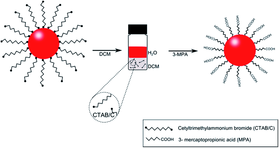 | ||
| Fig. 1 Schematic illustration of gold nanoparticle surface modification via a two-step ligand exchange reaction. | ||
In this work, we introduce a new, solvent extraction based ligand exchange method to remove cetyl trimethylammonium bromide (CTAB) and cetyl trimethylammonium chloride (CTAC) surface ligands from as-synthesised AuNPs with 3-mercaptopropionic acid (3-MPA) ligand molecules. With this method, aggregation was prevented and complete ligand exchange achieved by stepwise replacement of the CTAB/C surfactants with 3-MPA. Our ligand exchange comprises two reactions; the primary reaction involves the extraction of CTAB/C ligands by mixing as-synthesised AuNPs with an immiscible solvent, in this case: dicholoromethane (DCM). In this solvent extraction process, the CTAB/C molecules were gradually removed from the upper phase (as-synthesised AuNPs in water) to the bottom phase (DCM). However, only incomplete CTAB/C removal occurs here to ensure that there is still enough CTAB/C present to maintain colloidal stability. The optimum amount of solvent extraction can be easily determined by dynamic light scattering. The subsequent reaction involves the complete replacement of CTAB/C ligands with 3-MPA via direct chemisorption. This strategy represents a versatile method for AuNPs surface modification whilst preserving their colloidal stability, size and shape of the particles.
To prepare CTAB/C capped AuNPs, a published seeded-growth method22 was modified and resulted in particle diameters of ∼11 nm, which is within the optimal size range for many applications. As-synthesised AuNPs feature CTAB/C surfactants, which afford colloidal stability through steric stabilisation. However, CTAB and CTAC's strong affinity to the AuNPs' surface and its lengthy hydrocarbon chain presents difficulties for further surface modification. The first step of this strategy includes removal of excess CTAB/C without compromising the colloidal stability of the AuNPs. The as-synthesised AuNP solution was washed with DCM, allowing the CTAB/C surfactants to be extracted from the colloid and into DCM. Repeated washing is beneficial for further removal of CTAB/C, however it is necessary to optimise the amount of ‘washing’, as excessive solvent extraction will result in a complete loss of surface ligands and therefore lead to aggregation.
Subsequently, to remove the remaining CTAB/C completely, the DCM-washed AuNPs were exposed to 0.19 M, 3-mercaptopropionic acid (3-MPA, pH 2.53) solution. High affinity of the gold surface to thiol groups provided by the 3-MPA leads to an instantaneous chemisorption reaction, allowing 3-MPA to spontaneously bind onto the AuNPs' surface and replace the remaining CTAB/C. Details of the synthesis procedure can be found in the ESI.†
To confirm the successful and complete ligand exchange reaction, Fourier transform infrared (FTIR) spectroscopy (Fig. 2) was carried out. Functional groups of as-synthesised AuNPs (black), DCM-washed AuNPs (blue) and 3-MPA-capped AuNPs (red) were identified by comparing the spectra. Two prominent peaks at 2858 cm−1 and 2931 cm−1 have been detected and can be assigned as the C–H stretching of the CTAB and CTAC's long aliphatic chain. In addition to these two peaks, the signal at 3400 cm−1 can be assigned to the O–H stretch in H-bonded water. The peak at 1319 cm−1 corresponds to the C–O stretch and the signatures at 1705 cm−1 and 1778 cm−1 to the C![[double bond, length as m-dash]](https://www.rsc.org/images/entities/char_e001.gif) O stretch of a carboxylic acid group. The latter two signals can be found in the as-synthesised particles due to the ascorbic acid used during synthesis. The spectra of DCM-treated AuNPs shows that the intensities of the CTAB/C peaks have been reduced, indicating that CTAB/C was largely stripped from the nanoparticles. Further treatment with 3-MPA resulted in a significant increase in intensity of the carboxylic C–O and CO stretches at 1319 cm−1, 1705 cm−1, 1778 cm−1 and the H-bonded water at 3400 cm−1. This finding indicates the successful bonding of 3-MPA onto the AuNPs' surface, allowing the system to stabilise electrostatically through the 3-MPA's carboxylic acids (–COOH). In addition, the intensity of the characteristic C–H stretching at 2931 cm−1 and 2858 cm−1 ascribed to the CTAB/C surfactant has significantly diminished (practically vanished).
O stretch of a carboxylic acid group. The latter two signals can be found in the as-synthesised particles due to the ascorbic acid used during synthesis. The spectra of DCM-treated AuNPs shows that the intensities of the CTAB/C peaks have been reduced, indicating that CTAB/C was largely stripped from the nanoparticles. Further treatment with 3-MPA resulted in a significant increase in intensity of the carboxylic C–O and CO stretches at 1319 cm−1, 1705 cm−1, 1778 cm−1 and the H-bonded water at 3400 cm−1. This finding indicates the successful bonding of 3-MPA onto the AuNPs' surface, allowing the system to stabilise electrostatically through the 3-MPA's carboxylic acids (–COOH). In addition, the intensity of the characteristic C–H stretching at 2931 cm−1 and 2858 cm−1 ascribed to the CTAB/C surfactant has significantly diminished (practically vanished).
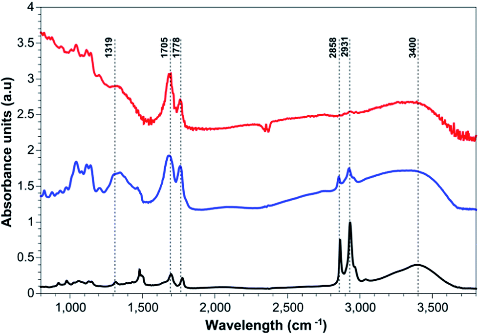 | ||
| Fig. 2 FTIR spectra of as-synthesised AuNPs (black), DCM washed-AuNPs (blue) and 3-MPA capped-AuNPs (red). | ||
Dynamic light scattering (DLS) and transmission electron microscopy (TEM) measurements have been carried out to confirm that our ligand exchange method does not cause significant changes in the particles' shape, size and colloidal stability. DLS measures the hydrodynamic diameters of particles in solution, whilst TEM micrographs depict the actual particle sizes and morphology. Fig. 3 shows the TEM micrographs and Fig. 4 DLS histograms of as-synthesised, DCM-washed and 3-MPA-coated AuNPs. The mean hydrodynamic diameter of AuNPs after DCM and 3-MPA ligand exchange was found to be 11 ± 0.6 nm and 15 ± 1.4 nm respectively whereas the as-synthesised AuNPs was found to be 13.1 ± 1.2 nm. The decrease in hydrodynamic diameter after solvent extraction is most likely due to partial removal of CTAB/C from the ligand shell. The increased hydrodynamic diameter after the ligand exchange is due to an increase in the solvation shell with the new ligand molecules (3-MPA) attached on the surface of the particles (hydrogen bonds allow for more solvent molecules to be “dragged” by the particle than in case of a non-polar shell). Interestingly, the same trend can be observed in the TEM micrographs, where the interparticle distance decreases after solvent extraction and remains very small in case of 3-MPA coated particles, because the solvation shell (water) is being removed in the ultrahigh vacuum environment of the TEM. Finally, we did not observe any formation of aggregates in the intensity distribution histograms of the DLS (data not shown here).
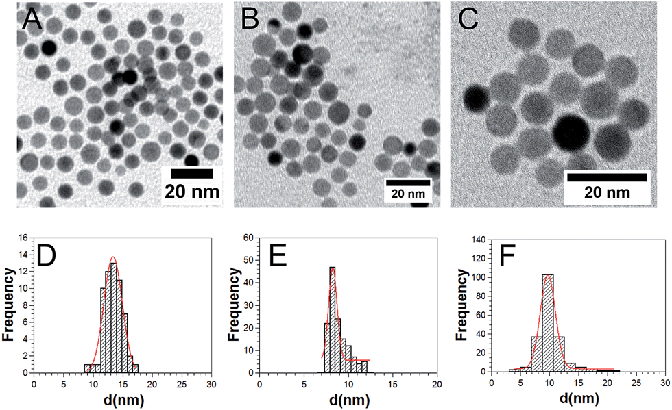 | ||
| Fig. 3 TEM micrographs of as-synthesised AuNPs (a), DCM washed-AuNPs (b) and 3-MPA capped-AuNPs (c); particle size distribution by TEM of as-synthesised AuNPs (d), DCM washed-AuNPs (e) and 3-MPA capped-AuNPs (f). | ||
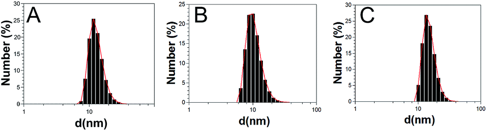 | ||
| Fig. 4 Particle size distribution by DLS of as-synthesised AuNPs (a), DCM washed-AuNPs (b) and 3-MPA capped-AuNPs (c). | ||
Fig. 5 shows the UV-vis spectra of as-synthesised, DCM-washed and 3-MPA capped AuNPs. Generally, spherical AuNP colloids in water display an absorption peak at approximately 520 nm, indicating the surface plasmon resonance (SPR). This SPR band in the as-synthesised AuNPs occurs at 521 nm and remains the same upon washing with DCM (no shift has been observed but a narrowing of the SPR peak). Further modification of DCM washed-AuNPs with 3-MPA results in a slight red-shift of 9 nm of the plasmon band. Since we did not observe any particle aggregation by means of DLS, we suspect that this red shift has been caused by electronic interaction of the 3-MPA ligands with the gold core of the particles.
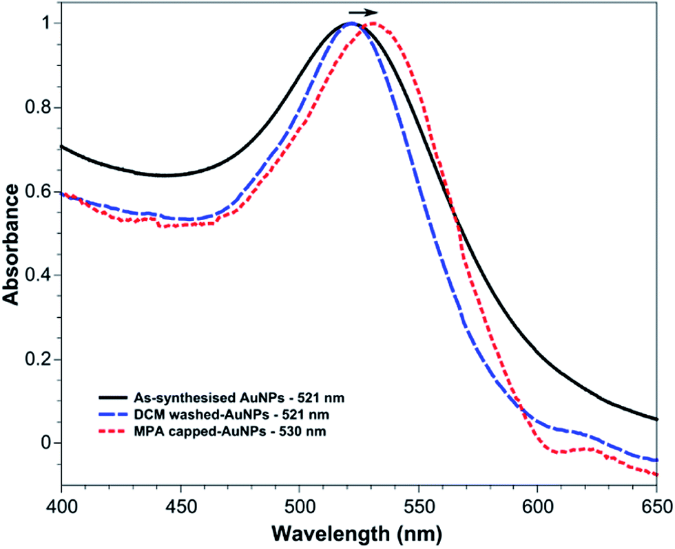 | ||
| Fig. 5 UV-vis spectra of as-synthesised AuNPs (black), DCM washed-AuNPs (blue) and 3-MPA capped-AuNPs (red). | ||
Conclusions
In this work, we present a simple method to perform AuNP surface modification via a two-step ligand exchange reaction without any noticeable aggregation of the particles. The CTAB/C surfactants could be easily removed via DCM solvent extraction whilst maintaining colloidal stability. Subsequent ligand replacement with 3-MPA resulted in a complete exchange of CTAB/C with 3-MPA. The resulting 3-MPA-capped AuNPs feature very small hydrodynamic diameters and exhibit excellent colloidal stability. We expect that this method is generally applicable for the complete ligand exchange on gold nanoparticles.Acknowledgements
This research was in part conducted and funded by the Australian Research Council Centre of Excellence in Convergent Bio-Nano Science and Technology (project number CE140100036).Notes and references
- L. N. Lewis, Chemical catalysis by colloids and clusters, Chem. Rev., 1993, 93, 2693–2730 CrossRef CAS.
- E. Dulkeith, et al., Gold Nanoparticles Quench Fluorescence by Phase Induced Radiative Rate Suppression, Nano Lett., 2005, 5, 585–589 CrossRef CAS PubMed.
- X. Liu, M. Atwater, J. Wang and Q. Huo, Extinction coefficient of gold nanoparticles with different sizes and different capping ligands, Colloids Surf., B, 2007, 58, 3–7 CrossRef CAS PubMed.
- S. Nie and S. R. Emory, Probing Single Molecules and Single Nanoparticles by Surface-Enhanced Raman Scattering, Science, 1997, 275, 1102–1106 CrossRef CAS PubMed.
- L. A. Dick, A. D. McFarland, C. L. Haynes and R. P. Van Duyne, Metal Film over Nanosphere (MFON) Electrodes for Surface-Enhanced Raman Spectroscopy (SERS):
![[thin space (1/6-em)]](https://www.rsc.org/images/entities/char_2009.gif) Improvements in Surface Nanostructure Stability and Suppression of Irreversible Loss, J. Phys. Chem. B, 2001, 106, 853–860 CrossRef.
Improvements in Surface Nanostructure Stability and Suppression of Irreversible Loss, J. Phys. Chem. B, 2001, 106, 853–860 CrossRef. - S. R. Nicewarner-Peña, et al., Submicrometer Metallic Barcodes, Science, 2001, 294, 137–141 CrossRef PubMed.
- S. A. Maier, et al., Plasmonics—A Route to Nanoscale Optical Devices, Adv. Mater., 2001, 13, 1501–1505 CrossRef CAS.
- J. Cheng, Y.-J. Gu, S. H. Cheng and W.-T. Wong, Surface Functionalized Gold Nanoparticles for Drug Delivery, J. Biomed. Nanotechnol., 2013, 9, 1362–1369 CrossRef CAS PubMed.
- G. Paciotti, et al., Colloidal Gold: A Novel Nanoparticle Vector for Tumor Directed Drug Delivery, Drug Delivery, 2004, 11, 169–183 CrossRef CAS PubMed.
- R. E. Messersmith, G. J. Nusz and S. M. Reed, Using the Localized Surface Plasmon Resonance of Gold Nanoparticles To Monitor Lipid Membrane Assembly and Protein Binding, J. Phys. Chem. C, 2013, 117, 26725–26733 CAS.
- M.-C. Daniel and D. Astruc, Gold Nanoparticles:Assembly, Supramolecular Chemistry, Quantum-Size-Related Properties, and Applications toward Biology, Catalysis, and Nanotechnology, Chem. Rev., 2003, 104, 293–346 CrossRef PubMed.
- X. Dai, G. G. Wildgoose, C. Salter, A. Crossley and R. G. Compton, Electroanalysis Using Macro-, Micro-, and Nanochemical Architectures on Electrode Surfaces. Bulk Surface Modification of Glassy Carbon Microspheres with Gold Nanoparticles and Their Electrical Wiring Using Carbon Nanotubes, Anal. Chem., 2006, 78, 6102–6108 CrossRef CAS PubMed.
- A. J. Viudez, R. Madueño, T. Pineda and M. Blázquez, Stabilization of Gold Nanoparticles by 6-Mercaptopurine Monolayers. Effects of the Solvent Properties, J. Phys. Chem. B, 2006, 110, 17840–17847 CrossRef CAS PubMed.
- C. Batchelor-McAuley, G. G. Wildgoose and R. G. Compton, The contrasting behaviour of polycrystalline bulk gold and gold nanoparticle modified electrodes towards the underpotential deposition of thallium, New J. Chem., 2008, 32, 941–946 RSC.
- J. R. G. Navarro, et al., Probing the interactions between disulfide-based ligands and gold nanoparticles using a functionalised fluorescent perylene-monoimide dye, Photochem. Photobiol. Sci., 2010, 9, 1042–1054 CAS.
- R. A. Sperling and W. J. Parak, Surface Modification, Functionalisation and Bioconjugation of Colloidal Inorganic Nanoparticles, Philos. Trans. R. Soc., A, 2010, 1333–1383 CrossRef CAS PubMed.
- Z. Li, R. Jin, C. A. Mirkin and R. L. Letsinger, Multiple thiol-anchor capped DNA–gold nanoparticle conjugates, Nucleic Acids Res., 2002, 30, 1558–1562 CrossRef CAS PubMed.
- J. Turkevich, P. C. Stevenson and J. Hillier, A Study of The Nucleation and Growth Processes in the Synthesis of Colloidal Gold, Discuss. Faraday Soc., 1951, 55–75 RSC.
- B. C. Rostro-Kohanloo, et al., The stabilization and targeting of surfactant-synthesized gold nanorods, Nanotechnology, 2009, 20, 434005 CrossRef PubMed.
- H. Liao and J. H. Hafner, Gold Nanorod Bioconjugates, Chem. Mater., 2005, 17, 4636–4641 CrossRef CAS.
- X. Chen, J. Lawrence, S. Parelkar and T. Emrick, Novel zwitterionic copolymers with dihydrolipoic acid: synthesis and preparation of nonfouling nanorods, Macromolecules, 2012, 46, 119–127 CrossRef.
- Y. Zheng, et al., Seed-Mediated Synthesis of Single-Crystal Gold Nanospheres with Controlled Diameters in the Range 5–30 nm and their Self-Assembly upon Dilution, Chem.–Asian J., 2013, 8, 792–799 CrossRef CAS PubMed.
Footnote |
| † Electronic supplementary information (ESI) available. See DOI: 10.1039/c4ra05035e |
| This journal is © The Royal Society of Chemistry 2014 |