DOI:
10.1039/C4RA04839C
(Paper)
RSC Adv., 2014,
4, 38169-38181
Shape-selective formation of MnWO4 nanomaterials on a DNA scaffold: magnetic, catalytic and supercapacitor studies†
Received
22nd May 2014
, Accepted 14th August 2014
First published on 15th August 2014
Abstract
A new route for the aqueous phase synthesis of single crystalline, shape-selective, magnetic MnWO4 nanomaterials on a DNA scaffold has been reported. The synthesis was done by the reaction of MnCl2·4H2O with Na2WO4 in DNA within five minutes of microwave heating. The process exclusively generates wire-like, flake-like and rice-like morphology just by tuning the DNA to Mn(II) salt and WO42− ion concentration and changing other reaction parameters. The field-cooled (FC) and zero-field-cooled (ZFC) magnetization study reveals that the flake-like structure shows the highest magnetization at 5 K compared to that of the wire-like and rice-like structures. The potential of the shape-selective MnWO4 nanomaterials has been tested in two different applications, firstly in a catalysis study for the decomposition of toxic KMnO4 and secondly in electrochemical supercapacitor applications. It was found that the MnWO4 nanomaterials showed different specific capacitance (SC) values for the various shapes and the order of the SC values is: wire-like > flake-like > rice-like. The highest SC of 34 F g−1 was observed for MnWO4 having wire-like shape. The yields of the products with uniform shapes have been found to be significantly high and the synthesized materials are stable for more than six months under ambient conditions. The present work will find a new platform for the generation of other mixed oxides using bio-molecules as scaffolds at low temperature and in short time scales. Moreover, the synthesized material might be useful for other potential applications in the fields of catalysis, sensors, energy storage materials and so on.
Introduction
Over the last few years considerable interest has been focused on the fabrication of nano/microstructured materials, mainly due to their unique electronic, optical, magnetic and catalytic properties and various applications.1–4 Among the different materials, the shape-selective synthesis of mixed metal oxide nanoparticles (NPs) has become one of the essential topics in nanoscience as their unique properties are generally not available in single metal oxide NPs. The shape-selective formation of mixed metal oxide NPs has gained much attention in the fields of electronics, optics, sensors, biology, magnetism, catalysis and luminescence studies.5,6 Moreover, they show fascinating color change in the UV-Vis region due to their close lying conduction and valence bands in which electrons can move freely. The specific shape of nanomaterials dictates their physicochemical properties because the number of active atom located at the edges/corners and exposed facets of the crystal.7 The fascinating properties of mixed metal oxide NPs depend not only on the complex morphology but also on the crystallinity of the particles in microarchitectures.8 As an examples, individual spherical superparticles of CdSe/CdS showed strong linear polarized emission but single disk spherical superlattices nano rods does not show any significant polarization.9 Therefore, shape-selective formation of single-crystalline mixed metal oxide NPs are highly desirable for tailoring their unique properties and for high performance in many potential applications.
Among the different mixed metal oxide studied so far, manganese tungstate (MnWO4) is one of the most promising material which shows high sensitivity in humidity change and have unique magnetic property.10,11 MnWO4 has a Wolframite type of structure having space group P2/C (no. 13) in which each Mn and W atom have an approximately octahedral coordination surrounded by six near oxygen atom sites. These oxygen atoms form a distorted hexagonal close pack structure with Mn and W atoms which occupying one-fourth of the octahedral interstices. The electrical conductivity of MnWO4 is highly sensitive for humidity sensor which is important in different areas due to their bulk electrical conductivity and relatively low melting point.12 Moreover, MnWO4 shows attraction in magnetism due to its anti-ferromagnetic spin structure.13
In recent years, different types of bio-molecules like amino acids, proteins, peptides have been used for the growth and stabilization of nanomaterials. Among the different bio-molecules, the deoxyribonucleic acid (DNA) is arguably the most remarkable molecular guide and used as a structural material rather than as a carrier of biological information because of the many possibilities it offers for creating defined nanostructures. DNA was a natural occurring material which can be readily obtained in high quantities from different sources and it is relatively less expensive compared to several other bio-molecules. In native state, DNA has the diameter ∼ 2–3 nm and has large persistence length in highly suitable for confining materials growth in two dimensions. Moreover, the length of DNA can be readily controlled with high degree of precision by changing the number of base pairs (bp) within the molecules. In DNA, the poly-anionic phosphate backbone is able to form electrostatic interaction with positively charged species while the aromatic base molecules are capable of coordinating with metal cations. All these interactions in DNA are important for the fabrication of DNA templated inorganic nanostructures.14–18 Au,14 Ag,15 Pd,16 Pt17or CdS4) nanowires has been fabricated using DNA as template. Although there is still remain a significant challenge for the synthesis of mixed metal oxide nanomaterials using DNA as scaffold.
In literature, there are several reports for the synthesis of MnWO4 nanomaterials including hydrothermal method,19 solvothermal method,20 sol–gel method,21 low-temperature molten salt method,22 surfactant assisted complexation–precipitation method,23 etc. Yu et al. fabricated 3D urchin-like MnWO4 microsphere using CTAB as a surfactant.24 Trong-On Do and co-workers synthesized different shaped MnWO4 nanomaterials in bi-functional amino acid media at high temperature (∼180 °C).25 Zhou and co-workers studied the variation of Mn/W molar ratio on the phase composition of MnWO4.26 Zheng et al. studied the synthesis of MnWO4 nanofiber using a surfactant assisted complexation–precipitation method.23 Priyadharsini et al. studied the polyaniline/MnWO4 composite for electrochemical applications.27 3D flower-like MnWO4 nanostructure prepared by Xing et al. recently.28 Although, most of the above reports usually required hazardous metal–organic precursor or relatively high annealing temperature and the synthesized product results a mixture of various shapes. There are few more limitations such as prolonged reaction time, poor reaction efficiency, addition of surfactant or even hazardous organic solvent as well as the necessity of pre-treating substrate. Recently, microwave assisted hydrothermal method has been used to overcome all these drawbacks which has several advantages like heating throughout the media, rapid and concentrated heating, fast reaction, high yield, excellent reproducibility, narrow particles size distribution, high purity and high-efficient energy transformation.29,30 Kundu and co-workers synthesized earlier different metal and metal oxide NPs using microwave heating method.31–34 It is important to note that most of the earlier reports of MnWO4 synthesis takes place at high temperature (>100 °C) using hydrothermal methods. To the best of our knowledge, there is no report for the ‘one-step’ fast synthesis of DNA based shape-selective MnWO4 nanomaterials using five minutes of microwave heating at low temperature (∼80 °C) in aqueous solution.
In the present study, we report a new approach for the aqueous phase synthesis of single crystalline shape-selective, magnetic MnWO4 nanomaterials within five minutes of microwave heating on DNA scaffold. The synthesis was done by the reaction of MnCl2·4H2O with Na2WO4 in presence of DNA and the process exclusively generates rice-like, flake-like and wire-like morphologies just by tuning the DNA to Mn(II) salt and WO42− ion concentration and changing the other reaction parameters. The FC and ZFC magnetization study reveals that the flake-like structure shows the highest magnetization at 5 K as compared to that of the wire-like and rice-like structure. The synthesized MnWO4 nanomaterials have been used in two different applications. Firstly, the catalytic decomposition of toxic KMnO4 has been studied using MnWO4 as catalyst. Secondly, the shape-selective MnWO4 nanomaterials have been utilized for the electrochemical supercapacitor applications and the orders of specific capacitance (SC) values are follows as: wire-like > flake-like > rice-like. The highest SC of 34 F g−1 was observed for MnWO4 having wire-like shapes. The synthesis process is very simple, fast, reproducible and cost effective. To the best of our knowledge, the low temperature and fast synthesis of DNA based shape-selective MnWO4 nanomaterials and their potential applications in catalysis and supercapacitor study is not exposed before.
Experimental section
Reagents and instruments
All the chemicals used in this present work were analytical reagent (AR) grade. De-oxyribonucleic acid (double-stranded) from herring testes with an average molecular weight ∼48,502 bp (base pair) having GC content ∼41.2% was purchased from Sigma-Aldrich and the calculated Tm (melting temperature) value was ∼98 °C. The manganese(II) chloride, tetra hydrate [MnCl2·4H2O] and sodium tungstate (Na2WO4) were obtained from Sigma-Aldrich and used as received. Polystyrene sulphonate (PSS) and adenosine tri-phosphate (ATP) was purchased from Sigma-Aldrich and used as received. Potassium permanganate (KMnO4) was also purchased from Sigma-Aldrich. Sodium sulfate (Na2SO4), poly vinyledene fluoride (PVDF), n-methyl pyrrolidinone (NMP) and Super P were procured from Alfa Aesar, India and were used without any further purification. Absolute ethanol was purchased from local Balaji Scientific Company, Karaikudi, Tamilnadu. De-ionized (DI) water was used for the entire synthesis and catalysis work. The shape-selective MnWO4 nanomaterials on DNA scaffold were characterized using several spectroscopic techniques like UV-Vis, TEM, EDS, XRD, LASER Raman, PL, thermal and FT-IR analyses and the specification details of all these characterizations were given in the ESI.†
Shape-selective synthesis of MnWO4 nanomaterials on DNA using microwave heating
MnWO4 nanomaterials with different morphologies have been synthesized by tuning the concentration of DNA to Mn(II) salt and WO42− ions concentration and also by changing other reaction parameters. For a typical synthesis, 4 mL of stock DNA solution (concentration ∼1.23 × 10⁻¹ M) was mixed with 64 mL of aqueous Mn(II) salt solution (0.1 M) and stirred well for through mixing. Then 64 mL of Na2WO4 solution (0.1 M) was added drop wise while continuous stirring and the resultant clear solution turns light white and finally white colloidal solution. With increasing time, the white color solution turned to light yellowish. The solution mixture was stirred for another 10 minutes for homogeneous mixing and then placed inside the microwave oven for total 300 seconds irradiations. It is important to note that during microwave heating we have given a pause of one minute in every 10 seconds heating to avoid the over-heating and to cool the reaction vessel. The reaction temperature of the solution mixture while heating via microwave was measured and it was maximum ∼80 °C after 5 minutes of heating. The solution become cooled, washed and centrifuged 3–4 times by water, ethanol and annealed at 80 °C for 1 hour in hot air oven. The solution was extensively contains MnWO4 nanomaterials having rice-like morphology. The other sets are prepared by changing the concentration of DNA and other reaction parameters. The details about the final concentration of all the reagents, reaction time, size and shape of the particles were summarized in Table 1. The formations of shape-selective MnWO4 nanomaterial were schematically shown in Scheme 1.
Table 1 The detailed final concentrations of all the reaction parameters, time of microwave irradiation, particles size, shape, etc. are summarized in table
| Set no. |
Final conc. of DNA (M) |
Final conc. of Mn(II) solution (M) |
Final conc. of Na2WO4 solution (M) |
Microwave irradiation time (min) |
Average particles size (nm) |
Shape of the particles |
| 1 |
2.7 × 10⁻² |
3.9 × 10⁻² |
3.9 × 10⁻² |
5 |
D = 75 ± 15 nm, L ≥ 700 nm |
Wire-like |
| 2 |
1.3 × 10⁻² |
4.4 × 10⁻² |
4.4 × 10⁻² |
5 |
L = 90–180 nm |
Flake like |
| 3 |
3.7 × 10⁻³ |
4.8 × 10⁻² |
4.84 × 10⁻² |
5 |
L = 90 ± 10 nm, D = 25 ± 5 nm |
Rice-like |
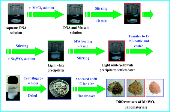 |
| | Scheme 1 The overall preparation process for the shape-selective synthesis of DNA–MnWO4 nanomaterials using microwave heating. | |
Preparation of sample for magnetic study
The dc magnetization measurements were carried out, using a vibrating sample magnetometer (Cryogenic, UK make), as a function of temperature and magnetic field. For the temperature dependent zero-field-cooled (ZFC) magnetization measurements, the sample was first cooled from room temperature down to 4 K in zero external magnetic field. After applying the magnetic field at 4 K, the magnetization was measured in the warming cycle with the field on. Whereas, for the temperature dependent field-cooled (FC) magnetization measurements, the sample was cooled in the same field (measuring field in the ZFC case) down to 4 K, and then the FC magnetization was measured in the warming cycle keeping the field on. Magnetic field dependence of magnetization was measured at 5 and 50 K after cooling the sample to the measurement temperature (5 and 50 K) under zero field.
Preparation of electrode for supercapacitor studies
The working electrodes were fabricated on a high purity stainless steel (SS) foil as a current collector. 75 wt% of MnWO4, 20 wt% of super P and 5 wt% of PVDF were ground in a mortar and few drops of NMP were added to form slurry. The slurry was coated on to the pre-treated SS which was already polished with successive grades of emery, cleaned with detergent, washed with doubly distilled water, rinsed with acetone and dried in air. The coated SS electrodes were dried at 100 °C under vacuum for 12 h. The electrochemical studies were carried out using a three electrode configuration with MnWO4 coated SS as the working electrode, Pt as the counter electrode and saturated calomel electrode (SCE) as the reference electrode. All potential values were reported against SCE. Discharge specific capacitance (SC) of MnWO4 was calculated using the formula,where, I is current in amps, t is time in seconds of discharge, m is mass in grams of the active material and ΔE is operating potential window in volt of charge or discharge.
Preparation of sample for other various characterizations
The shape-selective MnWO4 nanomaterials on DNA scaffold were characterized using UV-Vis, TEM, photoluminescence (PL), EDS, XRD, Raman, FT-IR, and thermal analysis studies. The DNA–MnWO4 nanostructures solution after successive centrifugation and annealing was used for the various spectroscopic measurements. For UV-Vis study a liquid solution was made by sonicating the solid MnWO4 powder for 30 min with DI water. The samples for TEM was prepared by placing a drop of the corresponding MnWO4 NPs solution onto a carbon coated Cu grid followed by slow evaporation of solvent at ambient conditions. The dispersed aqueous MnWO4 nanostructures were used for the PL measurement. For EDS, XRD, Laser Raman, and FT-IR analysis, glass slides were used as substrates for thin film preparation. The slides were cleaned thoroughly in acetone and sonicated for about 30 minutes. The cleaned substrates were covered with the MnWO4 nanostructures solution and then dried in air. After the first layer was deposited, subsequent layers were deposited by repeatedly adding more MnWO4 nanostructures solution and drying. Final samples were obtained after 5–6 times depositions and then analyzed using the above techniques. For TGA/DTA analysis, the as synthesized MnWO4 nano powders were used for the measurement. For catalysis study, MnWO4 nano powder was directly used and those details were given in the catalysis section.
Results and discussion
UV-Vis spectroscopic analysis
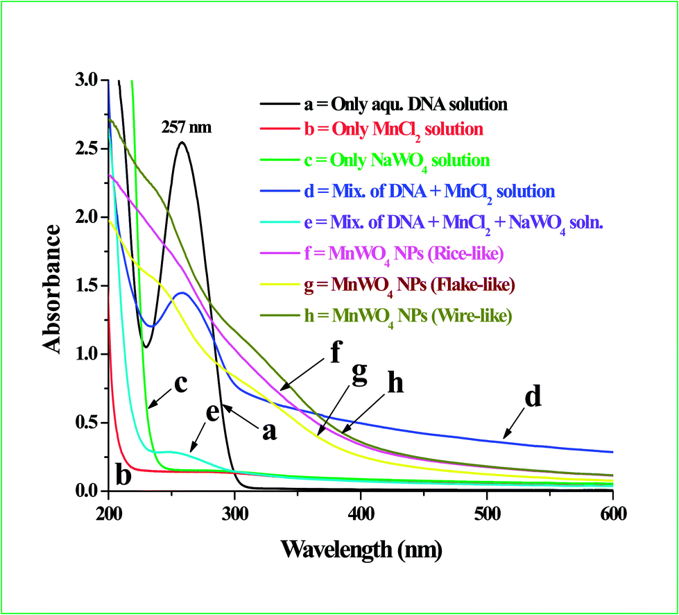
Shape-selective MnWO4 nanomaterials have been synthesized on DNA scaffold by the reaction of Mn(II) salt with Na2WO4 under microwave heating for five minutes. Fig. 1 shows the UV-Vis absorption spectra of the solution mixture at the different stages of the synthesis process. Curve a, Fig. 1 shows the absorption band of aqueous DNA solution which has a λmax at 257 nm due to presence of aromatic base molecules on its skeleton.3 An aqueous Mn(II) salt solution has no specific absorption band but a small hump at 281 nm due to ligand to metal charge transfer (LMCT) spectra (curve b, Fig. 1) having similarities with aqueous HAuCl4 solutions.35 Curve c, Fig. 1 shows the absorption band of aqueous Na2WO4 solution which has no specific absorption band in the UV-Vis region. Curve d, Fig. 1 shows the absorption band of a mixture of aqueous DNA and aqueous Mn(II) salt solution which shows a band at 260 nm. There is a shifting of 3 nm was observed compared to original DNA absorption band (at 257 nm) and the curve become broaden indicates the interaction or absorption of Mn2+ ions on DNA. Curve e, Fig. 1 shows the absorption band of a mixture of DNA, Mn(II) salt and Na2WO4 (before microwave heating) which shows a broad absorption band in the region 240 to 300 nm due to interaction of DNA with Mn(II) ions and WO42− ions. The absorption value and the peak maxima also reduced significantly. Now, after continuous stirring and microwave heating of the solution mixture containing DNA, Mn(II) salt and Na2WO4, the light white color solution turns to light yellowish which indicates the formation of MnWO4 nanomaterials in the solution. The resultant solution color changes little bit depending upon the concentration of the reagents and due to formation of different morphologies of MnWO4. Curve f, g and h shows the absorption spectra of the MnWO4 nanostructures having rice-like, flake-like and wire-like morphology (as confirmed from TEM analysis as described below). The absorption band of the three different morphologies shows a small hump in the UV region at 260 nm (for rice-like), 243 nm (for flake-like) and 237 nm (for wire-like) respectively with a broad band at 280 to 390 nm in the UV-visible region. These small humps appeared for three different morphology is due to charge-transfer transition between oxygen 2p orbital and empty d orbital of the central W ion.36 The broadening of absorption band for three different shapes might be due to result of the smaller particle size of the product owing to quantum confinement effect.37 It is commonly accepted that the optical properties of metal tungstate materials are strongly dependent on their morphology and crystallinity.26,38 To understand how the overall reaction taking place, the UV-Vis spectra of the aqueous dispersion is taken (as seen in Fig. 1) and in addition, the diffuse reflectance spectra of solid MnWO4 for three different morphology is also done and placed in the ESI section as Fig. S-1.† With the change of morphologies of the particles, the electronic structure can also change which leads to the change in optical properties. So the concentration of the DNA and the other reagents show great effect on the final morphology of MnWO4.
 |
| | Fig. 1 The UV-Vis absorption spectrum of the different solution mixtures for the preparation of DNA–MnWO4 nanomaterials using microwave heating. (a) Show the absorption band of only aqueous DNA solution; (b) shows the absorption band of aqueous of MnCl2·4H2O salt solution; (c) shows the absorption band of only Na2WO4 solution; (d) shows the absorption band of the mixture of DNA with Mn(II) salt solution; (e) shows the absorption band of the mixture of DNA, Mn(II) salt and Na2WO4 solution; (f)–(h) shows the absorption spectra for MnWO4 nanostructures having rice-like, flake-like and wire-like morphology. | |
Transmission electron microscopy (TEM) analysis
Fig. 2 shows the transmission electron microscopy (TEM) images of the shape-selective MnWO4 nanomaterials at various magnifications. Fig. 2A and B shows the low and high magnified TEM image of the MnWO4 nanomaterials having rice-like morphology. From the image the average length and diameter of the rice-like morphology was ∼90 ± 10 nm and 25 ± 5 nm respectively. The inset of Fig. 2A shows the corresponding much higher magnified image where the lattice planes are visible. The inset of Fig. 2B shows the corresponding selected area electron diffraction (SAED) pattern which says the particles are single crystalline in nature. Fig. 2C and D shows the low and high magnified TEM image of the MnWO4 nanomaterials having flake-like structure. The average size of the nano flakes varies 150 ± 50 nm ranges. Fig. 2E and F shows the TEM image at low and high magnification of the wire-like MnWO4 nanomaterials. The average diameter of the MnWO4 particles in the wire-like structure are 75 ± 15 nm and the average length of the wires were ≥700 nm. The inset of Fig. 2D and F shows the corresponding SAED pattern which says that the particles are crystalline in nature. A very low and a very high magnified image of wire-like structure are provided in ESI section as Fig. S-2, A and B† respectively. Moreover, it was also observed that in case of rice-like and wire-like morphology, almost all the particles formed uniform shapes whereas the flake-like morphology generates >85% particles with uniform morphology. From Table 1 we can see that at low DNA concentration only rice-like structure are formed while at high DNA concentration wire-like structure are formed and at intermediate DNA concentration flake-like structure are formed. We believed that the flake-like structures are formed via the specific growth of rice-like structure so a certain % of rice-like structure is always present with the flake-like structure as observed from TEM images in Fig. 2. So from the above TEM analysis, it is confirmed that we were able to synthesized uniform MnWO4 nanomaterials of various shapes and sizes by altering the reaction parameters and the resulted particles shows crystalline in nature.
 |
| | Fig. 2 The transmission electron microscopy (TEM) images of shape-selective DNA–MnWO4 nanomaterials. (A) and (B) Show the low and magnified TEM images of MnWO4 nanomaterials having rice-like morphology. The inset of (A) shows the corresponding higher magnified image and inset of (B) show the selected area electron diffraction (SAED) pattern. (C) and (D) Show the low and high magnified TEM images of MnWO4 nanomaterials having flake-like morphology. The inset of (D) show the SAED pattern. (E) and (F) Shows the low and high magnified TEM image of MnWO4 nanomaterials having wire-like shapes. The inset of (F) shows the corresponding SAED pattern. | |
Energy dispersive X-ray spectroscopy (EDS) and X-ray diffraction (XRD) analysis
Fig. S-3 (ESI†) showed the energy dispersive X-ray spectroscopic (EDS) analysis of the rice-like MnWO4 nanomaterials on DNA scaffold and the spectrum was consists of the different peaks corresponding to Mn, O, W, P and Na. The more details about EDS analysis is given in SI section. The X-ray diffraction (XRD) pattern of the shape-selective MnWO4 nanomaterials are shown in Fig. 3 and the peaks observed in the range 10–60° confirmed the formation of MnWO4 nanomaterials having monoclinic structure. All the reflection peaks were observed at various 2θ values from the lattice plane of (010), (100), (011), (110), (−111), (111), (002), (200), (−102), (121), (112), (−211), (211), (022), (220), (130), (−202), (202) and (103) planes of monoclinic MnWO4 which are consistent with reported values for Joint Committee on powder Diffraction Standard (JCPDS), File no.: 80-0134.23–25,28 The broadening and intense diffraction peak indicates that small size and high crystallinity of the particles. From the XRD analysis, no other phases either single manganese oxide or tungstate oxide is detected in the MnWO4 nanomaterials indicates that a pure monoclinic phase is obtained by the synthetic approach. Curves A, B and C in Fig. 3 are indicates the XRD pattern for rice-like, flake-like and wire-like morphology respectively. All the different morphologies generate the similar diffraction pattern although the relative intensity of various diffraction peaks are varies. In all the three different morphology, the intensity of (100) diffraction peak is narrow and sharp which indicates a growth orientation along the (100) planes in the synthesized nanomaterials.
 |
| | Fig. 3 The X-ray diffraction pattern of three different shaped DNA–MnWO4 nanomaterials. Curve A, B and C are the diffraction pattern for rice-like, flake-like and wire-like morphology respectively. | |
Photoluminescence (PL) and LASER Raman study
Photoluminescence (PL) study was carried out to investigate the optical properties of the synthesized shape-selective MnWO4 nanomaterials on DNA. Fig. 4 shows the room temperature PL emission spectra of the as-synthesized MnWO4 nanomaterials with different morphology using an excitation wavelength 374 nm. Curve a, b and c in Fig. 4 shows the PL spectra for rice-like, flake-like and wire-like morphologies respectively. The PL emission spectra of the three different morphology shows almost similar spectral feature and the same blue emission band located at 390, 429, 462, 486, 523, 568 and 599 nm respectively under 374 nm excitation. These emission bands corresponding to the transition from 1A1 ground state to the high vibration level of 1T2 and from the low vibration level of 1T2 to the 1A1 ground state within the tetragonal WO42− group.39 This is attributed due to the excitation of WO42− group from charge transfer p–d transition. Due to this, the observed band positions were not strongly depends on the morphologies of the MnWO4 lattices. A similar type of PL emission band was observed by Trong-On Do and co-workers during the synthesis of 3D MnWO4 microspheres.25 Fig. S-4 (ESI†) showed the LASER Raman spectra of the shape-selective MnWO4 nanomaterials on DNA. The Raman spectra found similarities with previous reports.40–42 The detailed analysis and discussion were elaborated in the ESI section.†
 |
| | Fig. 4 The room temperature PL emission spectra of the MnWO4 nanomaterials using an excitation wavelength 374 nm. Curve a, b and c shows the spectra for rice-like, flake-like and wire-like morphology respectively. | |
Thermal analysis of the MnWO4 nanomaterials on DNA scaffold
Thermogravimetric analysis (TGA) and differential thermal analysis (DTA) was conducted to check the thermal stability and crystalline condition of the as prepared MnWO4 nanomaterials on DNA. The MnWO4 sample was heated from RT to 1000 °C with a successive increment of 10 °C per minute in air. Fig. 5, curve A and B shows the TGA and DTA curve of MnWO4 nanomaterials on DNA. Here, we conducted the analysis taking rice-like MnWO4 nanomaterials although other morphology also expected to generate similar types of spectra feature as all are prepared by using the DNA molecule. From curve A, the first weight loss takes place from 135 °C to 204 °C due to removal of moisture or adsorbed water molecule in the sample. The second weight loss took place after 200 °C to 380 °C probably due to removal of excess DNA molecule from the MnWO4 particles. Above 400 °C to 1000 °C the curve is flat which signify that there is no more weight loss in that range. We have started initially with 3.26 mg of sample and after analysis up to 1000 °C the amount of sample remaining is 3.09 mg. So the % of weight loss is 5.2%. The DTA analysis in curve B shows two endothermic peaks located at 186 °C and 333 °C.
 |
| | Fig. 5 Curve A and B shows the TGA and DTA curve of MnWO4 nanomaterials on DNA. | |
Fourier transforms infrared (FT-IR) spectroscopic analysis
Fig. 6, curve A and B shows the Fourier-transform infrared (FT-IR) spectra of only DNA and DNA–MnWO4 nanomaterials in the wavenumber range 4000–400 cm−1. As an example, we used the rice-like MnWO4 nanomaterials for the FT-IR studies although other morphologies of MnWO4 were tested and observed similar types of spectral pattern as expected. A detailed comparison study between both the spectrums supported the presence of DNA in the MnWO4 particles surface and clarified the nature of interaction takes place between them. In DNA, the organic base molecules and phosphate back bone is the main candidate for stabilization of the MnWO4 nanomaterials. In curve A, three peaks at the lower wave number region at 550 cm−1, 804 cm−1 and 961 cm−1 is observed for DNA which is due to stretching vibration of phosphate group in DNA molecule. Those specific peaks are shifted to the higher wave number region in case of DNA bound MnWO4 (curve B). The symmetric and asymmetric PO2− vibration arising from the DNA–phosphate backbone is reported at 1097 cm−1 and 1246 cm−1 respectively. The DNA backbone P–O/C–O stretching vibration is reported at 1071 cm−1 in literature which is shifted to 1084 cm−1 in case of DNA bound MnWO4 sample signifying the interaction of MnWO4 with DNA back bone. Three intense sharp peak at 581 cm−1, 698 cm−1 and 823 cm−1 was observed for DNA bound MnWO4 samples (curve B) is due to absorption of W–O bonds. The W–O bond absorption generally took place in the lower wave number region as observed from earlier reports.43 The region 1300–1800 cm−1 is assigned to stretching vibration of carbonyl group. Two peaks at 1635 cm−1 and 1681 cm−1 for pure DNA was due to carbonyl stretching is shifted to 1636 cm−1 and 1689 cm−1 in case of DNA bound MnWO4 samples. The stretching vibration appeared above 2850 cm−1 to 3600 cm−1 is due to hydroxyl group. The OH stretching peak at 3435 cm−1 for DNA is shifted to 3407 cm−1 for DNA–MnWO4. The C–H stretching vibration for DNA appeared at 2921 cm−1 which shifted to 2915 cm−1 for DNA bound MnWO4 samples. These spectral changes and specific shifting indicates that there is specific interaction taking place between the poly-anionic backbone of the DNA and MnWO4 nanomaterials. The interaction between the MnWO4 and DNA bases were also evident from several changes in the IR bands attributed to nucleo base vibration as observed in the lower wavenumber region below 1800 cm−1. The phosphodiester bond in DNA helps for binding the MnWO4 with DNA. Moreover, due to binding of MnWO4 with the PO2− group, the polarity of the nearby carbonyl group increases that generates a change in absorption intensity. Table T-1 (ESI†) shows the details about the different FT-IR bands reported for DNA, the experimentally bands we observed and the corresponding bond assignments. From Table T-1,† it is clear that all the peaks observed in DNA bound MnWO4 sample is either matching with pure DNA or slightly shifted which indicates the interaction and stabilization action of DNA on the surface of MnWO4 particles.
 |
| | Fig. 6 The Fourier-transform infrared (FT-IR) spectra of bare DNA (curve A) and DNA–MnWO4 (curve B) nanomaterials in the wave number range 400–4000 cm−1. | |
Magnetic study of the shape-selective MnWO4 nanomaterials
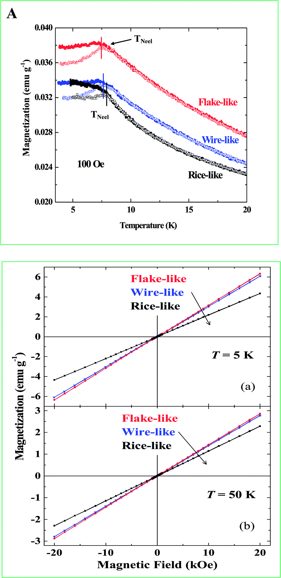
Fig. 7-A shows the temperature dependence of ZFC and FC magnetization under 100 Oe field for the shape-selective MnWO4 nanomaterials having rice-like, flake-like and wire-like structures. A clear peak has been observed at lower temperatures, around 7.6 K for the flake-like structure. However, a marginal shift of the peak temperature (Tpeak) towards a higher temperature was found for the rice-like structures. An anti-ferromagneic type ordering is, therefore, evident for all the three structures at T ≤ Tpeak, the Neel temperature TNeel. The TNeel decreased with decreasing the size/dimension of the samples i.e., from rice-like to wire-like to flake-like structures as labeled in the Fig. 7-A. As the samples were changed from rice-like to wire-like and then to flake-like structures, the fraction of atoms at surface increased and the exchange interaction became weaker due to the lower coordination number. This results into the reduction of Neel temperature. A branching in the ZFC and FC magnetization (at T ≤ Tpeak) has been observed for all the three structures. The magnetic field dependence of magnetization at 5 K (a) and 50 K (b) for all the three samples is shown in Fig. 7-B. The magnetization shows linear field dependence at both 5 and 50 K. The linear field dependence of magnetization at 50 K arises due to the paramagnetic nature of the samples at this temperature and due to creation of more uncompensated spins. Whereas, the observed linear field dependence of magnetization at 5 K (without any hysteresis) is a typical signature of the anti-ferromagnetic nature of these samples at T ≤ Tpeak. An increasing trend of magnetization is found in both Fig. 7-A and B with decreasing the size/dimension of the samples i.e., from rice-like, to wire-like to flake-like structures. As size/dimension of the samples is reduced, the magnitude of magnetization increases due to the creation of more uncompensated spins. The surface to volume ratio increases from rice-like to wire-like to flake-like structures, as a result of that the flake-like structure shows the highest magnetization at 5 K as compared to that of the wire-like and rice-like structures. We do not observe any ferromagnetic-like ordering for any one of these nanostructures.
 |
| | Fig. 7 (A) Temperature dependence of ZFC (solid symbols) and FC (open symbols) magnetization under 100 Oe for rice-like, flake-like and wire-like MnWO4 nanomaterials. (B) Magnetic field dependence of magnetization at 5 K (a) and 50 K (b) for the three samples. | |
Mechanisms for the formation of shape-selective MnWO4 nanomaterials on DNA scaffold
The shape-selective MnWO4 nanomaterials are formed by the reaction of Mn(II) salt with Na2WO4 in the presence of DNA under five minutes of microwave heating. We conducted few control experiments to check the effect of different reaction parameters on the formation of shape-selective MnWO4 nanomaterials on DNA. The specific morphology of rice-like, flake-like and wire-like shapes was formed at a particular concentration those are given in Table 1. The details about control experiments and the related TEM images (Fig. S-5, ESI†) at various reaction conditions were given in ESI.† The presence of DNA is extremely important for the formation, growth and stabilization of the MnWO4 NPs. As discussed earlier, in the absence of DNA, the MnWO4 particles formed aggregated structure due to absence of any specific stabilizer. From the FT-IR analysis (Fig. 6), it was observed that the base molecules and the phosphate backbone on DNA are mainly binds with the MnWO4 particles and control their growth in different shapes. Scheme 2 shows the formation mechanism and binding of Mn(II) ions to DNA for the generation of different morphology. Initially, after addition of Mn(II) salt on DNA solution, the Mn2+ ions are binds on the DNA chain due to electrostatic interaction of the oppositely charged ions. This interaction is also observed from the shifting of the UV-Vis spectral bands after addition of Mn(II) salt with DNA as compared with pure DNA as seen in Fig. 1. Now once the WO42− ions are added, it reacts with the Mn2+ ions and generates the MnWO4 nuclei. These MnWO4 nuclei grow on the adjacent area and finally generate a notable registry of the particles along the DNA chains or they aggregated depending upon the availability of the precursor molecules in the reaction medium. At low DNA concentration, the MnWO4 particles are homogeneously grow along the DNA chain and generate the rice-like morphology. At moderate DNA concentration the MnWO4 particles are formed flake-like morphology whereas at high DNA concentration they formed wire-like morphology as seen in Scheme 2 and from the TEM images in Fig. 2. In all the cases, we heated the solution mixture for five minutes using microwave. We assume that the phosphate backbone and the amino moiety present in the aromatic base molecules are the favorable target to feed the NPs growth. We assume that a certain % of the Mn2+ ions are directly bonded to the different functionalities present of DNA once we add Mn(II) salt with DNA. So during the process of direct bonding, that sites where the Mn2+ ions are absorbed can acts as a crystallization cores and the nanocrystals are located at that specific sites. So a certain % of the Mn2+ ions are attached to the DNA and the rest % of Mn2+ ions are used for crystal growth where the WO42− ions are mixed. So, after the addition of Na2WO4 solution, the WO42− ions reacts with the Mn2+ ions to generate the MnWO4 nuclei. Then the growth of the particles takes place in multistep on the DNA which is already present in the solution. Initially, the smaller size MnWO4 nuclei crystallize in the preferential direction, aggregate and stabilize on DNA and formed the different morphologies. During the microwave heating, small MnWO4 nuclei grow to big cluster and that cluster grow on the adjacent areas at certain regime and results in a notable registry of the particles either along the DNA chain (wire-like morphology) or they aggregated further or break down to generate the flake-like or the rice-like morphology. The specific role of DNA for the shape-selective synthesis of MnWO4 nanomaterials was further confirmed by other control experiments by doing the same reaction taking polyanions instead of DNA. We used negatively charged polymer, poly(styrene) sulphonate (PSS) or negatively charged bio-molecule, adenosine tri-phosphate (ATP) separately instead of DNA but the reaction generates only spherical and agglomerated MnWO4 particles (TEM not shown here) keeping same reaction conditions due to their different structural morphology. This indirectly proves that DNA has some specific role for the growth and formation of shape-selective MnWO4 particles which was not observed by using other polyanions. So we believe that the phosphate group and the base molecules not only help for the stabilizations after formation of the particles in the aqueous solution but also play an important role during the growth process of different morphology. Although, it is important to mentioned that the formation of detailed mechanism is premature at this stage but it is reasonable to assume that the phosphate backbone and the base molecule present on DNA act as a key role and thanks to their strong absorption in the UV and further investigation will be necessary to prove it. The shape-selective DNA–MnWO4 nanomaterials have been utilized for real applications in two different fields. Firstly, we checked the catalytic decomposition of toxic KMnO4 solution using MnWO4 as catalyst and then we used the shape-selective MnWO4 nanomaterials as a potential material for electrochemical supercapacitor applications as discussed below.
 |
| | Scheme 2 The formation mechanism and binding of Mn(II) ions to DNA for the generation of shape-selective MnWO4 nanomaterials. | |
Catalytic decomposition of toxic KMnO4 using shape-selective MnWO4 nanomaterials as catalyst
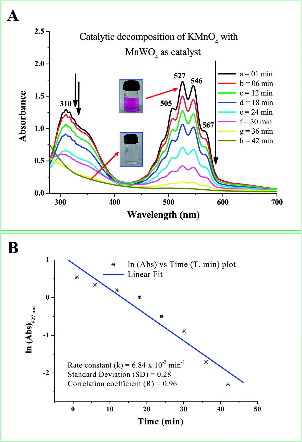
The potentiality of the shape-selective MnWO4 nanomaterials was tested in detail for the catalytic decomposition of aqueous KMnO4 solution. For the catalysis study, the synthesized MnWO4 nanomaterials were centrifuged and washed several times to remove any excess DNA or any other impurity from the nanomaterials and finally dried to get solid mass and directly used for the catalysis study. KMnO4 is an inorganic material and its salt contains K+ and MnO4− ions. It is used widely as strong oxidizing agent and after dissolve in water it gives intense pink color or purple color solution. It is reported in literature that KMnO4 is potentially toxic to human and other organisms including several fish species but not included in regulatory categories.44,45 For a typical catalysis reaction, 80 mL of 0.5 × 10⁻³ M aqueous KMnO4 solution was taken and mixed with 100 mg of dry MnWO4 powder and stirred vigorously for 10 minutes. The entire catalysis work was done with rice-like morphology although other two morphologies were also examined as preliminary experiment. After stirring the solution became colloidal and kept for settling at room temperature under dark. Now from the resultant colloidal solution, 3 mL solution was taken out and centrifuged for two minutes at 10![[thin space (1/6-em)]](https://www.rsc.org/images/entities/char_2009.gif) 000 rpm. After centrifugation, the MnWO4 particles are settled down at the bottom of the centrifuged tube and upper clear solutions was separated out and measure the absorbance value using UV-Vis spectrophotometer. Each five minute interval we did the same procedure and measure the absorbance value of the upper solution after centrifugation. It is important to be note that we did two control experiments where we check the KMnO4 catalysis study just using native DNA solution and keeping the KMnO4 solution under room light. There is no color bleaching or decomposition of KMnO4 was observed even keeping the solution for 2–3 days. We measured the pH value of the KMnO4 solution, MnWO4 solution (after mixing with water), and KMnO4 + MnWO4 powder after mixing in aqueous solution. The pH values are 7.02 for only KMnO4 solution, 6.12 for only MnWO4 solution and 5.51 for the mixture of KMnO4 + MnWO4 solution. It is observed that just mixing MnWO4 powder with KMnO4 solution, the catalysis reaction does not take place significantly without any stirring which signifies that for fast decomposition the catalyst particles needs in contact with the KMnO4 solution. The progress of the catalysis reaction or the decomposition of KMnO4 was monitor using UV-Vis spectrophotometer by measuring the changes in the specified absorption maxima. Fig. 8-A shows the UV-Vis spectral evolution for the catalytic decomposition of KMnO4 solution appeared at 310 and 350 nm (shoulder peak) and a set of other intense absorption peaks at 505 nm, 527 nm, 546 nm and 567 nm respectively. All these intense bands appeared for KMnO4 solution is arises due to metal to ligand charge transfer (MLCT) transition and here the transition takes place from oxygen to MnVII.46 The progress of the reaction was monitored via the steady decrease of absorbance value of all the specified peaks at a certain time interval. From Fig. 8-A, it is seen that the overall catalysis reaction was completed within 42 min and the pink color KMnO4 solution become colorless. The whole catalysis reaction was studied at room temperature at 31 °C temperature. The inset of Fig. 8-A shows two camera images, one is pink color KMnO4 solution and the other is the final clear solution after complete decomposition of KMnO4. Fig. 8-B shows the ln(Abs)527 nm vs. time (T, min) plot for the decomposition of KMnO4 with DNA–MnWO4 nanomaterials. From the linear plot we calculate the rate constant value which is 6.84 × 10⁻² min−1. The correlation coefficient and standard deviation value for the measurement was 0.96 and 0.28 respectively. As discussed earlier, the detailed catalysis work was conducted taking rice-like morphology although the effects of other particles were also tested as preliminary study. The preliminary experiment speaks that the catalysis decomposition of KMnO4 takes place in all the cases when we change the particles morphology from rice-like to flake-like to wire-like shapes. The BET surface area analysis for rice-like, flake-like and wire-like MnWO4 nanomaterials was conducted and the surface area values were 98.62, 146.13 and 49.95 m2 g−1 respectively. In all the cases the KMnO4 decomposition completed below 50 minutes time. It is well known that in heterogeneous catalysis reaction, the catalytic rate mostly depends upon the availability of the active surface area of the catalyst, number of catalyst particles, reaction temperature, etc. A detailed analysis taking different morphology on the catalysis reaction considering BET surface area will be conducted in near future to investigate the morphology effect on the catalysis reaction.
000 rpm. After centrifugation, the MnWO4 particles are settled down at the bottom of the centrifuged tube and upper clear solutions was separated out and measure the absorbance value using UV-Vis spectrophotometer. Each five minute interval we did the same procedure and measure the absorbance value of the upper solution after centrifugation. It is important to be note that we did two control experiments where we check the KMnO4 catalysis study just using native DNA solution and keeping the KMnO4 solution under room light. There is no color bleaching or decomposition of KMnO4 was observed even keeping the solution for 2–3 days. We measured the pH value of the KMnO4 solution, MnWO4 solution (after mixing with water), and KMnO4 + MnWO4 powder after mixing in aqueous solution. The pH values are 7.02 for only KMnO4 solution, 6.12 for only MnWO4 solution and 5.51 for the mixture of KMnO4 + MnWO4 solution. It is observed that just mixing MnWO4 powder with KMnO4 solution, the catalysis reaction does not take place significantly without any stirring which signifies that for fast decomposition the catalyst particles needs in contact with the KMnO4 solution. The progress of the catalysis reaction or the decomposition of KMnO4 was monitor using UV-Vis spectrophotometer by measuring the changes in the specified absorption maxima. Fig. 8-A shows the UV-Vis spectral evolution for the catalytic decomposition of KMnO4 solution appeared at 310 and 350 nm (shoulder peak) and a set of other intense absorption peaks at 505 nm, 527 nm, 546 nm and 567 nm respectively. All these intense bands appeared for KMnO4 solution is arises due to metal to ligand charge transfer (MLCT) transition and here the transition takes place from oxygen to MnVII.46 The progress of the reaction was monitored via the steady decrease of absorbance value of all the specified peaks at a certain time interval. From Fig. 8-A, it is seen that the overall catalysis reaction was completed within 42 min and the pink color KMnO4 solution become colorless. The whole catalysis reaction was studied at room temperature at 31 °C temperature. The inset of Fig. 8-A shows two camera images, one is pink color KMnO4 solution and the other is the final clear solution after complete decomposition of KMnO4. Fig. 8-B shows the ln(Abs)527 nm vs. time (T, min) plot for the decomposition of KMnO4 with DNA–MnWO4 nanomaterials. From the linear plot we calculate the rate constant value which is 6.84 × 10⁻² min−1. The correlation coefficient and standard deviation value for the measurement was 0.96 and 0.28 respectively. As discussed earlier, the detailed catalysis work was conducted taking rice-like morphology although the effects of other particles were also tested as preliminary study. The preliminary experiment speaks that the catalysis decomposition of KMnO4 takes place in all the cases when we change the particles morphology from rice-like to flake-like to wire-like shapes. The BET surface area analysis for rice-like, flake-like and wire-like MnWO4 nanomaterials was conducted and the surface area values were 98.62, 146.13 and 49.95 m2 g−1 respectively. In all the cases the KMnO4 decomposition completed below 50 minutes time. It is well known that in heterogeneous catalysis reaction, the catalytic rate mostly depends upon the availability of the active surface area of the catalyst, number of catalyst particles, reaction temperature, etc. A detailed analysis taking different morphology on the catalysis reaction considering BET surface area will be conducted in near future to investigate the morphology effect on the catalysis reaction.
 |
| | Fig. 8 (A) Shows the UV-Vis spectra evolutions for the catalytic decomposition of KMnO4 using DNA–MnWO4 nanomaterials; (B) shows the ln(Abs)527 nm vs. time (T, min) plot for the successive decomposition of KMnO4 solution. | |
Supercapacitor application of the DNA–MnWO4 nanomaterials
With an aim to assess the electrochemical supercapacitor behavior of the DNA stabilized MnWO4 electrodes, the cyclic voltammogram (CV) and charge–discharge cycling experiments were performed in aqueous electrolyte. It is important to be mentioned here that the detailed study was done with the MnWO4 nanomaterials having wire-like morphology although other two morphologies (rice-like and flake-like) were also tested as preliminary experiment to compare the shape-effect on the SC behavior of MnWO4 nanomaterials. Cyclic voltammetry is well recognized as ideal experimental technique to study the capacitive behavior of any material. An ideal capacitor47 shows large magnitude of current and rectangular type of voltammogram, which is symmetric in anodic and cathodic directions. Fig. 9-A depicts the typical voltammogram recorded for the as synthesized MnWO4 in 0.1 M Na2SO4 electrolyte at different scan rates within a potential range of 0 to 1.0 V. It is clear from the figure that there are no sharp redox peaks in the potential range recorded. The rectangular voltammograms exhibit mirror-image of the cathodic and anodic parts, thus suggesting an ideal capacitive behavior and high reversibility nature of MnWO4 electrodes. Variation of voltammetric cathodic current density at 0.5 V with sweep rate is shown in Fig. 9-B. There is a linear increase in current density with an increase in sweep rate confirming the capacitive behavior of the MnWO4 nanomaterials. The rectangular shape of the voltammetry remains same even at high scan rate indicating the excellent capacitive behavior of MnWO4. The electrodes made with MnWO4 were subjected to galvanostatic charge–discharge cycling between 0 and 1.0 V in 0.1 M Na2SO4 at a current density 0.5 mA cm−2. The typical charge–discharge plot in Fig. 9-C exhibit a linear variation of the potential during both charging and discharging a process, which again becomes another criterion for capacitance behavior of a material in addition to exhibiting rectangular voltammograms. It is observed that MnWO4 prepared in the present work exhibit excellent capacitor properties. The same charging and discharging time implies high reversibility and also the columbic efficiency of charge–discharge cycling close to unity. Specific capacitance (SC) values are calculated from galvanostatic charge–discharge cycling using eqn (1) (given in the Experimental section). The value of SC obtained from the Fig. 9-A is about 34 F g−1. Charge–discharge cycling was recorded at different current density at the range of 0.5–20 mA cm−2. Variation of SC of the electrodes with current density is shown in Fig. 9-D, showing a decrease in SC with increase in current density. This is might be due to the decrease in efficiency for the utilization of active material at high current density. Moreover, it is well known that at low current density the ions from the electrolyte can utilize all the available sites in the inner and outer surface of the electrode material that leads to the higher capacitance and vice versa.48,49 The value of SC obtained for the different shaped MnWO4 nanomaterials are as follows: rice-like ∼14 F g−1, flake-like 33 F g−1 and wire-like 34 F g−1. The observed SC values for flake-like and wire-like morphologies are found higher compared to previous report on MnWO4 nanorods.50 The high SC value observed for wire-like and flake-like shapes compared to rice-like shape probably due to morphology which leading to effective penetration of the electrolyte in the entire matrix. Although, it is important to note that the insight reason for the different SC values for shape-selective MnWO4 nanomaterials is premature at this stage but it is reasonable to assume that the materials utilization is the prime factor and further investigation will be necessary to prove it.
 |
| | Fig. 9 (A) A typical cyclic voltammogram (CV) for the synthesized MnWO4 nanomaterials (wire-like shape) in 0.1 M Na2SO4 electrolyte at different scan rates range from 0 to 1.0 V. (B) The variation of SC of the electrodes with current density at 0.5 V. (C) The typical charge–discharge plot exhibit a linear variation of the potential during both charging and discharging process, (D) variation of SC of the electrodes with current density, showing a decrease in SC with increase in current density. | |
Conclusion
In summary, shape-selective MnWO4 nanomaterials were synthesized for the first time at low temperature using DNA as scaffold in aqueous solution. The MnWO4 nanomaterials were synthesized by the reaction of Mn(II) salt with Na2WO4 in presence of DNA under microwave heating. Different shapes like rice-like, flake-like and wire-like were formed by tuning the different reaction parameters. The formation and probable mechanism of shape-selective MnWO4 synthesis was elaborated. We suppose that the phosphate and possibly the amino moiety of DNA binds with the nanomaterials to feed the NPs growth, formation of MnWO4 cluster and finally grow the cluster in adjacent areas to generate the specific morphologies. The FC and ZFC magnetization study reveals that the flake-like structure shows the highest magnetization at 5 K as compared to that of the wire-like and rice-like structures. The shape-selective DNA–MnWO4 nanomaterials have been tested for two different potential applications; such as the catalytic decomposition of toxic KMnO4 and as an active electrode material for electrochemical supercapacitor studies. It was observed that MnWO4 nanomaterials having wire-like shape acts as the best electrode material and the specific capacitance (SC) values for different shapes are follows as: wire-like > flake-like > rice-like. The highest SC value of 34 F g−1 was observed for MnWO4 having wire-like shapes. In futuristic, the overall result will give researchers a new platform for the generation of other mixed oxides using DNA as scaffold in low temperature and in a short time scale and could be used for many other potential applications in the field of catalysis, sensor, and energy storage devices and so on.
Acknowledgements
S. Kundu wishes to acknowledge Dr Vijayamohanan K. Pillai, Director, and Dr M. Jayachandran, HOD, ECMS division, CSIR-CECRI for their continuous support and encouragement. U. Nithiyanantham wishes to thank CSIR-CECRI for research internship fellowship and S. R. Ede wish to acknowledge council for scientific and industrial research (CSIR) for JRF fellowship. The institute start up funding (Project number IHP 0067, DU No 5, old number OLP-0067), support from the Central Instrumental Facility (CIF) and help from Mr A. Rathishkumar (TEM in-charge, CIF), CSIR-CECRI, Karaikudi are greatly appreciated.
References
- H. Yang, N. Coombs and G. A. Ozin, Nature, 1997, 386, 692 CrossRef CAS.
- V. K. Pustovalov and V. A. Babenko, Laser Phys. Lett., 2004, 1, 516 CrossRef CAS PubMed.
- S. Kundu and H. Liang, Langmuir, 2008, 24, 9668 CrossRef CAS PubMed.
- S. Kundu and H. Liang, Adv. Mater., 2008, 20, 826 CrossRef CAS PubMed.
- Z. Nie, A. Petukhoval and E. Kumacheval, Nat. Nanotechnol., 2010, 5, 15 CrossRef CAS PubMed.
- A. V. Blaaderen, Nature, 2009, 461, 892 CrossRef PubMed.
- H. G. Yang, C. H. Sun, S. Z. Qiao, J. Zou, G. Liu, S. C. Smith, H. M. Cheng and G. Q. Lu, Nature, 2008, 453, 638 CrossRef CAS PubMed.
- W. Fan, M. A. Synder, S. Kumar, P. S. Lee, W. C. Yoo, A. V. Mccormick, R. L. Penn, A. Stein and M. Tsapatsis, Nat. Mater., 2008, 7, 984 CrossRef CAS PubMed.
- J. Zhuang, A. D. Shaller, J. Lynch, H. Wu, O. Chen, A. D. Q. Li, Y. C. Cao and J. J. Am, J. Am. Chem. Soc., 2009, 131, 6084 CrossRef CAS PubMed.
- K. Taniguchi, N. Abe, T. Takenobu, Y. Iwasa and T. Arima, Phys. Rev. Lett., 2006, 97, 097203 CrossRef CAS.
- A. H. Arkenbout, T. T. M. Palstra, T. Siegrist and T. Kimura, Phys. Rev. B: Condens. Matter Mater. Phys., 2006, 74, 184431 CrossRef.
- R. Bharati, R. A. Singh and B. M. Wankel, J. Phys. Chem. Solids, 1982, 43, 641 CrossRef CAS.
- H. Ehrenberg, H. Weitzel, H. Fuess and B. Hennion, J. Phys.: Condens. Matter, 1999, 11, 2649 CrossRef CAS.
- C. A. Mirkin, Inorg. Chem., 2000, 39, 2258 CrossRef CAS.
- L. Berti, A. Alessandrini and P. Facci, J. Am. Chem. Soc., 2005, 127, 11216 CrossRef CAS PubMed.
- C. F. Monson and A. T. Woolley, Nano Lett., 2003, 3, 359 CrossRef CAS.
- K. Keren, M. Krueger, R. Gilad, G. Ben-Yoseph, U. Sivan and E. Braun, Science, 2002, 297, 72 CrossRef CAS PubMed.
- D. Sarker and M. J. Mandal, J. Phys. Chem. C, 2012, 116, 3227 Search PubMed.
- W. Q. Wu, W. H. Qin, Y. M. He, Y. Wu and T. H. Wu, J. Exp. Nanosci., 2012, 7, 390 CrossRef CAS.
- S. J. Chen, X. T. Chen, Z. L. Xue, J. H. Zhou, J. Li, J. M. Hong and X. Z. You, J. Mater. Chem., 2003, 13, 1132 RSC.
- W. Qu, W. Wlodarski and J. U. Meyer, Sens. Actuators, B, 2000, 64, 76 CrossRef CAS.
- Z. W. Song, X. Y. Li and J. S. Shi, Adv. Mater. Res., 2011, 284–286, 722 CrossRef CAS.
- S. J. Lei, K. Tang, Z. Fang, Y. H. Huang and H. G. Zheng, Nanotechnology, 2005, 16, 2407 CrossRef CAS PubMed.
- Y.-X. Zhou, Q. Zhang, J.-Y. Gong and S.-H. Yu, J. Phys. Chem. C, 2008, 112, 13383 CAS.
- T.-D. Nguyen, D. Mrabet, T.-T.-D. Vu, C.-T. Dinh and T.-O. Do, CrystEngComm, 2011, 13, 1450 RSC.
- S. Zhou, J. Huang, T. Zhang, H. Ouyang, A. Li and Z. Zhang, Ceram. Int., 2013, 39, 5195 Search PubMed.
- S. Saranya, R. K. Selvan and N. Priyadharsini, Appl. Surf. Sci., 2012, 258, 4881 CrossRef CAS PubMed.
- Y. Xing, S. Song, J. Feng, Y. Lei, M. Li and H. Zhang, Solid State Sci., 2008, 10, 1299 CrossRef CAS PubMed.
- S. Komarneni, R. Roy and Q. H. Li, Mater. Res. Bull., 1992, 27, 1393 CrossRef CAS.
- X. C. Xu, Y. Bao, C. S. Song, W. S. Yang, J. Liu and L. W. Lin, Microporous Mesoporous Mater., 2004, 75, 173 CrossRef CAS PubMed.
- S. Kundu, V. Maheshwari, S. Niu and R. F. Saraf, Nanotechnology, 2008, 19, 65604 CrossRef PubMed.
- S. Kundu, L. Peng and H. Liang, Inorg. Chem., 2008, 47, 6344 CrossRef CAS PubMed.
- S. Kundu, K. Wang and H. Liang, J. Phys. Chem. C, 2009, 113, 134 CAS.
- S. Kundu, K. Wang and H. Liang, J. Phys. Chem. C, 2009, 113, 5157 CAS.
- W. R. Mason III and H. B. Gray, Inorg. Chem., 1968, 7, 55 CrossRef.
- Q. Zhang, X. Chen, Y. Zhou, G. Zhang and S. H. Yu, J. Phys. Chem. C, 2007, 111, 3927 CAS.
- M. A. El-Sayed, Acc. Chem. Res., 2004, 37, 326 CrossRef CAS PubMed.
- L. Xu, J. Shen, C. Lu, Y. Chen and W. Hou, Cryst. Growth Des., 2009, 9, 3129 CAS.
- F. Zhang, Y. Yiu, M. C. Aronson and S. S. Wong, J. Phys. Chem. C, 2008, 112, 14816 CAS.
- J. T. Kloprogge, M. L. Weier, L. V. Duong and R. L. Frost, Mater. Chem. Phys., 2004, 88, 438 CrossRef CAS PubMed.
- M. Daturi, G. Busca, M. M. Borel, A. Leclaire and P. Piaggio, J. Phys. Chem. B, 1997, 101, 4358 CrossRef CAS.
- S. Thongtem, S. Wannapop, A. Phuruangrat and T. Thongtem, Mater. Lett., 2009, 63, 833 CrossRef CAS PubMed.
- M. Regragui, M. Addou, A. Outzourhi, J. C. Bernede, E. E. Idrissi, E. Benseddik and A. Kachouane, Thin Solid Films, 2000, 358, 40 CrossRef CAS.
- E. R. Cruz and C. T. Tamse, Bull. Environ. Contam. Toxicol., 1989, 43, 785 CrossRef CAS.
- L. L. Marking and T. D. Bills, Trans. Am. Fish. Soc., 1975, 104, 579 CrossRef CAS.
- A. Dandapat and G. De, J. Mater. Chem., 2010, 20, 3890 RSC.
- B. E. Conway, Electrochemical Supercapacitors, Kluwer Academic publishers/Plenum Press, New York, 1999 Search PubMed.
- Y. Munaiah, B. G. S. Raj, T. Prem Kumar and P. Ragupathy, J. Mater. Chem. A, 2013, 1, 4300 CAS.
- P. Ragupathy, H. N. Vasana and N. Munichandraiah, J. Electrochem. Soc., 2008, 155, A34 CrossRef CAS PubMed.
- S. Saranya, S. T. Senthilkumar, K. V. Sankar and R. K. Selvan, J. Electroceram., 2012, 28, 220 CrossRef CAS.
Footnote |
| † Electronic supplementary information (ESI) available: The detailed discussions and the related Fig. (S-1 to S-5) for UV-Vis study, TEM analysis, energy dispersive X-ray spectroscopy (EDS), LASER Raman and study with other reaction parameters (TEM images) are given. The datas related to FT-IR analysis are provided in Table T-1. See DOI: 10.1039/c4ra04839c |
|
| This journal is © The Royal Society of Chemistry 2014 |
Click here to see how this site uses Cookies. View our privacy policy here.