Md Palashuddin Ska and
Arun Chattopadhyay*ab
aDepartment of Chemistry, Indian Institute of Technology Guwahati, Guwahati-39, Assam, India. E-mail: arun@iitg.ernet.in; Fax: +91 361 258 2349; Tel: +91 361 258 2304/28
bCentre for Nanotechnology, Indian Institute of Technology Guwahati, Guwahati-39, Assam, India
First published on 18th June 2014
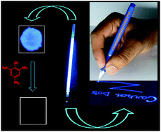
We report a new and facile method for the synthesis of highly fluorescent carbon dots (Cdots) using a commercially available induction coil heater. An aqueous solution of citric acid and a diamine compound was heated at 100 °C for 12–15 min, upon which the Cdots were produced. The Cdots, with an average size of less than 5 nm (produced when ethylenediamine was used), emitted a blue light with a high quantum yield when excited under UV light. The quantum yield was dependent on the nature of diamine and was as high as 73.5% for ethylenediamine. The as-prepared Cdots could be easily converted into a gel by mixing with chitosan biopolymer. The gel could be used for filling up the refill of a ball-point pen and can be used for UV-active marking, for sensing of explosive compounds (such as picric acid and 2,4-dinitrophenol) with high efficiency and for other fluorescence based applications. The use of a commercial induction coil heater, scalability and high chance of commercial viability make the method particularly appealing.
Recent reports suggest that some nanomaterials are in use by the greater population as food items. For example, the caramels of bread, biscuits, corn flakes and jaggery have been shown to contain fluorescent carbon nanoparticles (CNPs).1 Interestingly, because these food items have been consumed by human civilization for centuries and there is no known adverse effects on human health following prolonged consumption, these materials can be considered safe for large-scale production and usage. However, the synthesis of CNPs at a large scale is still challenging and requires investigation, especially particles with important optical and chemical properties.
In addition, recent results suggest that CNPs, in general, could compete with some aspects of the optical properties of quantum dots (Qdots). Importantly, the toxicity of a majority of Qdots makes a strong case for further investigating their properties. This is especially true with respect to their photoluminescence properties, which are generally observed to be wavelength-tunable. The key challenge, though, is to produce large quantities of CNPs with high quantum yield (QY) for photoluminescence. This is where newer methods can bring novelty and ease of production of CNPs.
Although carbohydrate and other polymer based molecules are known to produce smaller fluorescent CNPs, more popularly known as carbon dots (Cdots), there are recent reports suggesting that smaller molecules, such as citric acid, could be potential candidates for producing Cdots with high fluorescent quantum yield (QY).2–5,23,24 In addition, the introduction of microwave-based methods for the synthesis of Cdots has brought new results in terms of the tuning of the wavelength of emission and also photoluminescence QY. However, the production of Cdots using these methods generally requires either harsh reaction conditions or time-consuming processes, which does not necessarily yield products with high QY. Further, the lack of opportunity for the use of solvents other than water, loss of materials during heating and energy efficiency become major hurdles for commercial scale production. Here, we report a new method for the synthesis of highly fluorescent Cdots from a mixture of citric acid and nitrogen containing a passivating agent such as ethylenediamine using a commercially available induction coil heater. Induction heating is faster, safer and energy efficient in comparison to other commonly used heating techniques for chemical reactions.6 The current method produced small Cdots with high fluorescent QY. In addition, the Cdots were coupled with a chitosan polymer to produce a gel that could be used for UV-active marking. Further, the fluorescence property of the Cdots was used for sensing nitroaromatic phenol explosive with high sensitivity.
For large scale synthesis, a mixture of 5.040 g (24 mM) citric acid, 1.064 mL (16 mM) ethylenediamine and 50 mL water was heated in the frying pan. The rest of the procedure was the same as above; however, the heating time was set at 15 min. The weight percentage of the yield was ca. 86% with respect to citric acid, as calculated prior to purification. Further, the weight percentage of the yield was ca. 70% with respect to citric acid and ethylenediamine, as calculated prior to purification.
We also performed the reaction at 130 °C (800 W) with 3 mM (630 mg) citric acid and 2 mM (134 μL) ethylenediamine. However, the heating time was set at 8–9 min. It was observed that if heating was performed at 130 °C or higher, a relatively lesser time was required for the formation of Cdots than that at 100 °C.
For sensing applications, Cdot ink (gel) was prepared by dissolving 100 mg chitosan in 20 mL water with further addition of 100 μL acetic acid and the concentration of Cdot was maintained at 0.01 mg mL−1. Only 4 μL of Cdot ink was used for making a spot on non-fluorescent paper and then the spot was dried for sensing or other applications.
The UV-vis spectrum of the dispersion consisted of two peaks—one at 242 nm, while the other appeared at 350 nm (Fig. 1d). The former is assigned to the π–π* transition, which could be due to a product with c–c π bonds.4 On the other hand, the peak at 350 nm is typical of Cdots, which upon excitation with light resulted in a strong blue emission at 454 nm (Fig. 1e). The emission is assigned to the surface states of Cdots.3,7 However, the presence of defect states cannot be ruled out.8 Because Cdots are known to have a wavelength tuneable emission, this was also pursued. It was observed that although the emission did have wavelength tunability, the extent was weak. For example, when the dispersion was excited by light of wavelength ranging from 325 to 400 nm, there was a considerable change in the intensity of the emission and no discernible change in the emission wavelength. On the other hand, excitation beyond 400 nm resulted in a weaker emission with no significant change in the wavelength of the emission. This could be due to the production of uniformed sized Cdots.9 Further, QY for the sample prepared from different passivating agents (in addition to citric acid as the precursor) indicated that the presence of ethylenediamine provided Cdots with QY as high as 73.5%. It may be mentioned here that Cdots with QY as high as 83% have been reported earlier.24 The large QY resulted in an easy observation of fluorescence from Cdots using an ordinary chromatographic UV-lamp at concentrations as low as 1.0 μg mL−1. All the other compounds resulted in products with lower or much lower QY. The details are available in Table S2 in the ESI.†
The time-resolved fluorescence decay profile of the Cdots dispersed in water could be fitted with a bi-exponential function. The average life time was calculated to be 13.3 ns (Fig. S2a, ESI†). Interestingly, when the Cdots were dispersed in dimethylformamide (DMF), the average life time was 9.3 ns, which was calculated using a bi-exponential function (Fig. S14 and Table S3, ESI†), and indicating possible solvent dependence. Further, photoluminescence studies indicated that Cdots were stable and remained highly luminescent in the presence of a high salt (KCl) concentration (Fig. S3, ESI†), rendering them useful for versatile applications, especially in the presence of high ionic strength. The studies on the pH-dependent fluorescence of Cdots revealed that the emission intensity was nearly constant in the pH range of 4 to 11 (Fig. S3, ESI†), confirming their potential utility in a wide pH range. Photostability experiments revealed that Cdots were more stable (with a decay rate of 0.022%) in comparison to a popular dye such as rhodamine 6G (having a decay rate of 0.45%, Fig. S2b, ESI†).
Transmission electron microscopy (TEM) revealed that the as-synthesized Cdots were nearly spherical with an average particle size of 4.6 ± 1.2 nm (Fig. 2a). The size distribution of the Cdots, as calculated from the TEM images, is shown in Fig. S5, ESI.† The amorphous nature of the particles was established by the lack of observation of clear lattice fringes in the high resolution TEM as well as any clear pattern in the selected area electron diffraction (Fig. S4, ESI†). The X-ray diffraction (XRD) pattern consisted of a broad peak at 2θ = 17°, suggesting the formation of an amorphous carbon structure (Fig. S6, ESI†). Elemental analysis confirmed that the Cdots contained 39.76% C, 6.31% H, 10.03% N and 43.90% O (as calculated). Further, 13C NMR spectroscopy (Fig. S9, ESI†) revealed that sp2 as well as sp3 carbons were present in the Cdots. The sp2 carbons were carbonyl in nature (with peaks in the range of 170–185 ppm), which could be due to the presence of carboxylic or amide groups. The peaks around 30–45 ppm are attributed to the presence of an aliphatic sp3 carbon atom. Another peak at 72 ppm suggested that alcoholic or C–O–C aliphatic sp3 carbon atoms were present. In addition, the Fourier transform infrared (FTIR) spectrum confirmed the presence of –OH, CO, C–N–C and –NH groups in the Cdots (Fig. S7, ESI†). The appearance of a peak at 1709 cm−1 instead of the peak of the carbonyl group of citric acid at 1731 cm−1 indicated the formation of an amide bond (Fig. S8, ESI†).10 Further, the nature of the bonding of carbon in the Cdots was confirmed by X-ray photoelectron spectroscopy (XPS) (Fig. 3).3,4,11 The XPS result of C1s is attributed to the presence of C–C or C
C (284.8 eV), oxygenated carbon or C–N (286.2 eV) and amide carbonyl group or C
O (287.9 eV) functional groups. The N1s spectrum of Cdots contained two peaks at 399.9 eV and 401.5 eV, which correspond to C–N–C and N–H, respectively. Further, the O1s spectrum showed two peaks at 531.6 eV (C
O) and 533.2 eV (C–O). Interestingly, electron paramagnetic resonance (EPR) spectroscopy studies (Fig. 2b) indicated the presence of one or more singly occupied electron orbitals in the ground state (with g = 2.012451), suggesting the electron donation and acceptance properties of Cdots.12–14
![]() | ||
Fig. 3 X-ray photoelectron spectrum of Cdots (a) and expanded views corresponding to (b) C1s, (c) N1s and (d) O1s peaks. |
It is important to mention here that the Cdots were not only dispersible in water but also in organic solvents such as in N,N-dimethylformamide and dimethylsulfoxide, and weakly in methanol. Further, it was observed that product formation could easily be scaled to a higher amount primarily by higher amounts of starting materials. The advantages of high photoluminescence QY, scalability and ease of dispersion in various solvents facilitate the better application prospects of Cdots. A versatile application could be in the form of UV-active ink, which would otherwise be undetectable under visible light. For this purpose, a gel was prepared using the as-synthesized Cdots and chitosan biopolymer. The gel ‘ink’ was then used to fill up an ordinary ball-point pen refill, the images of which—under visible and UV light—are shown in Fig. 4a–c. The refill could be used to sketch images of different sizes and shapes on non-fluorescent currency notes. A few of these images, which were recorded in the presence of UV light, are shown in Fig. 4b. Importantly, the ink effectively adhered to the paper such that even after washing with water and detergent solution, the imprint was still clearly discernible (Fig. 4c). Further, as shown in Fig. 4f, the gel could be converted into a highly fluorescent film. An important application of the water dispersible Cdots could be to follow the water flow in fields. For this, gram was incubated in the dispersion for 24 h, following which it was exposed to UV-light for imaging. As is clear from Fig. 4d, the Cdots percolated into the gram with high efficiency and gave rise to a clear fluorescent image in comparison to the control sample kept in water.
Finally, the highly fluorescent gel was also used for the efficient detection of nitroaromatic phenols such as picric acid and 2, 4-dinitrophenol, the primary constituents of explosives. The Stern–Volmer binding constants (Ksv) for picric acid and 2,4-dinitrophenol were found to be 3.72 × 10⁴ M−1 and 3.23 × 10⁴ M−1, respectively, which are comparable with the previously reported values.14–22 The results are shown in Fig. S11 and S12, ESI.† The detection limit of picric acid was observed to be 75.6 ppb. We were also interested in finding the nature of the quenching of the luminescence of Cdots by picric acid. This was pursued by time-resolved photoluminescence (TRPL) studies revealing that there was no change in the average life time of Cdots in DMF upon the addition of picric acid (conc. range from 0 to 100 μM), suggesting static quenching (Fig. S14 & Table S3, ESI†). Additionally, we observed that the quenching efficiencies of picric acid and 2,4-dinitrophenol were much more than benzoquinone or 4-methoxy benzoic acid, indicating the selectivity of the process (Fig. 4e and S13, ESI†). This is evident from the difference in the luminescence intensities of the spots of Cdots in the presence of the above reagents, as shown in Fig. 4e.
Footnote |
† Electronic supplementary information (ESI) available: Quantum yield calculations, Fig. S1–S14 and Tables are included. See DOI: 10.1039/c4ra04264f |
This journal is © The Royal Society of Chemistry 2014 |