DOI:
10.1039/C4RA04237A
(Paper)
RSC Adv., 2014,
4, 29193-29201
Combinatorial bio-conjugation of gemcitabine and curcumin enables dual drug delivery with synergistic anticancer efficacy and reduced toxicity†
Received
7th May 2014
, Accepted 20th June 2014
First published on 20th June 2014
Abstract
The present study seeks to exploit a novel bio-conjugation strategy for improving the biopharmaceutical properties of gemcitabine (GEM) while enhancing its anticancer efficacy. A macromolecular bio-conjugate comprised of GEM and curcumin (CUR), was synthesized and authenticated. The effect of bio-conjugation was estimated on the physicochemical properties and stability in different buffers and plasma. MTT assay on human breast adenocarcinoma MCF-7 cell lines, apoptosis assay and DNA damage assay were performed to access the in vitro cell cytotoxicity. The conjugate was further tested in vivo for tumor growth inhibition studies in DMBA breast cancer induced SD rats and toxicity in Swiss mice. Covalent conjugation of GEM with CUR via a PEG spacer transformed the solubility profile of GEM–PEG–CUR and significantly improved the stability in aqueous buffers and plasma. GEM–PEG–CUR exhibited significantly higher cell cytotoxicity in comparison with free drug congeners (viz. free CUR/GEM), their combination and PEGylated congeners. Significantly higher tumor growth inhibition and lower toxicity further established the superiority of GEM–PEG–CUR conjugate over other pharmaceutical preparations in terms of both efficacy and safety. The results clearly indicate that the dual drug conjugation is an effective means to synergize the therapeutic potential of drug candidates while alleviating drug-associated toxicity.
1. Introduction
Multidrug resistance (MDR) is one of the key mechanisms by which cancer cells develop resistance to chemotherapeutic drugs.1 Tumors usually comprise of mixed populations of malignant cells, while drug-sensitive cells are killed by chemotherapy, it also leaves a huge population of drug resistant cells behind. These resistant cells show nominal response against chemotherapy when the tumor regrows. Combined therapy with two or more drugs has emerged as a promising strategy to suppress cancer drug resistance, as different drug molecules can exercise their therapeutic effects at varying stages of the growth cycles, thereby leading to synergistic anticancer response.2 In addition, synergistic combinations of two or more agents also aids in overcoming the deleterious side effects associated with high doses of single drugs by either counteracting biological compensation, sparing doses on each compound, or accessing context-specific multi-target mechanisms.3–5 The concept of “combination therapy” has evolved in such a scientific panorama and slowly made its way from bench to clinics. As evident from clinical trials, combination of multiple drugs has been proven to induce synergistic therapeutic response while preventing disease recurrence.6–8 Despite these positive attributes of combination therapy, the strategy is not free from pitfalls. All drug molecules constituting a particular combination have their own characteristic pharmacokinetic and activity profiles. Unifying the pharmacokinetics and cellular uptake of various drug molecules is a critical challenge, which, needs to be addressed in order to allow precise control of the dosage and scheduling of the multiple drugs for maximization of combinatorial effects. As of now, an array of nanocarriers-based systems have been used to co-deliver multiple drug molecules to their site of action.9–11 Nevertheless, fine-controlling the comparative loading yield and release kinetics of multiple drug payloads in a single system still remains an unmet need. In this regard, it will be particularly interesting if two or more drug molecules are chemically conjugated to each other via cleavable linkers. Such cleavable linkers will allow the therapeutic activity of the individual drugs to be resumed after the drug conjugates are delivered into their target cells. Since combinatorial conjugation of two or more drug molecules leads to the formation of a new chemical entity, it holds tremendous potential to circumvent most of the solubility and/or pharmacokinetic-related problems associated with the constituent drug molecules while presenting a wide variety of beneficial properties and functions, crucial for synergism.
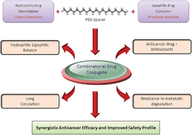
In the present work we hypothesized to assess the potential of combination of anticancer drug, GEM and antioxidant, curcumin (CUR) on promising platform of polymer drug conjugates. GEM is highly hydrophilic (solubility ∼ 83 mg ml−1)12 anticancer agent with very short plasma half-life (∼45 min) whereas, curcumin (CUR) is a potent antioxidant with anticancer properties but extremely poor aqueous solubility (11 ng ml−1)13 which poses severe formulation challenges. Therefore, it was hypothesized that the conjugation of GEM with CUR via a PEG spacer will introduce the necessary hydrophilic–hydrophobic balance in the structure of the dual drug conjugate (GEM–PEG–CUR) which will eliminate the solubility and stability related issues. Furthermore, the GEM–PEG–CUR conjugate was supposed to work as a bi-pill with improved efficacy and reduced toxicity due to the synergistic effect of GEM as anticancer agent and CUR as potent antioxidant.
2. Materials and methods
2.1 Materials
GEM and CUR (95%) were obtained as gift sample from Fresenius Kabi Oncology Limited, Gurgaon, India and Natural Remedies, India, respectively. O-(2-Aminoethyl)-O′-(2-carboxymethyl)polyethylene 3500 hydrochloride (NH2-PEG-COOH) and O-[(N-succinimidyl)succinyl-aminoethyl]-O′-methylpolyethylene glycol (m-PEG-NHS) were procured from JenKem Technology, USA. Succinic anhydride, anhydrous solvents (dimethyl sulphoxide, dimethyl formamide and benzene) and dialysis membrane (1 kD MWCO), minimum essential medium (MEM), fetal bovine serum (FBS), antibiotic–antimycotic solution, 3-(4,5-dimethylthiazol-2-yl)-2,5-diphenyltetrazolium bromide (MTT), Triton X-100, ethylenediaminetetraacetic acid (EDTA), 7,12-dimethylbenz[α]-anthracene (DMBA) were purchased from Sigma-Aldrich Chemical Co., St. Louis, MO, USA. Dicyclohexylcarbodiimide (DCC) and N-hydroxysuccinimide (NHS) were purchased from Fluka. Pyridine, dichloromethane and diethyl ether were obtained from Merck, India. Hydrochloric acid, sodium bicarbonate, disodium hydrogen phosphate and sodium acetate were procured from Loba Chemie Pvt. Ltd., Mumbai, India. Dichloromethane and pyridine were dried over phosphorus pentoxide and potassium hydroxide respectively and distilled prior to use. MCF-7 cell lines were obtained from the cell repository facility of National Centre for Cell Sciences (NCCS), Pune, India. All other solvents and reagents, unless otherwise stated, were of analytical grade and procured from local suppliers.
2.2 Synthesis and spectral characterization of GEM–PEG–CUR
The GEM–PEG–CUR was synthesized in three steps: (i) preparation of active NHS ester of CUR; (ii) PEGylation of the activated ester with heterobifunctional NH2-PEG-COOH; (iii) covalent conjugation of GEM·HCl with activated ester of PEG-derived CUR by using standard carbodiimide chemistry. All synthesized compounds including final products and intermediates were characterized by UV, FTIR, NMR and mass spectroscopy. (Refer ESI† for detailed synthesis and characterization.)
2.3 Physico-chemical characterization of synthesized bio-conjugate (GEM–PEG–CUR)
2.3.1 Melting temperature. The melting point of free GEM and its bio-conjugates was recorded using differential scanning calorimetry (DSC; Perkin Elmer MicroDSC). The instrument was calibrated for temperature and heat flow accuracy by using melting of pure indium (MP 156.6 °C and ΔH of 25.45 J g−1). Samples were placed on non-hermetic aluminum pan and analysis was carried out at temperature range of 20–180 °C at a rate of 10 °C min−1 with nitrogen flow rate at 50 ml min−1.
2.3.2 Solubility. For solubility determination, excess sample was added in triple distilled water and incubated at 37 °C for 24 h with gentle shaking at 80 rpm in a shaker bath followed by centrifugation at 80 rpm in a shaker bath followed by centrifugation at 13
000 rpm for 5 min. The supernatant was collected and analyzed by using UV spectrophotometer.
2.4 In vitro hydrolysis in simulated media
In vitro hydrolysis of GEM–PEG–CUR was assessed by monitoring the rate of GEM release in phosphate buffered saline (PBS) of pH 5.5 and pH 7.4.14–16 Additionally, the effect of crude protease on in vitro hydrolysis was also studied. GEM released from the conjugate was assessed by dissolving 100 μg ml−1 of conjugate in PBS at pH 5.5 and 7.4, respectively for 24 h. At scheduled time intervals, aliquots were withdrawn and analyzed by HPLC to quantify the amount of GEM released from the conjugates.
2.5 Plasma stability study
In vitro plasma stability of conjugate was assessed by rate of degradation of conjugate with time in presence of rat plasma. Unmodified GEM and its degradation product 2′,2′-difluorodeoxyuridine (dFdU) were measured by incubating the free GEM and GEM–PEG–CUR conjugate in plasma (pH 7.4) for 24 h. At scheduled time intervals, samples were withdrawn and analyzed by validated bioanalytical method using HPLC.17
2.6 In vitro cytotoxicity assay
The cell cytotoxicity of GEM–PEG–CUR, free drug counterparts and PEGylated congeners was determined using MTT assay in MCF-7 (human breast adenocarcinoma) cell lines by following our previously reported protocol.18 Briefly, cells were grown in Minimum Essential Medium Eagle (MEM, Sigma), accompanied with Earle's salts, L-glutamine, nonessential amino acids, sodium bicarbonate, sodium pyruvate, 10% FBS, 100 U ml−1 penicillin, and 100 μg ml−1 streptomycin (PAA Laboratories GmbH, Austria) and maintained under 5% CO2 atmosphere at 37 °C. Cells were harvested by using 0.25% w/v trypsin–EDTA solution (Sigma, USA), sub-cultured in 96-well culture plate (Costars, Corning Inc., NY, USA) at a density of 10
000 cells per well and incubated with different equivalent concentrations (0.001, 0.01, 0.1 and 1.0 μM) of GEM. The extent of viability of the cells is indicated by conversion of MTT to purple colored formazan by metabolically active cells. The cells were then solubilized with DMSO and optical density of the released, solubilized formazan reagent was measured at 540 nm spectrophotometrically. The cell viability was evaluated by following equation:
2.7 Apoptosis assay
The cell cytotoxicity potential of the GEM and its bio-conjugates was further assessed as a function of their capability to induce apoptosis in MCF-7 cells. Standard phosphatidyl serine externalization assay based on Annexin V binding was monitored to estimate the apoptosis.19 Briefly, MCF-7 cells were seeded at a density of 105 cells per well in the six-well tissue culture plate (Costars, Corning Inc., NY, USA) and allowed to attach overnight at 37 °C and 5% CO2. The media was aspirated and cells were exposed fresh media containing various GEM bio-conjugates, equivalent to 1 μg ml−1 and incubated for 6 h. Post incubation the cells were washed twice with PBS and double stained with Annexin V Cy3.18 conjugate (AnnCy3) and 6-carboxyfluorescein diacetate (6-CFDA) following the manufacturer's protocol (Annexin V-Cy3™ Apoptosis Detection Kit, Sigma, USA). The cells were then visualized under confocal laser scanning microscopy (CLSM) under green (for 6-CFDA) and red (for AnnCy3) channels. The cells stained with only green fluorescence were considered as live; those stained with both red and green were regarded as apoptotic, while cells stained only with red were considered as necrotic. Apoptosis index, ratio of the fluorescence intensity from the red fluorescence (originated from the Annexin V Cy3.18 conjugate, measure of apoptosis) normalized to that of green fluorescence (originated from the 6-carboxyfluorescein, measure of viable cells) was also calculated for the developed formulations. The quantitative measure of fluorescence within the images could be assessed by processing images with Image J software (U. S. National Institutes of Health, Bethesda, Maryland, USA, http://imagej.nih.gov/ij/).
2.8 DNA damage assay
The DNA damage potential of the synthesized conjugate was assessed as a function of alterations in the levels of 8-hydroxyguanosine (8-OHdG), which is considered as a marker of DNA damage. Briefly, cultured MCF-7 cells were exposed to varying concentrations of GEM, GEM–PEG–CUR and GEM + CUR (0.01, 0.1, 1 and 10 μg ml−1 equivalent to GEM) for 12 h. Post incubation, cells were washed twice with ice-cold Hank's Balanced Salt solution (HBSS), without calcium and magnesium. 8-OHdG within the cells was estimated following protocol reported earlier.19,20 Briefly, cells were detached using trypsin–EDTA solution (Sigma, USA), pelletized and digested under anaerobic conditions. The levels of 8-OHdG were analyzed within the samples using ELISA kit (OxiSelect Oxidative DNA Damage ELISA Kit, STA-320) following the manufacturer's instructions (Cells Biolabs. Inc. San diego, CA). DMSO treated cells were employed as negative control.
2.9 In vivo evaluation
2.9.1 In vivo antitumor efficacy. Female Sprague Dawley (SD) rats 180–200 g were used to evaluate the efficacy of the free GEM and the developed bio-conjugates. All animal study protocols were duly approved by the Institutional Animal Ethics Committee (IAEC) of National Institute of Pharmaceutical Education & Research (NIPER), India. The animals were acclimatized at temperature of 25 ± 2 °C and relative humidity of 50–60% under natural light/dark cycles for one week before experimentation. Chemical-induced breast cancer model was used for the study. Briefly, DMBA in soya bean oil was administered orally to animals at a dose of 45 mg kg−1 at weekly intervals for three consecutive weeks.21,22 Tumor bearing animals were separated and divided randomly into different treatment groups comprising of control (untreated group), free GEM, free CUR, mixture of free CUR with GEM, and GEM–PEG–CUR. Additionally, GEM–PEG and CUR–PEG were also given to determine the advantage of dual drug conjugate over the PEGylated counterparts. Each group (n = 8) was intravenously injected with three repeated doses (on 0, 4 and 8th day) of corresponding formulation at a dose/dose equivalent to 5 mg kg−1.23 The tumor dimensions were measured regularly using an electronic digital caliper up to 30 days. In addition behavioral changes were noticed and change in body weight with time was also recorded during the treatment period. Furthermore survival of the animals treated with different formulations was observed up to 30 days, and the data was analyzed by the Kaplan–Meier survival plot.24
2.9.2 In vivo hepato- and nephro-toxicity. The toxicity study was carried out on female Swiss mice (20–25 g). The animals were divided into different treatment groups, each containing five animals (n = 5). Free GEM (10 mg kg−1), Free CUR (10 mg kg−1), mixture of CUR and GEM (10 mg kg−1), GEM–PEG (dose equivalent to 10 mg kg−1 of free GEM), CUR–PEG (dose equivalent to 10 mg kg−1 of free CUR), and GEM–PEG–CUR (dose equivalent to 10 mg kg−1 of free GEM) conjugate were administered through a single dose of intravenous injection via tail vein. After 7 days, animals were humanely sacrificed and blood was collected by cardiac puncture. Levels of aspartate transaminase (AST), alanine transaminase (ALT) for hepatotoxicity and blood urea nitrogen (BUN) and plasma creatinine level for nephrotoxicity were determined by using commercially available kits (Accurex, Biomedical Pvt. Ltd). Oxidative stress parameter, malondialdehyde (MDA), was estimated in liver tissue homogenate using our earlier reported protocols.22,24
2.10 Statistical analysis
All data have been specified are expressed as mean ± SD. Statistical analysis was performed with Graph Pad Prism software (version 4.03, USA) using one-way ANOVA, followed by Tukey–Kramer multiple comparison test. p < 0.05 was considered as statistically significant.
3. Results
3.1 Physicochemical characterization
3.1.1 Melting temperature. Table 1 presents the melting point of GEM bio-conjugate compared to individual components.
Table 1 Melting point of synthesized conjugate and individual constituents
Sample |
Melting point (°C) |
GEM |
290 |
CUR |
183 |
GEM–PEG–CUR |
43–46 |
As evident from the data, the melting temperature of the conjugate is markedly different from either GEM or CUR. It was interesting to observe that melting point of the synthesized conjugate was found to be approximately 46 °C, which is quite similar to that of PEG.
3.1.2 Solubility. To determine the hydrophilic–lipophilic balance of the conjugate, aqueous solubility of GEM bio-conjugate compared to individual constituent was determined. Table 2 presents the solubility profile of synthesized conjugate compared to free drug.
Table 2 Solubility of bio-conjugate and individual constituents
Samples |
Solubility (mg ml−1) |
CUR |
0.011 × 10⁻³ |
GEM |
75.05 ± 4.23 |
GEM–PEG |
85.43 ± 0.36 |
CUR–PEG |
0.976 ± 0.04 |
GEM–CUR |
0.58 ± 0.02 |
GEM–PEG–CUR |
62.47 ± 3.92 |
The solubility of free GEM is 75.05 mg ml−1 at 25 °C however it was 62 mg ml−1 in case of GEM–PEG–CUR.
3.2 In vitro hydrolysis in simulated media
The in vitro hydrolysis of GEM–PEG–CUR conjugate was evaluated under two different pH conditions. As depicted in earlier reports,14–16 nominal amount of GEM was released at pH 5.5 however, this was increased at pH 7.4 (Fig. 1). Of note, addition of enzymes (crude protease) in medium hardly influenced the rate of GEM release.
 |
| Fig. 1 In vitro stability profile of GEM–PEG–CUR at pH (A) 5.5 and (B) 7.4. | |
3.3 Plasma stability studies
The % of intact GEM and its metabolite (dFdU) with time in case of free GEM and GEM–PEG–CUR are shown in Fig. 2.
 |
| Fig. 2 Time dependent estimation of GEM and DFDU in rat plasma (pH 7.4) at 37 °C. | |
In vitro plasma stability study demonstrated controlled release of GEM from GEM–PEG–CUR conjugate which was approximately 20% in 24 h with respect to initial GEM content in plasma. GEM released from the conjugate was eventually converted to dFdU in presence of cytidine deaminase, present in the plasma. At 24 h the amount of dFdU detected was approx 17% with respect to initial GEM content while free GEM was degraded at much higher rate as evident by presence of almost 60% dFdU in 24 h.
3.4 In vitro cytotoxicity and apoptosis assay
In vitro cell cytotoxicity of GEM-bio-conjugates was evaluated against MCF-7 cells (human breast adenocarcinoma cell lines) which revealed significantly higher cytotoxicity of developed bio-conjugates at tested time points as compared to that of free drugs alone and their combination (Table 3).
Table 3 In vitro cytotoxicity of various GEM-bio-conjugates against MCF-7 cell lines upon incubation at various time points
Drug/bio-conjugate |
IC50 values (μM) |
24 h |
48 h |
72 h |
Recovery modea |
Recovery experiments were executed in terms of 6 h exposure of drugs followed by incubation with fresh media for next 18 h, total incubation period being 24 h. |
GEM |
37.12 |
10.18 |
5.88 |
77.81 |
CUR |
243 914.14 |
1697.93 |
235.17 |
>100 000 |
GEM–PEG |
15.31 |
9.08 |
5.39 |
27.96 |
CUR–PEG |
2354.83 |
1272.61 |
215.20 |
2828.71 |
GEM + CUR |
31.80 |
9.23 |
5.54 |
73.51 |
GEM–PEG–CUR |
18.50 |
8.24 |
5.12 |
19.45 |
Both time and concentration dependent cell cytotoxicity patterns were noted. The PEGylated versions of bio-actives exhibited higher cell cytotoxicity as compared to the plain counterparts and drastic improvement in the order of ∼100 fold was noted for CUR–PEG while the appreciation was marginal in case of GEM–PEG. A dominant time dependent appreciation in cell cytotoxicity was observed in case of GEM–PEG–CUR. At initial time points, the cell cytotoxicity of PEGylated GEM surpassed that of GEM–PEG–CUR however, at later time points, >48 h, the effectiveness of GEM–PEG–CUR increased significantly (p < 0.05). Furthermore, the cell cytotoxicity potential of the developed bio-conjugates was also noted in recovery experiments which revealed 3.77-fold increase in cell cytotoxicity of GEM–PEG–CUR as compared to that of GEM + CUR. The observed results were further confirmed by the apoptosis assay. The apoptotic index was found to be remarkably higher in case of GEM–PEG–CUR as compared to that of free drugs alone and in combination (Fig. 3).
 |
| Fig. 3 Apoptosis assay of developed bioconjugate against MCF-7 cells; green channel depicts the fluorescence from carboxy fluorescein (cell viability marker dye); red channel depicts fluorescence from Annexin Cy3.18 conjugate (cell apoptosis marker dye) third channel represents the overlay image whereas fourth window depicts the differential contrast image of representative cells. The apoptosis index measured as ratio fluorescence intensity from the red channel to that of green channel. The fluorescence intensities of the images were measured using Image J software, U. S. National Institutes of Health, Bethesda, Maryland, USA, http://imagej.nih.gov/ij/. | |
3.5 DNA damage assay
The DNA damage assay further revealed that observed cytotoxicity of the GEM, GEM + CUR and GEM–PEG–CUR conjugate is mediated by DNA damage. Significantly higher levels (p < 0.001) of 8-OHdG were noted in case GEM and GEM + CUR as compared to that of control (Fig. 4). Interestingly, these levels were even higher in case of GEM–PEG–CUR (p < 0.001) corroborating with results of cell cytotoxicity experiments. Notably, insignificant differences were observed among 8-OHdG levels of cells treated with GEM alone and physical mixture of GEM + CUR.
 |
| Fig. 4 8-OHdG levels in cells treated with GEM and its bio-conjugate in MCF-7 cells. | |
3.6 In vivo antitumor efficacy
Tumor-growth inhibition profile of rats treated with free GEM, free CUR, combination of GEM + CUR, PEGylated counterparts of GEM and CUR and GEM–PEG–CUR conjugate is shown in Fig. 5.
 |
| Fig. 5 In vivo anticancer efficacy of GEM–PEG–CUR bio-conjugate as compared to free GEM, free CUR, PEGylated counterparts and combination of CUR and GEM. Plot indicates the percentage tumor volume remaining with time [a w.r.t control, b w.r.t free GEM, c w.r.t. free CUR and d w.r.t physical mixture of CUR and GEM (CUR + GEM) (***p < 0.001, **p < 0.01)]. | |
Amongst all the treatment groups, animals treated with GEM–PEG–CUR showed significantly higher tumor growth inhibition as compared to those treated with free GEM (p < 0.001), free CUR (p < 0.001), combination of CUR and GEM (p < 0.001), GEM–PEG (p < 0.01) and CUR–PEG (p < 0.001). After 1st dosing, the tumor burden in GEM–PEG–CUR treated animals was remarkably lower than those treated with free GEM, free CUR, combination of CUR and GEM, GEM–PEG and CUR–PEG. No significant change was observed in case of free CUR in comparison to control. Although significant reduction (p < 0.001) in tumor volume was observed in case of free GEM, and its combination with free CUR in comparison with control, however toxicity produced in both the cases was so acute that all the animals in these two groups died within 10 days. Statistically insignificant difference (p > 0.05) in antitumor efficacy was observed between GEM and combination of GEM + CUR.
To have an idea, that the death observed in case of GEM and GEM + CUR treated animals, was the result of severe toxicity, another parameter, loss in body weight with time was also estimated. In line with our hypothesis severe toxicity in rats was well evident from the significant loss in body weight throughout the treatment course, especially after second and third dosing as presented in Fig. 6.
 |
| Fig. 6 Decrease in body weight of animals after administration of different formulations. Plot indicates the body weight remaining (%) with respect to time. | |
In visual observation animals treated with GEM became lethargic within a day of first intravenous dosing and were seen to congregate at the corner of the cage. However, animals, treated with the combination of GEM + CUR, were in good condition which could be attributed to the antioxidant effect of CUR. On the contrary, no significant weight loss was detected for animals treated with GEM–PEG–CUR. In fact, all subjects treated with free GEM and its combination with CUR died within 10 days of first dosing (Fig. 7).
 |
| Fig. 7 Kaplan–Meier survival plot for evaluation of animal's survival during antitumor efficacy study. | |
Furthermore, survival analysis revealed 50% survival within 5 days of first dosing in case of GEM and its combination with CUR while the survival was 100% in case of animals treated with GEM–PEG–CUR. Additionally, no behavioral changes were observed in animals treated with GEM–PEG–CUR even after a week of third dosing and appeared lively without presenting any obvious alterations in vocalization, labored breathing, difficulties in movement, hunching and interactions with cage mates.
3.7 In vivo hepato- and nephrotoxicity
The levels of different biochemical parameters at 7 days post-exposure with free drugs, combination, PEGylated counterparts and conjugate is presented in Fig. 8. In all the cases, mice treated with GEM–PEG–CUR presented no significant change with respect to control, suggesting that dual drug conjugate induced minimal hepato- and nephro-toxicity in mice. Notably, the AST and ALT levels in mice treated with free GEM and GEM–PEG were significantly higher than that of control (Fig. 8A and B). In line with the results of AST and ALT, MDA level of conjugate treated group showed insignificant difference form the control, signifying that dual drug-conjugate induced minimal oxidative stresses (Fig. 8C). However, the combination of GEM and CUR led to lower the oxidative stress up to some extent yet it was significantly higher (p < 0.001) in comparison to GEM–PEG–CUR conjugate. In contrary to the hepatotoxicity results, none of the treatment group exhibited nephrotoxicity as no significant change (p > 0.05) in BUN and creatinine levels was observed in any of the treatment group (Fig. 8D and E).
 |
| Fig. 8 Biochemical markers (A) AST (B) ALT (D) BUN (E) creatinine levels in plasma and (C) MDA level in liver homogenate after 7 days following administration of free GEM, free CUR, PEGylated counterparts, combination of GEM and CUR and GEM–PEG–CUR conjugate as compared to control [(a) w.r.t control, (b) w.r.t free GEM, (c) w.r.t. free CUR and (d) w.r.t physical mixture of CUR and GEM (CUR + GEM) (***p < 0.001)]. | |
4. Discussion
The present study was designed with an aim to enhance the therapeutic efficacy of intrinsically hydrophilic drug molecules with inherently poor pharmacokinetic profile. In line with that idea, a novel bio-conjugate was designed in which GEM formed one of the key constituents of the combination to combine with an antioxidant (viz. CUR). The CUR was selected strategically as CUR has extremely low water solubility and was supposed to provide proper HLB balance to the desired conjugate. Furthermore, potent antioxidant and antitumor property of CUR was envisaged to exhibit synergistic efficacy with reduced toxicity following conjugation with GEM. Keeping in mind the importance of long-half life for any intravenous formulation, we considered conjugating both GEM and CUR to the distal ends of a PEG spacer via amide linkages. Although cleavage of amide bonds in presence of cellular enzymes is reported, these bonds are chemoenzymatically more stable than their ester, acetal, ketal, imine or hydrazone counterparts. Furthermore, GEM is highly prone to metabolism in presence of deoxycytidine deaminase. This enzyme, principally located in blood, liver and kidney, catalyzes the deamination of GEM into a completely inactive uracil derivative.25 In this context, the use of an amide linkage is particularly useful because such bio-conjugates have been reported to show superior anticancer activity over the parent drug due to their enhanced stability against metabolism and extended plasma half-life.14–16,26 To achieve the desired objectives, the said conjugate was synthesized by standard carbodiimide chemistry and authenticated by different analytical techniques. After authentication, our next target was to elucidate the change in different physical properties of the synthesized conjugate such as melting point and solubility as both these parameters can majorly affect the physiological performance.
The thermal behavior observed in case of GEM–PEG–CUR is easily interpretable if we tally the relative proportion (%w/w) of the individual component in the synthesized conjugate. In the synthesized conjugate, PEG comprises of approximately 75–80% of the total mass of the conjugate and hence it is expected that the physical properties of the conjugate will be dominated by PEG. Similarly, the synthesized conjugate was freely soluble in water yet the solubility was considerably different from either free GEM or CUR. To further confirm whether the higher solubility of GEM–PEG–CUR is the result of PEG spacer, we also synthesized the individual PEGylated congeners and GEM–CUR conjugate without PEG spacer. In line with our hypothesis a remarkable increase in solubility of PEGylated congeners was observed in comparison with free drugs. However, the solubility of GEM–CUR without PEG spacer was still extremely lower in comparison with GEM–PEG–CUR which further supported our hypothesis of bio-conjugation by using PEG spacer as a useful strategy to mitigate the problems of poor solubility (Table 2). The results indicated that appropriate molecular engineering can significantly transform the physicochemical properties of drug molecules which in turn may be beneficial in terms of their pharmacological activity. Such modifications also pave the ways to fine-tune the pharmacokinetics and subsequent therapeutic activity of medicinally active compounds, especially clinically established drugs with recognized problems. Furthermore in vitro hydrolysis study was designed to establish the release behavior of the GEM form the GEM–PEG–CUR at two different pH 7.4 and 5.5, to simulate the conditions of systemic circulation and tumor microenvironment, respectively.12 A controlled release up to 24 h observed at both the pH conditions could be attributed to the controlled cleavage of the amide bond formed between the GEM and activated CUR (Fig. 1).
Free GEM has been widely reported to show drastic conversion to its metabolite 2′,2′-difluorodeoxyuridine (dFdU) on incubation with plasma.15 To verify whether the synthesized conjugate was stable enough to resist enzymatic degradation, we incubated free GEM and GEM–PEG–CUR with plasma. The findings clearly demonstrated the ability of GEM–PEG–CUR to protect bound GEM against deoxycytidine deaminase mediated degradation in plasma (Fig. 2). The results suggest that the release of GEM from the conjugate in plasma is a controlled process which critically depends on the cleavage of amide bond formed between GEM and activated CUR. Our findings are consistent with previous reports in which conjugation of GEM has been reported to provide plasma stability for extended period of time.27
Cell culture experiments revealed significantly higher cell cytotoxicity of the developed GEM bio-conjugates as compared to the combination of individual free drug. Drastic improvement in the cell cytotoxicity in case of CUR–PEG while marginal appreciation in case of GEM–PEG could be correlated with the modulation of the physicochemical properties after PEGylation. The results provide insights on careful selection of components that should be considered for the rationalized development of bio-conjugates. Interestingly, the effectiveness of GEM–PEG–CUR also surpassed that of PEGylated bio-actives in a time dependent manner. This could be attributed to relatively rapid internalization, retention and sustained release of the developed bio-conjugate within the cells as compared to that of free drugs which might be effluxed by transmembrane efflux proteins such as P-gp.28 The results of the cell cytotoxicity in recovery mode were further corroborated by intracellular levels of GEM which revealed about ∼5-fold and 6-fold higher concentration in case of GEM–PEG–CUR as compared to that of GEM + CUR and GEM, respectively (data not shown). The DNA damage potential of the developed GEM bio-conjugates was also assessed in separate set of experiments and corroborated with the cell cytotoxicity results observed with MTT assay (Fig. 3). The drastic increase in the levels of 8-OHdG and subsequent synergism in case of synthesized conjugate could be attributed to rapid internalization and retention of actives within the cells as compared to that of plain drug alone or in combination. In vivo antitumor efficacy was determined to further support our hypothesis of improved pharmacodynamics of dual drug conjugate. The GEM–PEG–CUR developed during the course of study revealed highest antitumor efficacy without significantly affecting the body weight and 100% survival during the course of study. The enhanced efficacy and safety of GEM–PEG–CUR over free GEM, free CUR, PEGylated counterparts of GEM and CUR, and combination of free GEM and CUR can be attributed to (i) the presence of a macromolecular PEG chain, which enhances localization of the conjugate at the tumor site through passive targeting (EPR effect); (ii) sustained release of GEM from the conjugate in the intracellular milieu via chemoenzymatic hydrolysis of amide bond; (iii) synergistic enhancement in anticancer efficacy and reduced toxicity due to combined effects of anticancer drug and antioxidant.9,11,19,29 Of note, the drastic improvement in the in vivo antitumor efficacy of GEM–PEG–CUR as compared to the GEM–PEG could be attributed to the prominent in vivo anticancer activity of curcumin via allied mechanisms such as inhibition of angiogenesis, anti inflammatory pathways, etc. Additional advantage associated with the dual drug bioconjugate approach is simultaneous co-delivery of therapeutics at the site of action which is usually not the case with co-administration of therapeutics. The hypothesis is testified by noting the fact that significantly higher in vivo antitumor efficacy has been noted for GEM–PEG–CUR as compared to the GEM–PEG + CUR–PEG. The results were in line with our previous reports on co-delivery of therapeutics by nanocarrier based approaches.9,30,31
Gemcitabine chemotherapy is often associated with fatal, cholestatic hepatotoxicity and oxidative stress; some cases of GEM-induced nephrotoxicity and hemolytic uremic syndrome are also reported.32,33 Therefore, it was necessary to examine the hepatotoxicity and nephrotoxicity of GEM–PEG–CUR in appropriate animal models. In contrast to antitumor activity, toxicity profiling was done in normal Swiss mice because mice model presents far better immunogenicity and immunosensitivity, as compared to rats.22 Having monitored the general toxicity of free drug/conjugate-treated mice, we sought to examine the various biochemical parameters, indicative of drug-induced hepatotoxicity or nephrotoxicity. Liver is the major site of accumulation and metabolism for most of the xenobiotics. Therefore, any changes in liver enzymes are indicative of liver's physiological state. Levels of serum AST, ALT and MDA in liver homogenate are important bio-chemical markers and any injury or damage to liver is associated with the elevation of these biochemical markers.22 Herein, GEM treatment led to increased AST and ALT levels and enhanced oxidative stress, which, however was not observed in case of GEM–PEG–CUR conjugate. In contrary, none of the treatment group exhibited short-term nephrotoxicity. These observations implied that combinatorial conjugation strategy is an effective mean to synergize the therapeutic index of potential drug candidates while alleviating the drug-associated toxicity. Although dose-dependent and long-term toxicity of the present conjugate need to be assessed following regulatory norms, the present results confirm that dual drug conjugate developed in course of the study are devoid of any obvious hepato- or nephrotoxicity.
5. Conclusions
We have successfully developed a novel macromolecular bipill constituted of two drug molecules of opposite lipophilicity viz. CUR and GEM interconnected through a hydrophilic PEG spacer. Combinatorial conjugation of GEM with CUR dramatically transformed the physicochemical and pharmacological properties of the constituent drug molecules. Our results clearly demonstrated the superiority of the GEM–PEG–CUR conjugate over its free drug counterparts as well as combination of individual components in conferring stability against enzymatic deamination by deoxycytidine deaminase. The result of efficacy and safety studies unambiguously supported our hypothesis of improved performance of dual drug conjugate over single drug or physical combination of two drugs. Further, pharmacokinetic evaluation can provide in depth understanding on how such bio-conjugation strategy can be useful tool for difficult to deliver drug molecules like GEM.
Acknowledgements
Authors are thankful to Director, NIPER for providing necessary infrastructure facilities. AKA, VK and KT are also thankful to Council of Scientific and Industrial Research (CSIR) for financial assistance. Technical support by Mr Rahul Mahajan is also duly acknowledged.
References
- A. Persidis, Nat. Biotechnol., 1999, 17, 94–95 CrossRef CAS PubMed.
- J. Lehar, A. S. Krueger, W. Avery, A. M. Heilbut, L. M. Johansen, E. R. Price, R. J. Rickles, G. F. Short III, J. E. Staunton and X. Jin, Nat. Biotechnol., 2009, 27, 659–666 CrossRef CAS PubMed.
- J. R. Sharom, D. S. Bellows and M. Tyers, Curr. Opin. Chem. Biol., 2004, 8, 81–90 CrossRef CAS PubMed.
- W. G. Kaelin, Jr, Nat. Rev. Cancer, 2005, 5, 689–698 CrossRef PubMed.
- C. T. Keith, A. A. Borisy and B. R. Stockwell, Nat. Rev. Drug Discovery, 2005, 4, 71–78 CrossRef CAS PubMed.
- F. Calabrò, V. Lorusso, G. Rosati, L. Manzione, L. Frassineti, T. Sava, E. D. Di Paula, S. Alonso and C. N. Sternberg, Cancer, 2009, 115, 2652–2659 CrossRef PubMed.
- L. A. Martello, H. M. McDaid, D. L. Regl, C. P. H. Yang, D. Meng, T. R. R. Pettus, M. D. Kaufman, H. Arimoto, S. J. Danishefsky and A. B. Smith III, Clin. Cancer Res., 2000, 6, 1978–1987 CAS.
- S. M. Tu, E. Hossan, R. Amato, R. Kilbourn and C. J. Logothetis, J. Urol., 1995, 154, 1719–1722 CrossRef CAS.
- A. K. Jain, K. Thanki and S. Jain, Mol. Pharmaceutics, 2013, 10, 3459–3474 CrossRef CAS PubMed.
- A. K. Jain, K. Thanki and S. Jain, Pharm. Res., 2014, 31, 923–945 CrossRef CAS PubMed.
- A. K. Jain, K. Thanki and S. Jain, Pharm. Res., 2014, 31, 946–958 CrossRef CAS PubMed.
- M. Das, R. Jain, A. K. Agrawal, K. Thanki and S. Jain, Bioconjugate Chem., 2014, 25, 501–509 CrossRef CAS PubMed.
- Z. Song, R. Feng, M. Sun, C. Guo, Y. Gao, L. Li and G. Zhai, J. Colloid Interface Sci., 2011, 354, 116–123 CrossRef CAS PubMed.
- G. Cavallaro, M. Licciardi, S. Salmaso, P. Caliceti and G. Gaetano, Int. J. Pharm., 2006, 307, 258–269 CrossRef CAS PubMed.
- M. L. Immordino, P. Brusa, F. Rocco, S. Arpicco, M. Ceruti and L. Cattel, J. Controlled Release, 2004, 100, 331–346 CrossRef CAS PubMed.
- G. Pasut, F. Canal, L. Dalla Via, S. Arpicco, F. M. Veronese and O. Schiavon, J. Controlled Release, 2008, 127, 239–248 CrossRef CAS PubMed.
- N.-M. Lin, S. Zeng, S.-L. Ma, Y. Fan, H.-J. Zhong and L. Fang, Acta Pharmacol. Sin., 2004, 25, 1584–1589 CAS.
- S. Jain, S. Kumar, A. K. Agrawal, K. Thanki and U. C. Banerjee, Mol. Pharmaceutics, 2013, 10, 2416–2425 CrossRef CAS PubMed.
- A. K. Jain, K. Thanki and S. Jain, Nanomed.: Nanotechnol., Biol. Med., 2014, 10, 959–969 CrossRef CAS PubMed.
- N. K. Swarnakar, A. K. Jain, R. P. Singh, C. Godugu, M. Das and S. Jain, Biomaterials, 2011, 32, 6860–6874 CrossRef CAS PubMed.
- M. Das, S. R. Datir, R. P. Singh and S. Jain, Mol. Pharmaceutics, 2013, 10, 2543–2557 CrossRef CAS PubMed.
- S. Jain, S. R. Patil, N. K. Swarnakar and A. K. Agrawal, Mol. Pharmaceutics, 2012, 9, 2626–2635 CrossRef CAS PubMed.
- L. H. Reddy, P. E. Marque, C. Dubernet, S. L. Mouelhi, D. Desmaele and P. Couvreur, J. Pharmacol. Exp. Ther., 2008, 325, 484–490 CrossRef CAS PubMed.
- S. Jain, D. Kumar, N. K. Swarnakar and K. Thanki, Biomaterials, 2012, 33, 6758–6768 CrossRef CAS PubMed.
- B. C. Kuenen, L. Rosen, E. F. Smit, M. R. Parson, M. Levi, R. Ruijter, H. Huisman, M. A. Kedde, P. Noordhuis, W. J. van der Vijgh, G. J. Peters, G. F. Cropp, P. Scigalla, K. Hoekman, H. M. Pinedo and G. Giaccone, J. Clin. Oncol., 2002, 20, 1657–1667 CrossRef CAS PubMed.
- L. H. Reddy, H. Khoury, A. Paci, A. Deroussent, H. Ferreira, C. Dubernet, X. Decleves, M. Besnard, H. Chacun, S. Lepetre-Mouelhi, D. Desmaele, B. Rousseau, C. Laugier, J. C. Cintrat, G. Vassal and P. Couvreur, Drug Metab. Dispos., 2008, 36, 1570–1577 CrossRef CAS PubMed.
- L. V. Kiew, S. K. Cheong, K. Sidik and L. Y. Chung, Int. J. Pharm., 2010, 391, 212–220 CrossRef CAS PubMed.
- S. Aryal, C. M. Hu and L. Zhang, Small, 2010, 6, 1442–1448 CrossRef CAS PubMed.
- N. K. Swarnakar, K. Thanki and S. Jain, RSC Adv., 2013, 3, 14671–14685 RSC.
- A. K. Jain, K. Thanki and S. Jain, Pharm. Res., 2014, 31, 923–945 CrossRef CAS PubMed.
- A. K. Jain, K. Thanki and S. Jain, Pharm. Res., 2014, 31, 946–958 CrossRef CAS PubMed.
- T. Brodowicz, S. Breiteneder, C. Wiltschke and C. C. Zielinski, J. Natl. Cancer Inst., 1997, 89, 1895–1896 CrossRef CAS PubMed.
- K. Robinson, L. Lambiase, J. Li, C. Monteiro and M. Schiff, Dig. Dis. Sci., 2003, 48, 1804–1808 CrossRef.
Footnote |
† Electronic supplementary information (ESI) available. See DOI: 10.1039/c4ra04237a |
|
This journal is © The Royal Society of Chemistry 2014 |
Click here to see how this site uses Cookies. View our privacy policy here.