MnO2 nanoflakes grown on 3D graphite network for enhanced electrocapacitive performance†
Xiuxia Suna,
Huanjing Wanga,
Zhibin Lei*a,
Zonghuai Liua and
Lingling Wei*b
aSchool of Materials Science and Engineering, Shaanxi Normal University, 199 South Chang'an Road, 620 West Chang'an Street, Xi'an, Shaanxi 710119, China. E-mail: zblei@snnu.edu.cn; Fax: +86-29-81530702; Tel: +86-29-81530810
bSchool of Chemistry and Chemical Engineering, Shaanxi Normal University, 620 West Chang'an Street, Xi'an, Shaanxi 710119, China. E-mail: weill@snnu.edu.cn
First published on 17th June 2014
Abstract
Free-standing 3D graphite (3DG) with an interconnected porous network and a highly conductive backbone was prepared by a chemical vapor deposition technique with Ni foam as the sacrificial template and styrene as the carbon precursor. The 3D graphite network serves as an excellent scaffold to grow MnO2 nanoflakes through a hydrothermal reaction between 3DG and KMnO4 aqueous solution. The interconnected and conductive 3D graphite network offers a pathway for fast electron and ion transport, while the MnO2 nanoflakes that emanate from the backbone surface of 3DG minimize the interfacial contact resistance between 3DG and MnO2, favoring the effective electron transport from the 3DG skeleton to MnO2 nanoflakes. As a result, the 3DG–MnO2 composite electrode with 13% MnO2 nanoflakes exhibited a specific capacitance of 210 F g−1 at a constant discharge current density of 2.0 A g−1, which still remained at 202 F g−1 at a high current density of 15 A g−1. Moreover, a good capacitance retention of 75% and an outstanding coulombic efficiency of 97.8% were achieved after 4000 cycles of galvanostatic charge–discharge. These superior capacitive properties make the 3DG–MnO2 composite one of the promising electrodes for electrochemical energy storage.
Introduction
Supercapacitors are of great interest as energy storage devices because of their unique properties including a high power density, fast charge–discharge rate, excellent cycling performance and low maintenance cost.1–5 These advantages make supercapacitors one of the most attractive power devices that have extensive applications in mobile electronics, hybrid electric vehicles and large industrial equipment.4,6,7 The carbon-based supercapacitor stores energy through fast ion adsorption on the electrode surface,5,8 whereas the conducting polymers and metal oxides accumulate charges by reversible electrochemical redox reactions at the electrode/electrolyte interface.9 Due to the involved electrochemical Faradic redox reaction, pseudocapacitors usually deliver a much higher capacitance than electric double-layer capacitors (EDLCs). Among various pseudocapacitive materials, MnO2 has received great attention because of its high theoretical capacitance, low cost and environmentally compatibility properties.10–13 However, MnO2-based electrodes usually suffer from poor cycling stability, low electrical conductivity and large volume expansion/shrinkage during the charge–discharge process,14,15 which greatly precludes their practical application in supercapacitors.Considering that MnO2 contributes to the capacitance through a fast surface Faradic redox reaction, an MnO2 electrode with a large electrochemically active surface area is expected to offer a much shorter ion diffusion pathway for effective electrolyte penetration, and thus to maximize the Faradic redox reaction.16,17 On the other hand, the rate performance of an MnO2 electrode also strongly depends on how fast the electron and ion can be transported in the interior of the electrode. A common strategy to promote electron transport is to incorporate the pseudocapacitive MnO2 electrode onto a highly conductive and electrochemically stable support with a large ion-accessible surface area.18–20 Having the excellent electrical conductivity and possessing a unique two-dimensional planar nanostructure, graphene holds great promise as an attractive matrix to accommodate various pseudpocapacitive materials for electrochemical energy storage.21–25 Moreover, the usual method for preparing a MnO2–graphene composite electrode involves the solution-based electrostatic self-assembly or physical combining of MnO2 with the chemically reduced graphene oxides (RGO).26–29 In such systems, the active MnO2 nanostructures are mostly in poor contact with the RGO matrixes. The large interfacial resistance largely affects the effective charge transfer from the graphene matrix to the active MnO2. Moreover, the relatively low electronic conductivity of RGO due to the substantial number of residual oxygen functional groups and the densely packed graphene layers, which block access to the electrolyte, make RGO not a preferable matrix to accommodate the pseudocapacitive MnO2 component.
In contrast, three-dimensional graphene with interconnected networks shows many attractive and competitive advantages as compared with RGO with respect to its excellent electronic conductivity, three-dimensional porous structure and good mechanical flexibility.23,25,30–38 Herein, we report the capacitive performance of MnO2 nanoflakes that were grown on the highly conductive three-dimensional graphite network (3DG). The 3D graphite network was prepared by a chemical vapor deposition method with commercially available Ni foam as the sacrificial template and styrene as the carbon precursor. The hydrothermal reaction between 3DG and aqueous KMnO4 allows the uniform growth of MnO2 nanoflakes on the surface of 3DG backbone to form a hybrid nanostructure (3DG–MnO2). 3DG with a seamlessly interconnected porous network serves as an excellent scaffold to enhance the electron and ion transport, while the intimate junction between the 3DG backbone and MnO2 minimizes the interfacial contact resistance and greatly promotes electron migration from the 3DG network to MnO2. Moreover, the uniform MnO2 nanoflakes offer a large electrochemically active surface area for electrolyte penetration. As a result, the composite electrode delivers a high specific capacitance of 210 F g−1 at 2.0 A g−1, and a high capacitance of 202 F g−1 persists at 15 A g−1. Importantly, a good capacitance retention of 75% and an outstanding coulombic efficiency of 97.8% were also achieved even after 4000 cycles of galvanostatic charge–discharge.
Experimental
Sample preparation
The 3D graphite networks were prepared by a chemical vapor deposition method with 3D Ni foam as the sacrificial template and styrene as the carbon precursor. Prior to the CVD process, the Ni foam (purchased from Changsha Lyrun New Material Co. Ltd in China with a density of 380 g m−2 and thickness of 1.5 mm) was cut into 1.2 cm × 1.5 cm sheets and then pressed to 0.6 mm thick. These Ni pieces were sequentially treated with 1.0 mol L−1 HCl for 10 min and rinsed with acetone and deionized water. The treated Ni foam pieces were loaded in the quartz boat, which was placed in the center of a quartz tube heated by a program-controlled electric furnace. After the temperature of the furnace was raised to 900 °C with a ramp rate of 10 °C min−1 under Ar gas flow, Ar gas saturated with styrene (50 sccm) was introduced in the furnace for 60 min. The Ni foam covered with graphene was naturally cooled to room temperature.To preserve the structural integration of the 3D graphite network, the 3DG–Ni foam was first immersed in poly(methyl methacrylate) (PMMA, average molecular weight of 996![[thin space (1/6-em)]](https://www.rsc.org/images/entities/char_2009.gif) 000) solution (4.5% PMMA dissolved in anisole) for 10 min. The 3DG–Ni foam loaded with PMMA was then baked at 160 °C for 30 min to solidify the composite, which was subsequently immersed in 6.0 mol L−1 HCl solution for 24 h to completely remove the Ni foam. Finally, the PMMA-coated 3D graphene was dissolved by immersing the composite in hot acetone for 2 h, followed by rinsing with copious water and ethanol and drying at 80 °C. The growth of MnO2 nanoflakes on 3D graphite was achieved by the following stoichiometric reaction between 3DG and KMnO4:26,39
000) solution (4.5% PMMA dissolved in anisole) for 10 min. The 3DG–Ni foam loaded with PMMA was then baked at 160 °C for 30 min to solidify the composite, which was subsequently immersed in 6.0 mol L−1 HCl solution for 24 h to completely remove the Ni foam. Finally, the PMMA-coated 3D graphene was dissolved by immersing the composite in hot acetone for 2 h, followed by rinsing with copious water and ethanol and drying at 80 °C. The growth of MnO2 nanoflakes on 3D graphite was achieved by the following stoichiometric reaction between 3DG and KMnO4:26,39
| 4MnO4− + 3C + H2O → 4MnO2 + CO32− + 2HCO3− |
In a typical procedure, 3DG was placed in an autoclave with 40 mL water containing the given amount of KMnO4. The hydrothermal reaction was conducted at 140 °C for 3 h. The final product was collected by rinsing with copious water and drying at 80 °C. The mass content of MnO2 in the 3DG–MnO2 composite was calculated by measuring the weight of each sample before and after hydrothermal reaction.
Characterization
X-ray diffraction (XRD) measurements were carried out using a D/Max-3c X-ray diffractometer with Cu Kα (λ = 0.154 nm), using an operation voltage and current of 40 kV and 30 mA, respectively. The morphology of the samples was observed by a field-emission scanning electron microscopy (FESEM) on FEI Quanta 600F equipped with an energy-dispersed X-ray spectrometer (EDX) analyzing system. The microstructure of the samples was examined on a JEOL-2100F HRTEM at an acceleration voltage of 200 kV. X-ray photoelectron spectroscopy (XPS) spectra were recorded on an AXIS ULTRA spectrometer (Kratos Analytical) using a monochromatized Al Ka X-ray source (1486.71 eV). Raman spectra were measured on a Renishaw inVia Raman microscope with an excitation wavelength of 514.5 nm. The electrical conductivity of the graphite network was measured by a four-point probe technique. The sample was pressed into a thin disk under a pressure of 15 Mpa.Electrochemical measurements
The electrochemical performance of the electrode materials was characterized by cyclic voltammetry (CV), galvanostatic charge–discharge and electrochemical impedance spectroscopy (EIS) on a CHI 660D (Chenhua, Shanghai) electrochemical workstation. The 3DG and 3DG–MnO2 were directly applied as the working electrode, and 1.0 mol L−1 Na2SO4 was used as the aqueous electrolyte. The mass loading for each electrode was typically 1.0 mg cm−2. In a three-electrode configuration, the Pt foil and the Ag/AgCl electrode were applied as the count electrode and reference electrode, respectively. The specific capacitance was calculated from the galvanostatic discharge process according to the following equation: C = I × Δt/(ΔV × m), where I is the discharge current (A), Δt is the discharge time (s), ΔV is the voltage change (V) excluding the IR drop during the discharge process, and m is the mass of the active material of MnO2 (g), excluding the 3DG due to its negligible contribution to the capacitance, which will be discussed later. In a two-electrode system, the capacitor was configured with 3DG–MnO2 as the symmetric electrodes and Celgard® 3501 membrane as the separator. The specific capacitance was calculated using C = 4 × I × Δt/(ΔV × m), where m is the total mass of the electrode. Electrochemical impedance spectroscopy (EIS) was performed with an amplitude of 5 mV at the frequency range of 0.01 to 100 kHz.Results and discussion
Fig. 1a schematically illustrates the fabrication procedures of 3DG with nickel foam as the sacrificial template and styrene as the carbon precursor. Compared to the commonly used gaseous carbon precursor like methane and ethanol,20,24,30,33 the use of styrene makes the present CVD process safer and more cost-effective. Fig. 1b shows digital photos of samples fabricated at each step. The color of the nickel foam changes from the initial shiny white into dark gray after the carbon deposition. Upon removing the nickel foam scaffold from the graphite–nickel foam composite, the macroscopic morphology of the nickel foam was well retained. Moreover, the free-standing 3DG with a good mechanical flexibility was also evident from Fig. 1c. Fig. 2a displays the XRD patterns of the 3DG and graphite–nickel foam composite. The growth of the graphite on the surface of 3D nickel foam was verified by the occurrence of a diffraction peak at 2θ = 26.52°, which can be assigned to the (002) diffractions of graphitic carbon. This peak became more intensive upon removing Ni foam with aqueous HCl, indicating a good crystallinity of the graphite backbone. Fig. 2b presents the typical Raman spectrum of 3DG. The high intensity of the G band (1581 cm−1) and an asymmetric 2D band (2730 cm−1) can be clearly seen, whereas the usual D band, which is characteristic of the disordered carbon in graphene, was nearly not observed. These observations agree well with the XRD patterns and reveal a high quality of the as-grown graphite.40 The intensity ratio of the 2D band to the G band (I2D/IG) of 0.45 along with the 60 cm−1 of FWHM of the 2D band indicate that the graphene layer number is about 4–6 layers in the 3DG backbone.41,42 | ||
| Fig. 1 Schematic illustration of the preparation procedure for MnO2 nanoflakes grown on a 3D graphite network (a), photograph of the samples fabricated in each step (b) and the obtained free-standing 3D graphite network with good flexibility (c). | ||
 | ||
| Fig. 2 Powder XRD patterns (a) and the Raman spectra (b) of 3DG and the 3DG–MnO2 composite. | ||
The morphology of the resulting 3D graphite was examined by SEM, and a representative SEM image is presented in Fig. 3a. It is clearly seen that the graphite network is composed of a continuous skeleton with interconnected micrometer-sized macropores, which resembles that of the initial 3D nickel foam (Fig. S1†). The identical macroscopic morphology as compared with the Ni foam template clearly demonstrates an effective replication of the CVD process. It is noteworthy that the seamlessly interconnected 3D graphite network largely minimizes the inter-sheet junctions and favors the rapid electron transport within the whole framework.30,32,36 The HRTEM image shown in Fig. 3b gives more detailed information about the graphitization of the graphite skeleton. It is seen that the skeleton is composed of graphene sheets with a thickness varying from 1.4 to 1.9 nm, corresponding to a layer number of 4–6. This observation is consistent with the result deduced from the Raman spectrum (Fig. 2b). Moreover, the selected area electron diffraction (inset in Fig. 3b) presents the characteristic hexagonal spot patterns, further verifying the high quality of the graphite network prepared by the present CVD method. The electrical conductivity of the 3DG was also measured by a four-point probe technique. A remarkably high electrical conductivity of 1.5 × 10³ S m−1 as compared with the typical 10–500 S m−1 for most of the chemically derived graphenes43,44 provides further evidence that 3DG is highly crystallized.
 | ||
| Fig. 3 SEM (a) and TEM images (b) of 3DG with the inset showing the selected area electron diffraction pattern. | ||
The 3D graphite network was used as the scaffold to grow MnO2 nanoflakes through a hydrothermal reaction between carbonaceous materials and the aqueous KMnO4 solution. The phase of manganese oxides in the composite was first identified by XRD (Fig. 2a). Apart from the strong (002) diffraction attributed to the graphite network, an additional weak peak occurring at 2θ = 12.63° could be indexed to the (001) reflection of birnessite-type MnO2 (JCPDS 42-1317) with a d (001) value of 0.70 nm, which is in good agreement with the that of the layered birnessite-type MnO2 prepared by a sol–gel route.45 The Raman spectrum of 3DG–MnO2 shows two clear bands at 560 and 632 cm−1 (Fig. 2b). The band of 560 cm−1 corresponds to the Mn–O stretching vibration in the basal plane of MnO2 sheet, whereas the band at 632 cm−1 is related to the symmetric stretching vibration of MnO2 groups.46 These observations confirm the formation of birnessite-type MnO2 as the result of the stoichiometric reaction of 3D graphite network with the aqueous KMnO4 solution.
The morphology of MnO2 on the 3D graphite network was characterized by SEM. In spite of the MnO2 content varying from 13% to 35%, the 3DG–MnO2 composite retained a similar morphology to that of pristine 3DG except that its skeleton surface became coarser as the result of the growth of MnO2 nanoflakes (Fig. 4a–c). At a low MnO2 loading of 13%, a part of the 3DG skeleton surface can still be seen (Fig. 4a). When the MnO2 loading was increased to over 23%, the 3DG surface was completely covered by the dense fiber-like MnO2 (Fig. 4b and c). The EDX spectrum (Fig. S2†) recorded in the selected area of Fig. 4d presents the elemental C, Mn, K and O, confirming the uniform dispersion of MnO2 on the backbone of 3DG. A close examination shows that MnO2 grown on the graphene network exhibits a uniform bundle-like structure (Fig. 4e). The higher magnification SEM image shown in Fig. 4f reveals that each bundle is actually composed of a great number of ultrathin MnO2 nanoflakes. Such a hierarchical structure is of great significance for maximizing the electrochemical redox reaction, because it not only offers a large contact area for effective electrolyte penetration but also provides a shortened ion diffusion pathway, allowing a fast migration of electrolyte ions at a very high current density
 | ||
| Fig. 4 SEM of 3DG–MnO2-13% (a), 3DG–MnO2-35% (b) and 3DG–MnO2-23% (c, e, and f) at different magnifications. (d) SEM images of 3DG–MnO2-23%. | ||
Fig. 5 shows the TEM images of the 3DG–MnO2 composite. It is clearly seen that the graphite skeleton was fully surrounded by uniform MnO2 nanoflakes (Fig. 5a and b). While ultrasonic treatment was used for TEM sample preparation, MnO2 was still firmly connected onto the graphene skeleton without notable peeling (Fig. 5b). Such an intimate junction indeed minimizes the interfacial contact resistance between the highly conductive graphite skeleton and the pseudocapacitive MnO2 and would greatly facilitate the electron transport at high discharge current density. The representative HRTEM images of the individual MnO2 nanoflake show a thickness of ∼12 nm with a lattice spacing of 0.653 nm (Fig. 5d), corresponding to the spacing of the (110) plane of birnessite-type MnO2,26,45 also consistent with the result calculated from the Bragg equation.
 | ||
| Fig. 5 TEM images of 3DG–MnO2-23% composite at low (a and b) and high magnification (c and d). | ||
The chemical composition of 3DG–MnO2 was characterized by XPS. The occurrence of elemental Mn, O, C and K is evident in the survey XPS (Fig. 6a), in accordance with the EDX result in Fig. S2.† As shown in Fig. 6b, the signals of elemental K at a binding energy of 292.5 (K 2p3/2) and 295.2 eV (K 2p1/2) can be assigned to the K+ that occupies the interlayer space of birnessite-type MnO2 to compensate for the lower oxidation state of Mn.16,45 Two characteristic peaks of the Mn 2p spectrum at 654.1 and 642.3 eV correspond to the Mn 2p3/2 and Mn 2p1/2, respectively. According to the relationship between the oxidation state of Mn and the separation of peak energy ΔE (Mn 3s),47 the ΔE of 4.9 eV means that the average oxidation state of elemental Mn in the 3DG–MnO2 composite is around ∼3.7 (Fig. 6d), showing the dominance of Mn4+ in the birnessite-type MnO2.
 | ||
| Fig. 6 XPS spectra of 3DG–MnO2-23% composite. (a) Survey XPS, (b) C 1s XPS spectra of 3DG and 3DG–MnO2-23%, and (c) Mn 2p and (d) Mn 3s XPS spectra. | ||
The capacitive performances of the 3DG and 3DG–MnO2 electrodes were evaluated by CV, galvanostatic charge–discharge and electrochemical impedance spectroscopy (EIS). Fig. 7a shows the CV behavior of 3DG electrodes measured in a three-electrode system with 1.0 mol L−1 Na2SO4 as the aqueous electrolyte. Even at a very high scan rate of 2 V s−1, CV of 3DG electrode still retained a quite good rectangular shape, demonstrating a high-rate electrochemical behavior as the result of fast electrostatic ion adsorption on the surface of the 3DG electrode. After growth of 13% MnO2, the composite electrode exhibits a dramatically increased current response as compared with that of the 3DG electrode, highlighting the significant role of MnO2 nanoflakes in dramatically improving the overall capacitance of the composite electrode (Fig. 7b). The much smaller integrated area of 3DG electrode implies a quite low ion-accessible surface area, which is presumably related to the hydrophobic surface nature of the 3DG electrode. Fig. 7c shows the CV profiles of the 3DG–MnO2-13% electrode at various scan rates. The nearly linear increase in current density with a scan rate up to 300 mV s−1 indicates an ideal capacitive behavior (Fig. S3†). The superior electrochemical performance clearly reveals the crucial role of the highly conductive 3DG skeleton, which significantly enhances the fast ion and electron migration throughout the 3DG–MnO2 composite electrode.
 | ||
| Fig. 7 CV profiles of 3DG at different scan rates (a), and CV curve of 3DG and 3DG–MnO2-13% composite at a scan rate of 50 mV s−1 (b). The CV of 3DG–MnO2-13% at various scan rates (c). The comparative galvanostatic charge–discharge curves of the 3DG and 3DG–MnO2-13% composite electrode (d). The capacitance retention (e) and Nyquist plots (f) of the 3DG and 3DG–MnO2 composite electrode with the inset showing the plots in the high-frequency region. | ||
The galvanostatic charge–discharge profiles of 3DG and 3DG–MnO2 were compared in Fig. 7d. The nearly symmetric charge–discharge curves of the 3DG electrode at different current densities is characterized by a double-layer charge storage mechanism, while the triangular profile of 3DG–MnO2-13% reveals the reversible pseudocapacitive reaction between Na+ and the MnO2 electrode.48 No distinct IR drop was observed for the composite electrode even at a very high current density of 5.0 A g−1, implying a rather low equivalent series resistance (ESR) as a result of the short ion diffusion pathway and excellent electrical conductivity of the underlying 3D skeleton. The high-rate performance of the composite electrode was also evidenced by increasing the MnO2 loading from 13%, 23% to 35%. As shown in Fig. 7e, the specific capacitance of the composite electrode reach 210, 182 and 173 F g−1 at 2.0 A g−1 when the MnO2 loading was 13%, 23% and 35%, respectively. Considering the typical mass loading of 1.0 mg cm−1 for each electrode, their corresponding area capacitances were calculated to be 0.21, 0.18 and 0.17 F cm−2, respectively. The much lower area capacitance as compared with 1.42 F cm−1 of the previous 3D graphene–MnO2 composite electrode24 may be due to the low mass loading of our samples (1.0 vs. 9.8 mg cm−1). Nevertheless, the capacitances remained at high values of 203, 151 and 150 F g−1 even at a high current density of 15 A g−1, corresponding to a capacitance retention of 97%, 83% and 87% for MnO2 loading of 13%, 23% and 35%, respectively. While the specific capacitance of the composite electrode is somewhat lower than that in previous results,20,49–51 the capacitance retention of the MnO2–RGO electrode is significantly higher that of the MnO2–RGO composite electrodes prepared by an electrostatic assembly or the physical combining method, such as 60–70% of birnessite-type MnO2 dispersed on a functionalized RGO electrode,27,28,52 54% on the MnO2–CNT–graphene electrode (from 0.2 to 5.0 A g−1),26 50% on the CNT–MnO2 thin film electrode (from 10 to 1000 mV s−1),53 45% on the graphene–MnO2–CNT film (from 10 to 500 mV s−1),54 and 30% on a highly conductive porous Au electrode with MnO2 deposited over it (from 0.3 to 20 A g−1).12 The remarkable high-rate performance of the composite electrode is directly related to the highly conductive 3DG network and the intimate junction between MnO2 nanoflakes and the backbone surface of the interconnected 3DG skeleton. Meanwhile, the individual MnO2 with ultrathin nanoflakes also greatly shortens the ion diffusion pathway for rapid access of Na+ to the electrode surface. These unique properties make the 3DG–MnO2 composite one of the most promising candidates for a high-rate supercapacitor electrode.
The electrochemical impedance spectroscopy of 3DG and the 3DG–MnO2 composite electrode was measured at a frequency range from 0.01 Hz to 100 kHz, and the typical Nyquist plots were shown in Fig. 7f. In the low frequency range, the 3DG electrode exhibits a nearly vertical curve along the imaginary axis, in accordance with its charge storage through an ion adsorption mechanism, whereas the 3DG–MnO2 electrode displays a more inclined profile, indicative of a significant contribution from a surface Faradic redox reaction. In the high frequency range, neither the semicircle nor the Warburg portion was observed for the 3DG electrode (inset in Fig. 7f), suggesting an extremely low ESR due to rapid ion and electron transport.55 In contrast, for the 3DG–MnO2 composite electrode, a semicircle intercepts the real axis at Rs (solution resistance) in the very high frequency, and Rs + Rct (charge transfer resistance) in the high frequency (inset in Fig. 7f). The progressive increase in the semicircle with MnO2 loading reflects a slight decrease of the charge transfer rate due to the incorporation of the poorly conductive MnO2 (10−5 to 10−6 S cm−1).16 Considering the MnO2 loading varied from 13% to 35%, the increase in Rct from 3.5 to 5.0 Ω is not prominent.
To test the long-term cycling performance, the composite electrode with a high MnO2 loading of 23% was measured by continuous galvanostatic charge–discharge at a constant current density of 4.0 A g−1 (Fig. 8). The 3DG–MnO2 electrode shows a slight decrease in specific capacitance over the initial 400 cycles and then retained at quite a stable capacitance value in the subsequent 3600 cycles. It has been proposed that electrode dissolution during cycling may be one of the crucial reasons for the capacitance decay of the MnO2-based electrode.56 In our cycling measurement, we noted that a small amount of MnO2 dissolved as the electrolyte turned from colorless initially into pale yellow. A plausible reason may be that the MnO2 bundles protruding out from the 3DG are vulnerable to the continuous insertion/deinsertion of Na+ into and out of the MnO2 nanoflakes during the charge–discharge process. Nevertheless, the 3DG–MnO2 electrode retained 75% of its initial capacitance after 4000 cycles of galvanostatic charge–discharge. The coulombic efficiency, an indicator of the reversibility of electrode materials, however, remains at quite a high value of 97.8%. This result reveals a highly reversible pseudocapacitance reaction between Na+ and MnO2 nanoflakes. The symmetric charge–discharge curves of the last 10 cycles (Fig. S5†) also support this electrochemical stability.
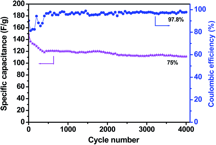 | ||
| Fig. 8 Cycle performance and coulombic efficiency of the 3DG–MnO2-23% composite electrode at a current density of 4.0 A g−1 in 1.0 mol L−1 aqueous Na2SO4 solution. | ||
A two-electrode capacitor with symmetric 3DG–MnO2-13% electrodes was configured, and its capacitive performance was also tested over a voltage window of 0–0.8 V. CV shows a quasi-rectangular profile over a wide range of scan rates (Fig. S5a†), demonstrating its excellent capacitive behavior. The galvanostatic charge–discharge also displays a symmetric profile at different current densities (Fig. S5b†). The specific capacitance based on the total mass of the two electrodes was measured to be 36 F g−1 at a constant current density of 0.5 A g−1, corresponding to 144 F g−1 for a single electrode when multiplying by a coefficient of 4. This observation followed the best practice method as described by Ruoff et al.57
Conclusions
We have successfully prepared a 3D graphite network by a chemical vapor deposition with 3D Ni foam as the sacrificial template and styrene as the carbon precursor. The seamlessly interconnected 3D network with high graphitization makes the 3DG an ideal scaffold to accommodate various pseudocapacitive components. A hydrothermal reaction between 3DG and aqueous KMnO4 yields a hierarchical nanostructure with MnO2 nanoflakes that were uniformly deposited on the surface of 3DG backbone. The highly conductive 3DG serves as an excellent network to greatly enhance the electron and ion transport, while the MnO2 nanoflakes emanating from the 3DG backbone offer a large electrochemically active surface area and a shortened diffusion pathway for effective electrolyte penetration. Consequently, the 3DG–MnO2 electrode delivers a specific capacitance of 210 F g−1 as compared with 1.3 F g−1 by the 3DG electrode at 2.0 A g−1 and retained a remarkably high-rate performance. Moreover, a reasonable cycling stability and an outstanding coulombic efficiency were simultaneously achieved after 4000 consecutive charge–discharge cycles, making the 3DG–MnO2 composite one of the promising electrodes for electrochemical energy storage. The cost-effective CVD method for preparing 3D graphite networks would also open up new opportunities to process 3D graphite-based functional materials for wide applications in biosensors, nanoelectronics, solar cells and so on.Acknowledgements
This work was financially supported by the National Nature Science Foundations of China (Grant no. 21373134, 51172137), Fundamental Research Funds for the Central Universities (Grant no. GK201403005, GK201101003, GK201301002), the foundation of returning overseas scholars, MOE, and the Program for Key Science & Technology Innovation Team (2012KCT-21) and Nature Science Foundation of Shaanxi Province (2013JM2001).References
- P. Simon and Y. Gogotsi, Acc. Chem. Res., 2013, 46, 1094–1103 CrossRef CAS PubMed.
- H. Jiang, P. S. Lee and C. Li, Energy Environ. Sci., 2013, 6, 41–53 CAS.
- L. Jiang and Z. Fan, Nanoscale, 2014, 6, 1922–1945 RSC.
- M. Beidaghi and Y. Gogotsi, Energy Environ. Sci., 2014, 7, 867–884 CAS.
- H. Ji, X. Zhao, Z. Qiao, J. Jung, Y. Zhu, Y. Lu, L. L. Zhang, A. H. MacDonald and R. S. Ruoff, Nat. Commun., 2014, 5, 3317 Search PubMed.
- J. R. Miller and P. Simon, Science, 2008, 321, 651–652 CrossRef CAS PubMed.
- J. Chmiola, C. Largeot, P. L. Taberna, P. Simon and Y. Gogotsi, Science, 2010, 328, 480–483 CrossRef CAS PubMed.
- L. L. Zhang, Y. Gu and X. S. Zhao, J. Mater. Chem. A, 2013, 1, 9395–9408 CAS.
- P. Simon and Y. Gogotsi, Nat. Mater., 2008, 7, 845–854 CrossRef CAS PubMed.
- W. F. Wei, X. W. Cui, W. X. Chen and D. G. Ivey, Chem. Soc. Rev., 2011, 40, 1697–1721 RSC.
- A. Sumboja, C. Y. Foo, X. Wang and P. S. Lee, Adv. Mater., 2013, 25, 2809–2815 CrossRef CAS PubMed.
- X. Lang, A. Hirata, T. Fujita and M. Chen, Nat. Nanotechnol., 2011, 6, 232–236 CrossRef CAS PubMed.
- Y. Zhang, M. Ma, J. Yang, W. Huang and X. Dong, RSC Adv., 2014, 4, 8466–8471 RSC.
- L. Athouël, F. Moser, R. Dugas, O. Crosnier, D. Bélanger and T. Brousse, J. Phys. Chem. C, 2008, 112, 7270–7277 Search PubMed.
- S.-L. Kuo and N.-L. Wu, J. Electrochem. Soc., 2006, 153, A1317–A1324 CrossRef CAS PubMed.
- Z. Lei, J. Zhang and X. S. Zhao, J. Mater. Chem., 2012, 22, 153–160 RSC.
- J. Liu, J. Jiang, C. Cheng, H. Li, J. Zhang, H. Gong and H. J. Fan, Adv. Mater., 2011, 23, 2076–2081 CrossRef CAS PubMed.
- X. Zhao, L. Zhang, S. Murali, M. D. Stoller, Q. Zhang, Y. Zhu and R. S. Ruoff, ACS Nano, 2012, 6, 5404–5412 CrossRef CAS PubMed.
- H. Jiang, J. Ma and C. Li, Adv. Mater., 2012, 24, 4197–4202 CrossRef CAS.
- X. Dong, X. Wang, J. Wang, H. Song, X. Li, L. Wang, M. B. Chan-Park, C. M. Li and P. Chen, Carbon, 2012, 50, 4865–4870 CrossRef CAS PubMed.
- C. Xu, B. Xu, Y. Gu, Z. Xiong, J. Sun and X. S. Zhao, Energy Environ. Sci., 2013, 6, 1388–1414 CAS.
- C. Wu, S. Deng, H. Wang, Y. Sun, J. Liu and H. Yan, ACS Appl. Mater. Interfaces, 2014, 6, 1106–1112 CAS.
- J. Ji, L. L. Zhang, H. Ji, Y. Li, X. Zhao, X. Bai, X. Fan, F. Zhang and R. S. Ruoff, ACS Nano, 2013, 7, 6237–6243 CrossRef CAS PubMed.
- Y. He, W. Chen, X. Li, Z. Zhang, J. Fu, C. Zhao and E. Xie, ACS Nano, 2013, 7, 174–182 CrossRef CAS PubMed.
- Y. Wang, X. Yang, L. Qiu and D. Li, Energy Environ. Sci., 2013, 6, 477–481 CAS.
- Z. B. Lei, F. Shi and L. Lu, ACS Appl. Mater. Interfaces, 2012, 4, 1058–1064 CAS.
- J. Zhang and X. S. Zhao, Carbon, 2013, 52, 1–9 CrossRef CAS PubMed.
- J. Zhu and J. He, ACS Appl. Mater. Interfaces, 2012, 4, 1770–1776 CAS.
- H. Chen, S. Zhou, M. Chen and L. Wu, J. Mater. Chem., 2012, 22, 25207–25216 RSC.
- Z. Chen, W. Ren, L. Gao, B. Liu, S. Pei and H.-M. Cheng, Nat. Mater., 2011, 10, 424–428 CrossRef CAS PubMed.
- B. G. Choi, M. Yang, W. H. Hong, J. W. Choi and Y. S. Huh, ACS Nano, 2012, 6, 4020–4028 CrossRef CAS PubMed.
- Z. Yan, L. Ma, Y. Zhu, I. Lahiri, M. G. Hahm, Z. Liu, S. Yang, C. Xiang, W. Lu, Z. Peng, Z. Sun, C. Kittrell, J. Lou, W. Choi, P. M. Ajayan and J. M. Tour, ACS Nano, 2013, 7, 58–64 CrossRef CAS PubMed.
- X. Cao, Y. Shi, W. Shi, G. Lu, X. Huang, Q. Yan, Q. Zhang and H. Zhang, Small, 2011, 7, 3163–3168 CrossRef CAS PubMed.
- M. F. El-Kady, V. Strong, S. Dubin and R. B. Kaner, Science, 2012, 335, 1326–1330 CrossRef CAS PubMed.
- J. H. Lee, N. Park, B. G. Kim, D. S. Jung, K. Im, J. Hur and J. W. Choi, ACS Nano, 2013, 7, 9366–9374 CrossRef CAS PubMed.
- X.-C. Dong, H. Xu, X.-W. Wang, Y.-X. Huang, M. B. Chan-Park, H. Zhang, L.-H. Wang, W. Huang and P. Chen, ACS Nano, 2012, 6, 3206–3213 CrossRef CAS PubMed.
- X. Cao, Z. Yin and H. Zhang, Energy Environ. Sci., 2014, 7, 1850–1865 CAS.
- S. Han, D. Wu, S. Li, F. Zhang and X. Feng, Adv. Mater., 2014, 26, 849–864 CrossRef CAS PubMed.
- X. Jin, W. Zhou, S. Zhang and G. Z. Chen, Small, 2007, 3, 1513–1517 CrossRef CAS PubMed.
- H. Ji, L. Zhang, M. T. Pettes, H. Li, S. Chen, L. Shi, R. Piner and R. S. Ruoff, Nano Lett., 2012, 12, 2446–2451 CrossRef CAS PubMed.
- A. C. Ferrari, J. C. Meyer, V. Scardaci, C. Casiraghi, M. Lazzeri, F. Mauri, S. Piscanec, D. Jiang, K. S. Novoselov, S. Roth and A. K. Geim, Phys. Rev. Lett., 2006, 97, 187401 CrossRef CAS.
- Y. Hao, Y. Wang, L. Wang, Z. Ni, Z. Wang, R. Wang, C. K. Koo, Z. Shen and J. T. L. Thong, Small, 2010, 6, 195–200 CrossRef CAS PubMed.
- D. C. Luo, G. X. Zhang, J. F. Liu and X. M. Sun, J. Phys. Chem. C, 2011, 115, 11327–11335 CAS.
- Z. Lei, L. Lu and X. S. Zhao, Energy Environ. Sci., 2012, 5, 6391–6399 CAS.
- S. Ching, D. J. Petrovay, M. L. Jorgensen and S. L. Suib, Inorg. Chem., 1997, 36, 883–890 CrossRef CAS.
- S. B. Ma, Y. H. Lee, K. Y. Ahn, C. M. Kim, K. H. Oh and K. B. Kim, J. Electrochem. Soc., 2006, 153, C27–C32 CrossRef CAS PubMed.
- M. Toupin, T. Brousse and D. Bélanger, Chem. Mater., 2002, 14, 3946–3952 CrossRef CAS.
- M. Toupin, T. Brousse and D. Bélanger, Chem. Mater., 2004, 16, 3184–3190 CrossRef CAS.
- L. Mao, K. Zhang, H. S. On Chan and J. Wu, J. Mater. Chem., 2012, 22, 1845–1851 RSC.
- S. Wu, W. Chen and L. Yan, J. Mater. Chem. A, 2014, 2, 2765–2772 CAS.
- U. M. Patil, J. S. Sohn, S. B. Kulkarni, H. G. Park, Y. Jung, K. V. Gurav, J. H. Kim and S. C. Jun, Mater. Lett., 2014, 119, 135–139 CrossRef CAS PubMed.
- J. T. Zhang, J. W. Jiang and X. S. Zhao, J. Phys. Chem. C, 2011, 115, 6448–6454 CAS.
- S. W. Lee, J. Kim, S. Chen, P. T. Hammond and Y. Shao-Horn, ACS Nano, 2010, 4, 3889–3896 CrossRef CAS PubMed.
- Y. Cheng, S. Lu, H. Zhang, C. V. Varanasi and J. Liu, Nano Lett., 2012, 12, 4206–4211 CrossRef CAS PubMed.
- L. L. Zhang, X. Zhao, M. D. Stoller, Y. Zhu, H. Ji, S. Murali, Y. Wu, S. Perales, B. Clevenger and R. S. Ruoff, Nano Lett., 2012, 12, 1806–1812 CrossRef CAS PubMed.
- D. Belanger, T. Brousse and J. W. Long, Electrochem. Soc. Interface, 2008, 49–52 CAS.
- M. D. Stoller and R. S. Ruoff, Energy Environ. Sci., 2010, 3, 1294–1301 CAS.
Footnote |
| † Electronic supplementary information (ESI) available: The SEM images of 3D Ni foam, EDX spectrum of MnO2–3DG-23%, the dependence of discharged current density on the scan rate for the 3DG–MnO2-13% electrode, the last 10 cycles of galvanostatic charge–discharge curves of 3DG–MnO2-23%, and the capacitive performance of the two-electrode capacitor with 3DG–MnO2-13% as symmetric electrodes. See DOI: 10.1039/c4ra03983a |
| This journal is © The Royal Society of Chemistry 2014 |