Reduction-triggered formation of EFK8 molecular hydrogel for 3D cell culture

Weiwei Wang a, Guoping Lia, Weiguang Zhanga, Jie Gaob, Jimin Zhanga, Chen Li*a, Dan Ding*b and Deling Kongab
aTianjin Key Laboratory of Biomaterial Research, Institute of Biomedical Engineering, Chinese Academy of Medical Science, Tianjin 300192, P. R. China. E-mail: cli0616826@gmail.com
bThe Key Laboratory of Bioactive Materials, Ministry of Education, College of Life Science, Nankai University, Tianjin 300071, China. E-mail: dingd@nankai.edu.cn; Fax: +86 22 23498775; Tel: +86 22 23502111

Received 24th April 2014 , Accepted 30th June 2014

First published on 1st July 2014


Abstract

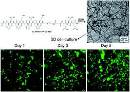
We report here a biocompatible strategy of EFK8-based molecular hydrogel formation via disulfide bond reduction in cell culture media under neutral conditions, which presents a promising approach in three-dimensional (3D) cell culture.


Recently, small molecular (SM) hydrogels have attracted ample research interest due to their inherent properties, including their formation via biocompatible self-assembly, their ability to incorporate multiple active units, and their responsiveness to external stimuli.1 These properties endow SM hydrogels with great potential in the applications of biosensing, controlled drug delivery, and especially the three-dimensional (3D) cell culture.2 In order to serve as an ideal 3D matrix for cell culture utilization, one SM hydrogel would possess several necessary qualities, which include (1) capability of forming hydrogels in the cell growth environment;3 (2) mild and biocompatible approaches to hydrogel formation;4 (3) maintaining the homogeneity5 and (4) causing minimal damage to the encapsulated cells during the formation of SM hydrogels.6 In recent years, bioactive molecule (e.g., enzyme, peptide) triggeration strategies have been developed to form SM hydrogels so as to meet the above-mentioned requirements,7 which open up a new avenue for the applications of SM hydrogels in the fields of 3D cell culture, tissue engineering and regenerative medicine. Among a variety of bioactive molecule triggeration strategies, a simple and biocompatible method through disulfide bond reduction has attracted considerable interest, as it can lead to the formation of SM hydrogels in a homogeneous and controllable manner. To date, Rao's,8 Nilsson's9 and Yang's5 groups have successfully prepared the homogeneous SM hydrogels, respectively, utilizing the strategy of disulfide bond reduction. These exciting results inspire us to use this simple and biocompatible strategy to further explore promising SM hydrogels for the advancement in the application of 3D cell culture.

EFK8 is one of the simplest pure peptide-based (not those capped with aromatic capping groups) SM hydrogelators, which shows strong hydrogelation ability as well as low cytotoxicity. However, EFK8 has not been widely used as 3D cell culture matrix development mainly due to its requirement for acidic environment during hydrogel formation.10 This is because the aromatic interaction between phenyl rings on Fs and electrostatic interaction between Es and Ks (Scheme 1) result in strong aggregation property of EFK8 at neutral pH.3,11 Although several rational designs of EFK8 peptide derivatives were developed to reduce its aggregation property in pure water solutions at neutral condition,12 new approaches to preparation of EFK8-based SM hydrogels in cell culture medium and in a homogeneous way remain in high demand for 3D cell culture.


image file: c4ra03760j-s1.tif
Scheme 1 Synthetic route to Ac-FEFKFEFK-CS-EEE and the cleavage of its disulfide bond by GSH. Conditions: (i) NaHCO3, succinic anhydride, dioxane/H2O, 0 °C; (ii) Fmoc-OSu, DMF, 0 °C; (iii) HBTU, DIPEA; (iv) acetic anhydride, HBTU, DIPEA.

In this contribution, we report on the biocompatible strategy of disulfide bond reduction to facilitate EFK8-based SM hydrogel formation in cell growth environment at neutral condition, which holds great promise for the application in 3D cell culture. The peptide of Ac-FEFKFEFK-CS-EEE was designed and synthesized as the precursor of SM hydrogelator. The hydrophilic EEE sequence is capable of rendering the peptide with good solubility in phosphate buffered saline (PBS) buffer or cell culture medium. The CS (Cystamine Succinate) part in the peptide contains a disulfide bond, which can be cleaved by reductants such as glutathione (GSH) and dithiothreitol (DTT). The disulfide bond reduction leads to self-assembly of peptide nanofibers and SM hydrogel formation. Finally, the application of the obtained EFK8-based hydrogels in 3D cell culture was evaluated by live-dead and CCK-8 assays and compared with conventional 2D cell culture.

The peptide of Ac-FEFKFEFK-CS-EEE was synthesized with detailed synthetic route in Scheme 1. The cystamine was firstly reacted with succinic anhydride and subsequently with Fmoc-OSu to yield Fmoc-Cystamine Succinate (Fmoc-CS).13 Fmoc-EEE was synthesized by standard Fmoc solid-phase peptide synthesis (SPPS), which was followed by reaction with Fmoc-CS to afford Fmoc-CS-EEE. Finally, Ac-FEFKFEFK-CS-EEE was obtained by SPPS using Fmoc-CS-EEE with other Fmoc-amino acids with side chains properly protected. The final product was afforded in a 45% total yield. The purity and identity of the final product were verified by 1H NMR, HRMS and FT-IR (Fig. S1–S3 in the ESI).

It is encouraging that the Ac-FEFKFEFK-CS-EEE peptide is able to form clear solution in PBS buffer or cell culture medium at pH 7.4. This finding suggests that the strong aggregation potency of EFK8 at neutral condition would be significantly reduced by the attachment of a hydrophilic peptide EEE through a disulfide bond bridge. As shown in Fig. 1A and B, upon addition of 4 equiv. of GSH to the PBS solution of Ac-FEFKFEFK-CS-EEE, the disulfide bond in the peptide can be cleaved and the residue peptide of Ac-FEFKFEFK-SH promotes the homogeneous SM hydrogel formation. The cleavage of disulfide bond in the peptide by the reductants was verified by LC-MS (Fig. S4 in the ESI). The minimum concentration needed for gelation for the precursor peptide was about 0.5 wt%.


image file: c4ra03760j-f1.tif
Fig. 1 Optical images showing (A) the clear solution of Ac-FEFKFEFK-CS-EEE (0.5 wt%) in PBS buffer (pH 7.4) and (B) the hydrogelation by addition of reductants to (A). (C) Dynamic time sweep and (D) dynamic frequency sweep of a PBS solution containing 0.5 wt% of Ac-FEFKFEFK-CS-EEE with 4 equiv. of GSH.

Rheological measurements with the mode of dynamic time sweep and dynamic frequency sweep, respectively, were performed for the PBS solution of Ac-FEFKFEFK-CS-EEE with 4 equiv. of GSH. As shown in Fig. 1C, after treated with GSH, self-supporting hydrogel is quickly formed. At the time point of 120 seconds, G′ crosses over G′′ and dominates in the following time points, which then reach the plateaus at around 20 minutes. This result suggests effective and viscoelastic hydrogels. In a frequency sweep, measurements were carried out at different oscillation frequencies with a constant oscillation amplitude and temperature (Fig. 1D). At higher frequency, the mechanical properties of the hydrogel are slightly influenced. Moreover, transmission electron microscopy (TEM) observations indicate that the Ac-FEFKFEFK-CS-EEE cannot form any inerratic nanostructrues in aqueous solution (Fig. 2A). After reductants were utilized to trigger the hydrogel formation, filamentous network structures with size of around 10 nm are observed in the hydrogel (Fig. 2B).


image file: c4ra03760j-f2.tif
Fig. 2 TEM images of (A) the clear solution of Ac-FEFKFEFK-CS-EEE (0.5 wt%) in PBS buffer (pH 7.4) and (B) the hydrogels formed by addition of reductants to (A).

The application of the obtained EFK8-based SM hydrogel in 3D cell culture was next investigated. In this experiment, Ac-FEFKFEFK-CS-EEE was firstly dissolved in cell culture medium (Dulbecco's Modified Eagle's Medium; DMEM). After addition of 4 equiv. of GSH, the resulting solution was mixed with equal volume of the DMEM containing 2 × 10⁶ per mL of NIH/3T3 fibroblast cells. The cell-gel construct could then form within 3 minutes with final peptide concentration of 0.5 wt% and cell density of 1 × 10⁶ per mL. As shown in Fig. 3A–C, The live-dead assay of the cell-gel construct after 1 day, 3 day, and 5 day culture at 37 °C, respectively, reveals that most of the NIH/3T3 cells are alive, as evidenced by the plenty of green dots (live cells are in green and dead ones are in red)14 throughout the images. The cell proliferation rate of NIH/3T3 cells in hydrogels was quantitatively determined by the CCK-8 assay as well. The NIH/3T3 cells incubated in cell culture medium only without addition of hydrogels (conventional 2D culture) were used as a control. As shown in Fig. 3D, the average OD value for the cell-gel construct is approximately 0.4, 0.7, and 2.0 upon 1 day, 3 day, and 5 day culture, respectively, which is significantly higher as compared to that of NIH/3T3 cells without hydrogel incorporation on day 3 and day 5. These results demonstrate that the amount of metabolically-active cells continuously increases during a 5 day culture period and our EFK8-based hydrogel could efficiently assist the NIH/3T3 cell proliferation.


image file: c4ra03760j-f3.tif
Fig. 3 Confocal laser scanning microscopy images indicate the live-dead assay of NIH/3T3 fibroblast cells cultured in hydrogel for (A) 1, (B) 3, and (C) 5 days. (D) Proliferation rates of NIH/3T3 cells cultured in hydrogel by CCK-8 assay. Data are presented as mean ± standard deviation (n = 5).

In summary, we report here a rationally designed EFK8-based peptide using the disulfide bond as a cleavable linker to control the molecular self-assembly and the formation of hydrogels at neutral conditions. The Ac-FEFKFEFK-CS-EEE forms clear solutions in PBS buffer or cell culture medium at pH 7.4 as a result of disulfide bond bridging to the hydrophilic peptide EEE. The addition of GSH results in disulfide bond reduction and thus the self-assembly of peptide nanofibers as well as the hydrogel formation. Subsequent 3D cell culture study supports the potential of the obtained hydrogel as 3D matrix to facilitate NIH/3T3 cell proliferation. As GSH is a well-known endogenous intracellular antioxidant, this study provides a simple and biocompatible strategy for SM hydrogel formation in a homogeneous and controllable manner. The study also provides fundamental guidelines to design and develop useful SM hydrogels for 3D cell culture and other relevant biomedical applications.

Experimental section

Hydrogel formation

Peptides of Ac-FEFKFEFK-CS-EEE were prepared in a stock solution at a concentration of 5 mg mL−1 in PBS (pH 7.4) or serum-free DMEM solutions. Hydrogels with desired concentrations would form after mixing with different volumes of reductants solution (4.0 equiv. of GSH to the peptide) within 2 min at room temperature in PBS or serum-free DMEM solutions, respectively.

Cell culture in hydrogels

NIH/3T3 (Mouse embryonic fibroblast cells) were used in this study. After the addition of GSH (4 equiv. to precursor) to serum-free DMEM solutions containing the precursor, the resulting solutions were mixed with equal volumes of serum-free DMEM solutions of NIH/3T3 cells suspensions. And then the resulting cells-peptide solutions were transferred to 96 well plates (50 μL per well). The final precursor concentrations were 0.5 wt%, cell density in cell-gel constructs was 1.0 × 10⁶ cells per mL. Half an hour later, 100 μL of complete DMEM medium supplemented with 10% FBS (fetal bovine serum), 100 units per mL of penicillin, and 100 μg mL−1 streptomycin was added to the cell-gel construct. Culture medium was changed every two days. The 96-well plate was maintained in a 37 °C/5% CO2 incubator.

Live/dead assay

Viability of the cells cultured in the hydrogels was tested by the Live/Dead assay according to the manufacturer's instruction. Specifically, the cell-gel constructs were washed twice with PBS for 20 min each. 100 μL of staining reagent containing 2 μM calcein AM and 4 μM EthD-1 was then added onto the cell-gel constructs. After 30 min incubation in a 37 °C/5% CO2 incubator, the cells were observed using a Nikon Eclipse TS100 inverted fluorescent microscope with excitation filters of 450–490 nm (green, Calcein AM) and 510–560 nm (red, EthE-1).

Determination of cell proliferation rate by CCK-8

To quantify cell proliferation inside the cell-gel constructs, the CCK-8 assay was performed at a series of time points. The 3D cell-gel constructs were made by encapsulating cells into hydrogels following the above 3D-culture procedure. To perform the CCK-8 assay, each cell-gel construct was incubated with 100 μL of 10% (v/v) CCK-8 in serum-free DMEM. The plates were then incubated in the 5% CO2 incubator for 4 h at 37 °C. The absorbance at 450 nm was subsequently determined by using the microplate reader (MultiskaniMark, Bio-Rad, USA). The experiments were conducted for five times and the standard deviation was determined. For the 2D cell culture, the NIH/3T3 cells were only cultured in DMEM medium containing 10% FBS, 100 units per mL of penicillin, and 100 μg mL−1 streptomycin, without addition of the hydrogels. The CCK-8 assay was then conducted at each time point following the same procedure.

Acknowledgements

This work is supported by National Natural Science Foundation of China (81301309, 31300732, 81301311 and 81220108015).

Notes and references

  1. L. A. Estroff and A. D. Hamilton, Chem. Rev., 2004, 104, 1201 CrossRef CAS PubMed; J. D. Badjić, A. Nelson, S. J. Cantrill, W. B. Tumbull and J. F. Stoddart, Acc. Chem. Res., 2005, 38, 723 CrossRef PubMed; R. V. Ulijn and A. M. Smith, Chem. Soc. Rev., 2008, 37, 664 RSC; Y. Gao, F. Zhao, Q. Wang, Y. Zhang and B. Xu, Chem. Soc. Rev., 2010, 39, 3425 RSC; J. H. Collier, J. S. Rudra, J. Z. Gasiorowski and J. P. Jung, Chem. Soc. Rev., 2010, 39, 3413 RSC; Y. Li, Y. Ding, M. Qin, Y. Cao and W. Wang, Chem. Commun., 2013, 49, 8653 RSC.
  2. V. Jayawarna, M. Ali, T. A. Jowitt, A. E. Miller, A. Saiani, J. E. Gough and R. V. Ulijn, Adv. Mater., 2006, 18, 611 CrossRef CAS; A. Wada, S. Tamaru, M. Ikeda and I. Hamachi, J. Am. Chem. Soc., 2009, 131, 5321 CrossRef PubMed; J. P. Jung, A. K. Nagaraj, E. K. Fox, J. S. Rudra, J. M. Devgun and J. H. Collier, Biomaterials, 2009, 30, 2400 CrossRef PubMed; S. Soukasene, D. J. Toft, T. J. Moyer, H. Lu, H. K. Lee, S. M. Standley, V. L. Cryns and S. I. Stupp, ACS Nano, 2011, 5, 9113 CrossRef PubMed.
  3. C. W. Ou, J. W. Zhang, Y. Shi, Z. Y. Wang, L. Wang, Z. M. Yang and M. S. Chen, RSC Adv., 2014, 4, 9229 RSC.
  4. Z. J. Qiu, H. T. Yu, J. B. Li, Y. Wang and Y. Zhang, Chem. Commun., 2009, 3342 RSC; I. M. Geisler and J. P. Schneider, Adv. Funct. Mater., 2012, 22, 529 CrossRef CAS; X. L. Zhang, X. L. Chu, L. Wang, H. M. Wang, G. L. Liang, J. X. Zhang, J. F. Long and Z. M. Yang, Angew. Chem., Int. Ed., 2012, 51, 4388 CrossRef PubMed.
  5. C. H. Ren, Z. J. Song, W. T. Zheng, X. M. Chen, L. Wang, D. L. Kong and Z. M. Yang, Chem. Commun., 2011, 47, 1619 RSC.
  6. J. B. Matson and S. I. Stupp, Chem. Commun., 2012, 48, 26 RSC; M. Zhou, A. M. Smith, A. K. Das, N. W. Hodson, R. F. Collins, R. V. Ulijn and J. E. Gough, Biomaterials, 2009, 30, 2523 CrossRef CAS PubMed.
  7. Z. M. Yang, G. L. Liang and B. Xu, Acc. Chem. Res., 2008, 41, 315 CrossRef CAS PubMed; R. J. Williams, A. M. Smith, R. Collins, N. Hodson, A. K. Das and R. V. Ulijn, Nat. Nanotechnol., 2009, 4, 19 CrossRef PubMed; D. Koda, T. Maruyama, N. Minakuchi, K. Nakashima and M. Goto, Chem. Commun., 2010, 46, 979 RSC.
  8. G. L. Liang, H. J. Ren and J. H. Rao, Nat. Chem., 2010, 2, 54 CrossRef CAS PubMed.
  9. C. J. Bowerman and B. L. Nilsson, J. Am. Chem. Soc., 2010, 132, 9526 CrossRef CAS PubMed.
  10. D. M. Marini, W. Hwang, D. A. Lauffenburger, S. Zhang and R. D. Kamm, Nano Lett., 2002, 2, 295 CrossRef CAS; S. G. Zhang, D. M. Marini, W. Hwang and S. Santoso, Curr. Opin. Chem. Biol., 2002, 6, 865 CrossRef; W. Hwang, D. M. Marini, R. D. Kamm and S. Zhang, J. Chem. Phys., 2003, 118, 389 CrossRef PubMed.
  11. X. Du, Z. D. Wu, J. F. Long and L. Wang, RSC Adv., 2013, 3, 18259 RSC.
  12. M. R. Caplan, E. M. Schwartzfarb, S. Zhang, R. D. Kamm and D. A. Lauffenburger, Biomaterials, 2002, 23, 219 CrossRef CAS; R. J. Swanekamp, J. T. M. DiMaio, C. J. Bowerman and B. L. Nilsson, J. Am. Chem. Soc., 2012, 134, 5556 CrossRef PubMed.
  13. C. H. Ren, Z. J. Song, W. T. Zheng, X. M.Chen, L. Wang, D. L. Kong and Z. M. Yang, Chem. Commun., 2011, 47, 1619 RSC.
  14. M. Lewicka, O. Hermanson and A. U. Rising, Biomaterials, 2012, 33, 7712 CrossRef CAS PubMed.

Footnotes

Electronic supplementary information (ESI) available: Peptide synthesis and characterization, 1H NMR, HRMS and FT-IR spectra of the peptide. See DOI: 10.1039/c4ra03760j
The authors pay equal contributions to this work.

This journal is © The Royal Society of Chemistry 2014
Click here to see how this site uses Cookies. View our privacy policy here.