Synthesis, spectroscopic characterization, pH dependent redox mechanism and DNA binding behavior of chlorohydroxyaniline derivatives

Suniya Shahzada, Afzal Shah*ab, Muhammad Sirajuddina, Bimalendu Adhikarib, Khurshid Ahmada, Usman Ali Ranac, Saqib Alia, Gul Shahzada Khand, Rumana Qureshia and Heinz-Bernhard Kraatzb
aDepartment of Chemistry, Quaid-i-Azam University, 45320, Islamabad, Pakistan. E-mail: afzal.shah@utoronto.ca; Tel: +92-416-208-2776
bDepartment of Physical and Environmental Sciences, University of Toronto, 1265 Military Trail, Toronto, M1C 1A4, Canada
cDeanship of Scientific Research, College of Engineering, King Saud University, Riyadh 11421, Saudi Arabia
dDepartment of Chemistry, Shaheed Benazir Bhutto University, Sheringal, District Dir (Upper), Khyber Pakhtunkhwa, Pakistan

Received 16th April 2014 , Accepted 12th May 2014

First published on 12th May 2014


Abstract

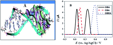
The derivatives of anilines are promisingly useful in rechargeable batteries, electrochromics and biosensors. Phenol and aniline based compounds are bestowed with strong antioxidant and anticancer activities. Based on these considerations three new chlorohydroxyanilines (CHAs) were synthesized and characterized by IR, 13C NMR, 1H NMR and UV-Vis spectroscopy. Cyclic, differential pulse and square wave voltammetry were used for the investigations of the electrochemical fate of these compounds in different pH media. Computational chemistry was used as a tool to verify the experimental outcomes. Two chlorohydroxyanilines were found to oxidize at a potential lower than the standard antioxidant, ascorbic acid. The pH dependent oxidation indicated the involvement of protons during electron transfer reactions. The quasi-reversible and irreversible nature of the first and second oxidation peak was evidenced by square wave voltammetry. The slope of peak potential vs. pH plot and the width at half peak height indicated 1e and 1H+ involvement in each oxidation step. Molecular docking and UV-Vis spectroscopy revealed that all chlorohydroxyanilines interact with DNA via an electrostatic binding mode. A sensitive differential pulse technique allowed the determination of a very low limit of detection.


1. Introduction

Oxidation is the primary reason for spoilage of foodstuffs, lipids and polymers.1 The unrestrained oxidation of DNA and protein leads to the progression of cancer and aging.2 Thus, to avoid the oxidation of biomolecules, antioxidants are used which undergo sacrificial oxidation and reduce the reactive free radicals by a mechanism involving the donation of electrons and/or protons. Antioxidants have lower oxidation potential and that's why these can act as stronger reducing agents. Electrochemistry is a suitable technique for the evaluation and comparison of the antioxidant activity of antioxidants. Hydroxy aniline based compounds (HACs) are bestowed with antioxidant activity and such compounds exert their antioxidant role by the donation of electrons and protons.3,4 Thus, the knowledge of the pH dependent redox mechanism of HACs is essential for a better understanding of their role in medicine and health sector. The research group of Bendary has recently reported that phenolic and anilines based compounds are strong antioxidants and their antioxidant activity depends and increases with the number of OH and NH2 groups.3 The position of these active groups also affect the antioxidant activity of the compounds. The molecules having such groups at ortho position show more activity due to intramolecular hydrogen bonding ability, followed by compounds with OH and NH2 groups present at para and meta position.3 Ferreira et al., evaluated the antioxidant activity of some dairy amines from their reducing power and related the antioxidant activity to the position of substituents at the aromatic ring.4 Dietary antioxidants such as polyphenolic compounds, vitamins C and E, and carotenoids have inverse relationship with the occurrence of inflammation, cardiovascular disease, cancer, Alzheimer's, and aging-related disorders.5,6 Although aniline and phenol have crucial toxic effects7,8 yet some of their derivatives have good antioxidant activity.3–6 Keeping the toxicity and antioxidant activity in consideration, the organic and medicinal chemists are trying to design such drugs which could have maximum therapeutic index and minimum toxicity. On the basis of these considerations we have synthesized and screened the antioxidant activity of three hydroxy aniline based compounds. This study is significantly important in the sense that the knowledge of antioxidant activity of aniline based compounds is imperative for their application as biomaterials.9

Compounds having amide functionality find useful applications as agrochemicals and pharmaceuticals.10,11 Molecules possessing amide functional group are used as drugs for the curing of cancer, cardiovascular disease, inflammation, pain, eating disorders, anxiety and depression.12–15 Moreover, these are used as complexation-agents for the discriminatory extraction of F-block elements.16 Amides also find use in the production of antioxidants for rubber industry and as intermediates for herbicides and pesticides.17 Hence, spurred on by the wide range applications of amide containing compounds we investigated three compounds containing this functionality.

Aniline is associated with advantageous physicochemical traits due to its specific electronic-spatial structure. This verity has attracted the interest of researchers to aniline derivatives. The oxidative polymerization of aniline in aqueous solution of HCl using ammonium peroxydisulfate has been reported by several research groups.18–20 The properties of conducting polymers greatly depend on their structural characteristics so a great deal of research has been done in recent decades on the synthesis of anilines to get the desirable characteristics for prospective applications in various fields, specifically in electrochemistry. Aniline based compounds have been found promisingly useful in rechargeable batteries, display devices, antistatic coating, electrochromics and biosensors.21 Due to broad range applications, the oxidation of anilines in organic solvents has been extensively studied, however; meager reports are available about their pH dependent oxidation in aqueous system. Hence, our work is an effort to fill this gap in literature. The chemical oxidation of substituted aniline produces a variety of degradation products depending upon the structure of aniline, reaction conditions and particularly the nature of oxidant. The commonly used waste disposal oxidation methods using horse radish peroxidase treatment is expensive and the solid waste is considered mutagenic. Oxidation of aniline using permanganate is inconvenient. To avoid such problems we have investigated the oxidation of aniline containing compounds by electrochemical techniques. The reasons for the use of electrochemical methods are their advantages like cost affectivity, sensitivity, specificity, operational comfortability, fast detection ability and consistency for the detection of electroactive compounds.

2. Experimental

2.1. Materials and reagents

4-(5-Chloro-2-hydroxyphenylamino)-4-oxobut-2-enoic acid (OBEA); 4-(5-chloro-2-hydroxyphenylamino)-4-oxobutanoic acid (OBA) and 2-((5-chloro-2-hydroxyphenyl)carbamoyl)benzoic acid (CBA) were synthesized. IR spectra in the range of 4000–100 cm−1 were obtained on a Thermo Nicolet-6700 FT-IR Spectrophotometer. Microanalysis was done using a Leco CHNS 932 apparatus. 1H and 13C NMR were recorded on a Bruker-300 MHz FT-NMR Spectrometer, using DMSO as an internal reference [1H (DMSO-d6) = 2.50 ppm and 13C (DMSO-d6) = 39.5 ppm].22 Chemical shifts and coupling constants (J) values are given in ppm and Hz. The multiplicities of signals in 1H NMR are given with chemical shifts; (s = singlet, d = doublet, t = triplet, m = multiplet).

For electrochemical measurements, stock solutions (2 mM) of the compounds were prepared in ethanol and kept at room temperature (25 ± 1 °C). Fresh working solutions of the analytes were prepared in 50% ethanol and 50% buffer. Britton Robinson Buffer (BRB) of pH 2–12 prepared according to literature reported method23 was used as supporting electrolyte. For pH measurements, INOLAB pH meter with model no. 720 was used. Microvolume measurements were done by EP-10 and EP-100 plus Motorized Microliter pipettes (Rainin Instrument Co. Inc., Woburn, USA). Salmon fish DNA was purchased from Sigma.

2.2. Equipments and measurements

Voltammetric experiments were done using μAutolab running with GPES 4.9 software, Eco-Chemie, Utrecht, The Netherlands. Glassy carbon electrode (GCE) was used as working electrode. A pt wire and Ag/AgCl (3 M KCl) were used as counter and reference electrodes. The surface of GCE was polished with diamond spray of 1 μm particle size followed by thorough rinsing by distilled water. All experiments were done in a high purity N2 atmosphere. The experimental conditions for differential pulse voltammetry (DPV) were pulse amplitude 50 mV, pulse width 70 ms and scan rate 5 mV s−1. For square wave voltammetry (SWV), the experimental conditions were 50 Hz frequency and 2 mV potential increments corresponding to an effective scan rate of 100 mV s−1. Molecular docking studies of the compounds were done by using MOE software. The interaction of the compounds with DNA was investigated by using UV 1700 spectrophotometer. The concentration of the stock solution of DNA was determined by UV-Vis spectrophotometry using the molar absorption coefficient of 6600 L mol−1 cm−1 at 260 nm. The stock solution was stored at 4 °C.

2.3. Synthesis of the compounds

For the preparation of compound OBA, a 5 mmol solution of succinic anhydride in 50 mL glacial acetic acid was added to 5 mmol solution of 5-chloro-2-hydroxyaniline in 50 mL glacial acetic acid. The mixture was stirred overnight at room temperature. The resulting solid product was subsequently filtered and washed with cold distilled water in order to remove the impurities and byproducts. The product thus obtained was recrystallized using acetone. The same method was used for the preparation of OBEA and CBA using maleic anhydride and phthalic anhydride. The synthetic procedure can be seen in Scheme 1.
image file: c4ra03434a-s1.tif
Scheme 1 Synthetic routes of the compounds.

3. Results and discussion

3.1. Characterization of the compounds

The compounds were characterized by several spectroscopic techniques. The details are given in the subsequent sections:
3.1.1. FT-IR spectra. FT-IR data of compounds OBEA, OBA and CBA are given in Table S1. The hydroxyl group gives a sharp peak of medium intensity at 3152, 3277, 3311 cm−1, respectively in all the compounds while the stretching frequency of NH peak appears at 3395, 3376, 3379 cm−1. The amide C[double bond, length as m-dash]O group gives a peak at 1748, 1748, 1712 cm−1, in compounds OBEA, OBA and CBA respectively. The values of Δνν = νasym(COO) − νsym(COO)] for OBEA, OBA and CBA are 333, 347, 315 cm−1, respectively.24
3.1.2. Multinuclear (1H, and 13C) NMR spectroscopy. The 1H and 13C NMR spectra of the synthesized compounds were recorded in DMSO using tetramethylsilane as the internal standard. The data are listed in Tables S2 and S3, respectively. The conclusion drawn from 1H NMR studies of the compounds provides further support to suggest the formation of CHAs. The characteristic peak corresponding to the hydrogen of OH group in the spectra of OBEA, OBA and CBA appears at 13.12, 11.17, 10.22 ppm, respectively, that confirms the formation of these compounds. The NH signal in the spectra of OBEA, OBA and CBA appears as a singlet at 10.24, 11.17, 9.27 ppm, respectively. The phenyl protons at position 7 (H7) appear as a doublet (d) while those at 9 (H9) as doublet of a doublet (dd) and 10 (H10) as doublet (d) in all the synthesized compounds. In CBA, additional peaks corresponding to the aromatic proton (a–d) appear doublet, multiplet, multiplet and doublet respectively. The 13C NMR data of the ligand are in agreement with the 1H NMR and FT-IR data of the synthesized compounds. The values were assigned to each carbon of the compounds on the basis of incremental method and on comparison with literature values. In compound CBA, additional peaks for the aromatic carbons (a–d) appear at 123.9, 143.3, 135.1, 130.7 ppm, respectively.

3.2. Cyclic voltammetry (CV)

As antioxidant activity is related to the electron donating ability of an anti-oxidant,25 so, cyclic and square wave voltammetry was carried out to investigate the oxidation of the selected compounds in N2 saturated solution. The use of N2 environment ensured the elimination of any possible peak due to atmospheric oxygen. The CVs and SWVs of 1 mM solution of chlorohydroxyanilines can be seen in Fig. 1. In the CVs of CBA and OBEA, a single irreversible oxidation peak at 0.21 and 0.63 V was observed while OBA displayed two oxidation signals at 0.19 and 0.58 V. Square wave voltammograms also showed the same features. The oxidation of OBA occurs at the lowest potential hence, it can act as the strongest reducing agent (antioxidant agent) among the three chlorohydroxyanilines. The lower oxidation potentials of both CBA and OBA indicate that these can act as stronger antioxidants than ascorbic acid having Epa around 0.4 V.26–28 Moreover, the two step oxidation of OBA suggests its preferred antioxidant candidature as compared to one step oxidation of CBA, OBEA and ascorbic acid.
image file: c4ra03434a-f1.tif
Fig. 1 Cyclic (A) and square wave voltammograms (B) of 0.5 mM OBA, OBEA and CBA obtained in pH 7.4 at 100 mV s−1.

3.3. Differential pulse voltammetry

Differential pulse voltammetric technique is associated with the ability of minimizing charging current and consequent enhanced sensitivity.29 Therefore, the redox behavior of CHAs was investigated by DPV. Literature survey reveals that antioxidants exert their role either by the transfer of electron or hydrogen.25,30 So for monitoring the protons involvement during electron transfer reactions and to probe the redox mechanistic pathways in detail, differential pulse voltammetry of all the three compounds was performed in a wide pH range of 2.0–12.0. The drift of Epa with pH of the solution provided information about the number of protons involved in the redox mechanism31 while the values of peak width at half peak height, W1/2, provided information about the number of electrons involved in oxidation processes.32

Like CV, the DPVs of OBA (Fig. 2A) showed two oxidation peaks in neutral and alkaline conditions. A 3rd oxidation peak designated as a3 appeared only under acidic conditions, indicating that the oxidation process corresponding to this peak is not feasible under basic and alkaline media. Shifting of all the three peaks towards less positive potentials with increase in pH indicates the ease of oxidation and protons involvement during electron transfer processes.33 The pH dependence of peaks a1, a2, is linear following the relationships Ep (V) = 0.5–0.056 pH, and Ep (V) = 0.8–0.056 pH, Fig. 2B. The slope of 56 mV per pH unit showed that the oxidation of OBA at GCE, involves the same number of electrons and protons.31–33 Based on the W1/2 values with a magnitude around 88 mV, the oxidation processes are concluded to involve the transfer of one-electron and one proton.34 The peak intensity is maximum in electrolytes of pH 6.0, so, the limits of detection and quantification of OBA were determined in solution of this pH. The displacement of peak potential stopped at the pKa value indicating chemical protonation–deprotonation of the oxidation process corresponding to a2. The pKa with a value of 9.43 was evaluated from the intersection of the two linear segments of Ep vs. pH plot. It has been documented in literature that organic compounds exhibiting pH dependent oxidation undergo deprotonation reaction during oxidation.34,35


image file: c4ra03434a-f2.tif
Fig. 2 (A) Differential pulse voltammograms of 1 mM OBA in different pH media (B) plots of Ep vs. pH for peak a1 and a2.

Differential pulse voltammograms of CBA displayed two oxidation signals (Fig. 3A). Peak a1 (attributable to the oxidation of OH group) appeared in the DPVs under all the studied pH conditions but signal a2 originated only in acidic media due to the possible oxidation of protonated NH group. Like OBA, the value of W1/2 and slope of Ep–pH plots (Fig. S1) showed both the oxidation steps of CBA to involve 1e and 1H+.32–34 The DPVs of OBEA shown in Fig. 3B demonstrate one step oxidation in the pH range 7.0–10.0. A 2nd oxidation peak emerging in 6 ≥ pH ≥ 11 at lower potential than the main peak indicate more facile oxidation of another oxidizable moiety of the compound. Like OBA and CBA, the oxidation peak of OBEA shifts to lower potentials with increasing pH owing to facile electron abstraction in media of higher pH values.


image file: c4ra03434a-f3.tif
Fig. 3 Differential pulse voltammograms of 1 mM CBA (A) and OBEA (B) recorded in different pH media at 5 mV s−1.

DP voltammograms of varying concentration of OBA, OBEA and CBA shown in Fig. 4A, S2A & B were recorded for the determination of limits of detection and quantification (LOD and LOQ). The peak current increased linearly with increase in concentration. With the help of LOD and LOQ, sensitivity of the differential pulse voltammetric proposed method was examined. LOD and LOQ were evaluated by using the equations; LOD = 3 × S/m and LOQ = 10 × S/m,36 where, S is standard deviation of the intercept and m the slope of current versus concentration plot. Regression parameters obtained are listed in Table 1. Repeatability tests showed relative standard deviations of <0.2%. The obtained LOD and LOQ values clearly indicate that very low concentrations of CHAs could be detected using differential pulse voltammetric method.


image file: c4ra03434a-f4.tif
Fig. 4 (A) DPVs showing concentration effect of OBA in pH 6.0 at 5 mV s−1 (B) SWVs showing If – forward and Ib – backward current components of It – total current in a medium of pH 6.0 and (C) consecutive SW voltammetric scans of 0.5 mM OBA in pH 6.0 at f = 20 Hz, ΔEs = 5 mV, νeff = 100 mV s−1, pulse amplitude 50 mV.
Table 1 Sensitivity, intercept, limit of detection, limit of quantification and other linear fit parameters calculated from calibration curves
<!—Col Count:8-->Comps Peaks Sensitivity (nA μM−1) S (nA) LOD (μM) LOQ (μM) R2 Linearity range (μM)
OBEA a1 11.20 ± 0.85 45.4 12.2 40.5 0.98 10–100
a2 9.04 ± 0.31 16.3 5.4 18.0 0.98 10–100
 
OBA a1 20.40 ± 0.99 77.4 11.4 37.9 0.99 8–100
a2 7.66 ± 0.29 22.7 8.9 29.6 0.99 8–100
 
CBA a1 4.90 ± 0.31 66.9 40.9 136.5 0.99 100–350
a2 4.39 ± 0.13 26.8 18.3 61.0 0.99 100–350


3.4. Square wave voltammetry

Square wave voltammetry is a fast and very sensitive electrochemical technique.37 It has the ability of recording the forward and backward current components of the total current in a single scan. Hence, the reversible, quasi-reversible or irreversible nature of the redox processes can be confirmed in only one scan. An examination of Fig. 4B reveals that the first oxidation peak of OBA is quasi-reversible as evidenced by the opposite directions of anodic and cathodic peaks with unequal current intensity. The second oxidation peak is irreversible in nature. Similar behavior was observed for OBEA and CBA (Fig. S3). Fig. 4C shows consecutive SW voltammograms of OBA obtained in the same solution without cleaning the electrode surface in between the scans. Decrease in peak current with successive scans indicates the adsorption of the oxidized product at the electrode surface thus reducing the sensing ability of the electrode. CBA and OBEA also showed similar voltammetric features.

3.5. Redox mechanism of compounds

Oxidation mechanism of OBA was suggested on the basis of differential pulse voltammetric experiments conducted in a wide pH range. Based upon half peak width and slope of Ep–pH plots, peaks a1 and a2 were attributed to 1e and 1H+ oxidation of –NH– and –OH moieties leading to the formation of cyclic peroxide as shown in Scheme 2. Cyclic peroxides are extensively used as antimalarial and antitumor agents.38–43 Thus, medicinally important peroxides can be synthesized by electrochemical techniques. A 3rd oxidation signal designated as a3 appeared in pH < 6.0 due to the possible oxidation of protonated –NH– group under acidic conditions.44 The oxidation corresponding to peak a3 occurred at negative potential as compared to a1 and a2 due to facile oxidation of –NH2+– group as compared to –NH– and –OH electropores. This was also confirmed by the computationally determined more negative EHOMO value of OBA as compared to EHOMO of its protonated form.
image file: c4ra03434a-s2.tif
Scheme 2 Oxidation mechanism of OBA.

Unlike the two peaks of OBA, the DPVs of CBA indicated the appearance of a single peak in media of pH > 6. The mechanism corresponding to this peak is portrayed in Scheme S1. However, the emergence of another oxidation peak in acidic conditions demonstrated CBA to oxidize in a manner similar to peak a3 of OBA. The half peak width and Ep–pH plots showed the loss of a single electron and proton in each step.45

The DPVs of OBEA displayed some different features than OBA and CBA. The two peaks came to sight in acidic and highly alkaline conditions. In the pH range 7–10 only one step oxidation was evidenced by a single peak. The mechanism based upon these voltammetric characteristics can be seen in Scheme S2. In spite of the same electropores of OBA, CBA and OBEA, their DPVs unequivocally demonstrate the fact that redox signatures are modulated by the attachment of different substituents (here different acidic groups attached to amide functionality).

3.6. DFT calculations of compounds

DFT calculations were carried out for all the three CHAs using 3-21G basis set for optimization and energy calculations. The values of charge densities and EHOMO were obtained which complemented the proposed mechanism of the compounds. The more negative EHOMO values correspond to difficult oxidation and hence more positive oxidation potential. Similarly more negative charge density of a functional group denotes facile oxidation. The EHOMO of all the three compounds are listed in Table 2. The values reveal that the ease of oxidation varies in the sequence: OBA > CBA > OBEA. Thus, DFT calculations support the order of oxidation as shown in Fig. 1B.
Table 2 Parameters obtained through molecular docking and DFT
<!—Col Count:10-->Comps Molecular docking DFT calculations
S (kcal mol−1) HBA HBD S[thin space (1/6-em)]log[thin space (1/6-em)]P NOR EHOMO eV ELUMO eV Dipole moment (D) Total energy a.u
CBA −1.180 4 4 −1.85 5 −0.23 −0.13 6.30 −1345.56
OBA −1.129 4 4 2.99 4 −0.22 −0.13 6.81 −1196.68
OBEA −1.126 4 4 1.62 4 −0.24 0.05 5.19 −1308.11


3.7. DNA Binding studies

Molecular docking and UV-Vis spectroscopy were used for the investigation of the interaction of CHAs with DNA. In molecular modeling, docking method helps in predicting the preferred orientation of a molecule interacting with another molecule. The information of preferred orientation could be useful to predict the strength of association between the two molecules. The relative interaction of two binding partners may affect the type of signal produced (e.g. agonism vs. antagonism). Therefore, docking offers information about the strength and type of signal produced.46,47 As cancer has turned out to be a global problem and many of the developed pharmacotherapeutic methods suffer from toxicity and drug-resistance problems, hence, discovery and development of effective, safer and novel cancer therapies are urgently demanded.48 Some aniline derivatives have been found potent in vitro inhibition of cell proliferation and growth.49–51 Recently, the structures of phenylacetamide and anilide derivatives are combined to design new anticancer agents.52 Based on these considerations we investigated the DNA binding behavior of three CHAs. The molecular docking studies revealed that CHAs act as ligands and interact with phosphate group and deoxyribose ring of DNA via hydrogen bond formation. The Lig plots and docked poses of the DNA binding CHAs can be seen in Fig. 5, S4 & S5. The negative binding energies shown in Table 2 reveal the spontaneous interaction of the compounds with DNA. CBA shows the strongest DNA binding affinity due to the possible greater number of rotatable bonds (NOR) that may lead to its favorable orientation for effective binding with DNA. These interactions can lead to inhibit the replication machinery of DNA (DNA of cancer cells). A large number of clinically important anticancer drugs are believed to exert their primary biological action by means of noncovalent interaction with DNA and subsequent inhibition of the DNA transcription and replication mechanism.53 Hence, the strong DNA binding propensity of CHAs indicates their potential candidature as anticancer drugs. We calculated their drug likeness through Lipinski's rule of five (RO5). Molecular descriptors were calculated and found to comply with Lipinski's cut off limits.
image file: c4ra03434a-f5.tif
Fig. 5 (A) Docked pose and (B) Lig plot of OBA.

The UV-Vis spectroscopic spectra of the compounds with and without DNA were obtained in a media of pH 7.4. The same amount of DNA solution was added in the reference and sample cells. Keeping both the concentration and the volume of CHAs solution constant, spectroscopic measurements were carried out for monitoring the system while varying the concentration of DNA. The solutions were allowed to equilibrate for at least 30 min before measurements were made. The peak intensity of the compounds was found to increase in the presence of increasing concentration of DNA. Based upon the variation in absorption maxima, some comments can be made about the DNA binding mode of the compounds. Hyperchromism (increase in absorbance) indicates electrostatic interaction of the compound with the anionic phosphate groups of DNA54,55 and hypochromism (decrease in absorbance) accompanied with bathochromic shift is suggestive of intercalation into the stacked base pairs pockets of DNA.56,57 The hyperchromic effect observed in UV-Vis spectra (Fig. 6) of OBA demonstrated the rise in local concentration of the compound presumably due to interaction with the negatively charged oxygen of the phosphate backbone of DNA.58 By using the equation, A/(AAo) = 1/Ao + 1/(K × Ao× [DNA]),59 the binding affinity values with magnitude 1.32 × 10⁴, 1.07 × 10⁴ and 1.00 × 10⁴ were evaluated for CBA, OBA and OBEA. This order is in very good agreement with that obtained from molecular docking studies.


image file: c4ra03434a-f6.tif
Fig. 6 UV-Vis absorption spectra of 40 μM OBA in the (a) absence and presence of (b) 5, (c) 10, (d) 20, (e) 30, (f) 40, (g) 50 and (h) 60 μM DNA. Inset shows a plot of Ao/AAo vs. 1/[DNA].

4. Conclusion

Three chlorohydroxyaniline derivatives; 4-(5-chloro-2-hydroxyphenylamino)-4-oxobut-2-enoic acid (OBEA), 4-(5-chloro-2-hydroxyphenylamino)-4-oxobutanoic acid (OBA) and 2-((5-chloro-2-hydroxyphenyl)carbamoyl)benzoic acid (CBA) were synthesized and characterized by spectroscopic and electrochemical techniques. Very low limits of detection and quantification of CHAs were evaluated by differential pulse voltammetry. CBA with a benzene ring substituent showed facile oxidation as compared to the other two compounds. OBA registered two oxidation peaks in neutral conditions. Square wave voltammetry (SWV) revealed the quasi-reversible nature of the first peak i.e., the one at lower potential and irreversible nature of the second. The same nature of both peaks of OBEA and CBA under acidic conditions was also evidenced by SWV. The lower oxidation potentials of both CBA and OBA revealed these compounds to act as stronger reducing agents (antioxidants) than natural antioxidant ascorbic acid. DFT calculations supported the order of electrochemical oxidation of CHAs. The molecular docking and UV-Vis spectroscopic results revealed all the three compounds to interact with DNA via phosphate binding mode. CBA was found to have stronger DNA binding affinity than the other two CHAs due to greater number of rotatable bonds that may lead to its favorable orientation for interaction with the phosphate backbone of DNA. The results obtained from molecular docking and UV-Vis spectroscopic studies were found in very good agreement.

Acknowledgements

The authors gratefully acknowledge the financial support of Higher Education Commission of Pakistan, Quaid-i-Azam University, the University of Toronto Scarborough, NSERC and Deanship of Scientific Research at King Saud University through the research group project no RGP-VPP-345.

References

  1. A. E. Ghaly, D. Dave, S. Budge and M. S. Brooks, Am. J. Appl. Sci., 2010, 7, 859–877 CrossRef CAS.
  2. D. Perrotti and P. Niviani, Lancet Oncol., 2013, 14, 229–238 CrossRef.
  3. E. Bendary, R. Francis, H. Ali, M. Sarwat and S. El Hady, Ann. Agric. Sci., 2013, 58, 173–181 Search PubMed.
  4. I. C. F. R. Ferreira, M. J. R. P. Queiroz, M. Vilas-Boas, L. c. M. Estevinho, A. Begouin and G. Kirsch, Bioorg. Med. Chem. Lett., 2006, 16, 1384–1387 CrossRef CAS PubMed.
  5. B. Halliwell, Annu. Rev. Nutr., 1996, 16, 33–50 CrossRef CAS PubMed.
  6. W. Willett, Eat, drink, and be healthy: the Harvard Medical School guide to healthy eating, Simon and Schuster, 2011 Search PubMed.
  7. A. Walpole, M. Williams and D. Roberts, Br. J. Ind. Med., 1952, 9, 255 CAS.
  8. V. Todorovi&cacute, Med. Pregl., 2003, 56(suppl 1), 37–41 Search PubMed.
  9. M. Gizdavic-Nikolaidis, J. Travas-Sejdic, P. A. Kilmartin, G. A. Bowmaker and R. P. Cooney, Curr. Appl. Phys., 2004, 4, 343–346 CrossRef PubMed.
  10. D. Hartley, H. Kidd, The Agrochemicals handbook, Royal Society of Chemistry, Nottingham, UK, 1984 Search PubMed.
  11. V. R. Pattabiraman and J. W. Bode, Nature, 2011, 480, 471–479 CrossRef CAS PubMed.
  12. V. Di Marzo, T. Bisogno and L. De Petrocellis, Chem. Biol., 2007, 14, 741–756 CrossRef CAS PubMed.
  13. K. Starowicz, S. Nigam and V. Di Marzo, Pharmacol. Ther., 2007, 114, 13–33 CrossRef CAS PubMed.
  14. D. A. Karanian and B. A. Bahr, Curr. Mol. Med., 2006, 6, 677–684 CrossRef CAS.
  15. C. C. Felder, A. K. Dickason-Chesterfield and S. A. Moore, Mol. Interventions, 2006, 6, 149 CrossRef CAS PubMed.
  16. O. Clement, B. Rapko and B. Hay, Coord. Chem. Rev., 1998, 170, 203–243 CrossRef CAS.
  17. W. Uter, G. Stropp, A. Schnuch and H. Lessmann, Ann. Occup. Hyg., 2007, 51, 219–226 CrossRef CAS PubMed.
  18. L. H. Mattoso, A. G. MacDiarmid and A. J. Epstein, Synth. Met., 1994, 68, 1–11 CrossRef CAS.
  19. A. MacDiarmid, J. Chiang, A. Richter and A. Epstein, Synth. Met., 1987, 18, 285–290 CrossRef CAS.
  20. A. Richter, A. Ray, K. Ramanathan, S. Manohar, G. Furst, S. Opella, A. MacDiarmid and A. Epstein, Synth. Met., 1989, 29, 243–249 CrossRef.
  21. F. D'Eramo, M. A. Zón, H. Fernández, L. Sereno and A. H. Arévalo, Electrochim. Acta, 2008, 53, 7182–7190 CrossRef CAS PubMed.
  22. H. E. Gottlieb, V. Kotlyar and A. Nudelman, J. Org. Chem., 1997, 62, 7512–7515 CrossRef CAS.
  23. H. T. S. Britton and R. A. Robinson, J. Chem. Soc., 1931, 1456–1462 RSC.
  24. F. A. Shah, M. Sirajuddin, S. Ali, S. M. Abbas, M. N. Tahir and C. Rizzoli, Inorg. Chim. Acta, 2013, 400, 159–168 CrossRef CAS PubMed.
  25. R. L. Prior, X. Wu and K. Schaich, J. Agric. Food Chem., 2005, 53, 4290–4302 CrossRef CAS PubMed.
  26. K. S. Ngai, W. T. Tan, Z. Zainal, R. M. Zawawi and M. Zidan, Int. J. Electrochem. Sci., 2013, 8, 10557–10567 CAS.
  27. D. Banan, W. Tan, Y. Sulaiman, M. Yusri, M. Zidan and S. A. Ghani, Int. J. Electrochem. Sci., 2013, 8, 12519–12530 CAS.
  28. S. Kumar and V. Vicente-Beckett, Beilstein J. Nanotechnol., 2012, 3, 388–396 CrossRef PubMed.
  29. A. C. Michael and L. M. Borland, Electrochemical methods for neuroscience, CRC Press/Taylor & Francis, Boca Raton, 2007 Search PubMed.
  30. C. Gamboa-Gomez, D. Hernandez-Saavedra, J. Gallegos-Infante, R. Gonzalez-Laredo, L. Manzocco and N. Rocha-Guzman, J. Med. Plants Res., 2013, 4, 2564–2573 Search PubMed.
  31. T. A. Enache and A. M. Oliveira-Brett, J. Electroanal. Chem., 2011, 655, 9–16 CrossRef CAS PubMed.
  32. E. Nosheen, A. Shah, A. Badshah, H. Hussain, R. Qureshi, S. Ali, M. Siddiq and A. M. Khan, Electrochim. Acta, 2012, 80, 108–117 CrossRef CAS PubMed.
  33. S. Munir, A. Shah, A. Rauf, A. Badshah, H. Hussain and Z. Ahmad, C. R. Chim., 2013, 16, 1140–1146 CrossRef CAS PubMed.
  34. A. M. Oliveira-Brett and M. E. Ghica, Electrochemical oxidation of quercetin, Electroanalysis, 2003, 15, 1745–1750 CrossRef.
  35. W. Sun, J. Han, Y. Ren and K. Jiao, J. Braz. Chem. Soc., 2006, 17, 510–517 CrossRef CAS PubMed.
  36. G. D. Christian, Analytical Chemistry, Wiley India Pvt. Limited, 6th edn, 2007 Search PubMed.
  37. S. Altınöz and B. Uyar, Anal. Methods, 2013, 5, 5709–5716 RSC.
  38. C. A. Morris, S. Duparc, I. Borghini-Fuhrer, D. Jung, C.-S. Shin and L. Fleckenstein, Malar. J., 2011, 10, 263 CrossRef CAS PubMed.
  39. H. Kaur, M. D. Green, D. M. Hostetler, F. M. Fernández and P. N. Newton, Therapy, 2010, 7, 49–57 CrossRef CAS.
  40. A. Gautam, T. Ahmed, V. Batra and J. Paliwal, Curr. Drug Metab., 2009, 10, 289–306 CrossRef CAS.
  41. D. Bradley, Drug Discovery Today, 2000, 5, 44–45 CrossRef CAS.
  42. R. Mohammed, J. Peng, M. Kelly, M. Yousaf, E. Winn, S. Odde, Z. Bie, A. Xie, R. J. Doerksen and M. T. Hamann, Aust. J. Chem., 2010, 63, 877–885 CrossRef CAS.
  43. D. M. Kasozi, S. Rahlfs and K. Becker, Apicomplexan Parasites: Molecular Approaches toward Targeted Drug Development, 2011, pp. 413–430 Search PubMed.
  44. M. Kumari and D. Sharma, J. Korean Chem. Soc., 2011, 55, 50–56 CrossRef CAS.
  45. A. Shah, A. Ullah, E. Nosheen, U. A. Rana, I. Shakir, A. Badshah, Z. Rehman and H. Hussain, J. Electrochem. Soc., 2013, 160, H765–H769 CrossRef CAS PubMed.
  46. T. Lengauer and M. Rarey, Curr. Opin. Struct. Biol., 1996, 6, 402–406 CrossRef CAS.
  47. D. B. Kitchen, H. Decornez, J. R. Furr and J. Bajorath, Nat. Rev. Drug Discovery, 2004, 3, 935–949 CrossRef CAS PubMed.
  48. W. Kemnitzer, J. Kuemmerle, S. Jiang, H. Z. Zhang, N. Sirisoma, S. Kasibhatla, C. Crogan-Grundy, B. Tseng, J. Drewe and S. X. Cai, Bioorg. Med. Chem. Lett., 2008, 18, 6259–6264 CrossRef CAS PubMed.
  49. W. P. Hu, H. S. Yu, Y. R. Chen, Y. M. Tsai, Y. K. Chen, C. C. Liao, L. S. Chang and J. J. Wang, Bioorg. Med. Chem., 2008, 16, 5295–5302 CrossRef CAS PubMed.
  50. S. X. Cai, B. Nguyen, S. Jia, J. Herich, J. Guastella, S. Reddy, B. Tseng, J. Drewe and S. Kasibhatla, J. Med. Chem., 2003, 46, 2474–2481 CrossRef CAS PubMed.
  51. A. Aliabadi, F. Shamsa, S. N. Ostad, S. Emami, A. Shafiee, J. Davoodi and A. Foroumadi, Eur. J. Med. Chem., 2010, 45, 5384–5389 CrossRef CAS PubMed.
  52. A. Aliabadi, S. Andisheh, Z. Tayarani-Najaran and M. Tayarani-Najaran, Iran. J. Pharm. Res., 2013, 12, 267 CAS.
  53. N. Li, Y. Ma, C. Yang, L. Guo and X. Yang, Biophys. Chem., 2005, 116, 199–205 CrossRef CAS PubMed.
  54. R. F. Pasternack, E. J. Gibbs and J. J. Villafranca, Biochemistry, 1983, 22, 5409–5417 CrossRef CAS.
  55. P. J. Cox, G. Psomas and C. A. Bolos, Bioorg. Med. Chem., 2009, 17, 6054–6062 CrossRef CAS PubMed.
  56. M. Kozurkova, D. Sabolova, L. Janovec, J. Mikes, J. Koval, J. Ungvarsky, M. Stefanisinova, P. Fedorocko, P. Kristian and J. Imrich, Bioorg. Med. Chem., 2008, 16, 3976–3984 CrossRef CAS PubMed.
  57. C. Wei, G. Jia, J. Yuan, Z. Feng and C. Li, Biochemistry, 2006, 45, 6681–6691 CrossRef CAS PubMed.
  58. F. Arjmand and S. Parveen, RSC Adv., 2012, 2, 6354–6362 RSC.
  59. M. Y. Nie, Y. Wang and H. L. Li, Pol. J. Chem., 1997, 71, 816–822 CAS.

Footnote

Electronic supplementary information (ESI) available. See DOI: 10.1039/c4ra03434a

This journal is © The Royal Society of Chemistry 2014
Click here to see how this site uses Cookies. View our privacy policy here.