A fluorescence assay for the trace detection of protamine and heparin

Shu Pang, Siyu Liu and Xingguang Su*
Department of Analytical Chemistry, College of Chemistry, Jilin University, Changchun, 130012, China. E-mail: suxg@jlu.edu.cn; Tel: +86-431-85168352

Received 2nd April 2014 , Accepted 30th May 2014

First published on 4th June 2014


Abstract

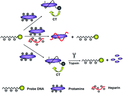
Here we designed a fluorescence sensing strategy for the determination of protamine, heparin and trypsin. A fluorescein isothiocyanate (FITC)-labeled DNA probe could wrap around protamine molecules tightly via electrostatic interactions accompanied by the fluorescence quenching of the DNA probe. In the presence of heparin, protamine preferred to bind to heparin instead of DNA due to the stronger affinity of heparin to protamine, and the fluorescence could be restored. Based on the fact that the protamine which is rich in arginine could be hydrolyzed by the addition of trypsin, the fluorescence intensity of the DNA/protamine complex could also be linked to the trypsin concentration. By measuring the fluorescence intensity changes, the concentrations of protamine, heparin and trypsin were determined. Under the optimized conditions, the linear response range was obtained from 2.5 to 17.5 ng mL−1, 0.156–1.875 ng mL−1 and 62.5 to 10[thin space (1/6-em)]000 ng mL−1 with the low detection limit of 2.2 ng mL−1, 0.078 ng mL−1 and 30.2 ng mL−1 for protamine, heparin and trypsin, respectively.


1. Introduction

Heparin is an anionic rodlike polysaccharide (copolymer of uronic/iduronic acids alternating with sulfated glucosamine residues) with an average molecular weight of approximately 15 kDa.1 The high content of negatively charged sulfo and carboxyl groups endows heparin with the highest negative charge density of any known biological macromolecule.2 Heparin can play a vital role in various normal physiological and pathological processes such as venous thromboembolism, lipid transport and metabolism, cell growth and differentiation, and blood coagulation.2–5 During anticoagulant therapy and surgery, heparin levels should be monitored closely to avoid complications such as hemorrhage or thrombocytopenia induced by heparin overdose.6–8 The therapeutic dosing level of heparin is 2–8 U mL−1 (17–67 μM) during cardiovascular surgery and 0.2–1.2 U mL−1 (1.7–10 μM) in postoperative and long-term care.9

Protamine is a mixture of proteins extracted from salmon roe. It is used in regular clinic as an excipient in insulin formulations10,11 and plays a biological role in binding DNA and providing a highly compact configuration of chromatin in the nucleus of the sperm.12,13 Protamine has a low molecular weight (ca. 4500 Da) and 20 positive charges (pI = 13.8) in physiological condition for its high content of basic arginine residues.14–16 The guanidinium groups of protamine can complex electrostatically with the sulfonate groups of heparin, and thus protamine is used as a well-known heparin antidote to reverse the anticoagulant effect of heparin near the end of most clinical procedures that use heparin for systematic anticoagulation.17

Therefore, it is important to determinate heparin and protamine especially during clinical procedures. To date, numerous methods have been established to monitor heparin and protamine levels. Guo's group reported on voltammetric detection of heparin at polarized 1,2-dichloroethane (1,2-DCE)/water or blood plasma interfaces formed at the tip of micropipette electrodes.18 Wang's group designed a surface-enhanced Raman scattering (SERS) platform for the detection of heparin based on antiaggregation of 4-mercaptopyridine (4-MPY) functionalized silver nanoparticles (AgNPs).19 Awotwe-Otoo's group developed a robust reverse phase-HPLC method to quantify protamine sulfate.20 However, some drawbacks such as complicated extraction, high cost, long operation time and low anti-interference ability existed in these methods limited their applications.

Recently, fluorescence measurements have attracted more attention owing to its low-cost, operational simplicity, high sensitivity, and real-time detection. Some research groups introduced the fluorescence measurements into the determination of protamine and heparin. Egawa et al. developed a fluorescent determination of heparin based on self-quenching of fluorescein-labeled protamine.21 Chem et al. developed a fluorescence turn-on method for selective and sensitive detection for protamine and heparin based on aggregation-induced emission enhancement.22 However, these reported fluorescence detections for protamine and heparin are mostly in the concentration range of μg mL−1.

In this paper, we utilized the FITC-labeled DNA sequence with negative charged as fluorescence probe that could effectively bind to protamine due to the electrostatic attraction.23 The addition of trace amount of protamine could effectively quench the fluorescence of the FITC-labeled DNA probe. In the presence of heparin, protamine preferred to bind to heparin instead of DNA due to the stronger affinity of heparin to protamine, and the fluorescence could be restored. Trypsin, as a serine protease, could cleave exclusively C-terminal to arginine and lysine residues. And the protamine which is rich in arginine could be hydrolyzed after the addition of trypsin to the mixture of DNA and protamine, thus the fluorescence could be recovered.24 Thus, we developed a fluorescence assay for the detection of protamine, heparin and trypsin.

2. Experimental section

2.1 Materials

DNA oligonucleotides were synthesized and purified by TAKARA Biotechnology (Dalian, China). Tris(hydroxymethyl)aminomethane (Tris) was purchased from Sinopharm Chemical Reagent Co. Ltd. (China). Fluorescein isothiocyanate (FITC), TE buffer (pH 8.0), trypsin (1[thin space (1/6-em)]:[thin space (1/6-em)]250) and heparin sodium were purchased from Beijing Dingguo Changsheng Biotechnology Co. Ltd. (Beijing, China). Hemoglobin (bovine erythrocytes, BR) was obtained from Shanghai Kayon Biological Technology Co. Ltd. Protamine sulfate, glucose oxidase (GOX), urease (jack bean), lactate dehydrogenase (LDH), alkaline phosphatase (bovine intestinal mucosa, ALP), bovine serum albumin (BSA) and human serum albumin (HSA) were obtained from Sigma-adrich. All chemicals used were at least of analytical reagent grade and used without further purification. The water used in all experiments had a resistivity higher than 18 MΩ cm−1.

Dye-labeled ssDNA sequence(P):

5′-FITC-TTTGGGTAGGGCGGGTTGGG-3′

AGRO100-FITC:

5′-FITC-GGTGGTGGTGGTTGTGGTGGTGGTGG-3′

The oligonucleotide sequences were dissolved in TE buffer to prepare 250 nM stock solution. All the protein solutions were stored at 4 °C before use.

2.2 Instrument

All fluorescence measurements and spectra were obtained on a Shimadzu RF-5301PC spectrofluorometer (Shimadzu Co., Kyoto, Japan). In this experiment, we used a 1 cm path length quartz cuvette to measure the fluorescence spectrum.

2.3 Fluorescent assays

An aliquot of 2 mL dye-labeled oligonucleotide sequence (P) in Tris–HCl buffer (20 mM, pH 8.2) was prepared to measure the initial fluorescence intensity in the quartz cuvette. The DNA solution was mixed with protamine and incubated for 5 min to record the quenched fluorescence intensity.

For the detection of heparin, different amounts of heparin solutions were mixed with P and a fixed amount of protamine. The fluorescence intensity was detected when the mixture has been incubated for 5 min.

For the detection of trypsin, P was mixed with a fixed amount of protamine and incubated for 5 min prior to the addition of different amounts of trypsin solutions. Fluorescence intensity was detected when the mixture has been incubated for 1 h. All operations were finished at room temperature (RT). The fluorescence intensity was obtained from the emission spectrum at the wavelength of 520 nm, the excitation wavelength was set at 480 nm. The slit widths for excitation and emission were both set at 10 nm.

3. Result and discussion

3.1 Design of the sensing strategy

Scheme 1 illustrated the sensing strategy for the determination of protamine, heparin and trypsin. Due to the electrostatic interaction between DNA sequence and protamine, after the addition of protamine to the DNA probe solution, the DNA chain will wrap around the protamine molecule to form a complex rapidly and such process will last until all of the protamine molecules have been involved in the complex.23 The tight binding between DNA and protamine offered the opportunity for electrons to transfer from FITC to protamine and such charge transfer (CT) process would lead to fluorescence quenching of the DNA probe.25 It is well known that protamine is a highly basic peptide that can form a more stable complex with heparin through electrostatic interactions.26,27 Thereby, protamine would prefer to bind to heparin instead of DNA after the addition of heparin, and the fluorescence of FITC was restored. Because trypsin can catalyze the hydrolysis of protamine,28 the addition of trypsin would reduce the amount of the DNA/protamine complex, and the fluorescence intensity of the solution could also be recovered.
image file: c4ra02936d-s1.tif
Scheme 1 Schematic representation of the fluorescent sensor for the determination of protamine, heparin and trypsin.

3.2 The effect of pH

The pH of solution might have effect on DNA probe and the interaction between DNA and protamine molecules. Thus we studied the effect of pH on the fluorescence quenching of P by protamine. As shown in Fig. 1, the fluorescence of P increased obviously with the increase of pH and reached the maximum at pH 8.6 (curve A), which was due to the pH-dependent fluorescence of FITC.29 In the presence of protamine, the fluorescence intensity of P increased gradually with the increase of pH (curve B). We compared the fluorescence intensity of P in the present and absence of protamine under different pH conditions (curve C), and obtained the maximum quenching efficiency at pH 8.2. So we chose pH 8.2 buffer in the further study.
image file: c4ra02936d-f1.tif
Fig. 1 The effect of pH on the normalized fluorescence intensity of (A) P, (B) P/protamine complex and (C) B/A. Conditions: Tris–HCl buffer (20 mmol L−1), 3.75 nmol L−1 P and 15 ng mL−1 protamine, 5 min for fluorescence quenching.

3.3 Detection of protamine

The formation of DNA/protamine complex was quite fast at room temperature, and the kinetic behavior of FITC-labeled DNA sequences in the presence of protamine (15 ng mL−1) was studied by monitoring fluorescence intensity as a function of time. Fig. 2 shows that in the presence of protamine, the fluorescence of P was quenched rapidly in 30 s. Then the fluorescence intensity decreased slowly and reached a platform after 2 min, indicating that DNA/protamine complex was formed and could be stable in the assay solution. In order to ensure all the protamine molecules bind to P, the mixture of P and protamine would be incubated for 5 min before the fluorescence intensity was recorded.
image file: c4ra02936d-f2.tif
Fig. 2 Fluorescence intensity changes of P in the presence of protamine (15 ng mL−1) with different incubation time. Conditions: Tris–HCl buffer (20 mmol L−1, pH 8.2), 3.75 nmol L−1 P. F and F0 were the fluorescence intensity of P in the presence and absence of protamine, respectively.

Individual DNA molecules exist in an elongated coil state. Upon the addition of protamine, the conformation of DNA molecules changes to a compact state through a partial globule as an intermediate state with an increasing protamine concentration.23 The increasing protamine molecules could make more DNA wrap around protamine tightly and thus assisted the electrons in transferring from P to protamine. Fig. 3 shows the fluorescence spectra changes of P in the presence of different protamine concentrations. The fluorescence intensity decreased linearly upon increasing the concentration of protamine from 2.5 ng mL−1 to 17.5 ng mL−1 (Fig. 3 Inset). The linear regression equation was

 
F/F0 = 1.1342 − 0.06009 [protamine], ng mL−1 (1)
(F and F0 were the fluorescence intensity of P in the presence and absence of protamine, respectively). The corresponding regression coefficient was 0.998 and the limit of detection (LOD) corresponding to the fluorescence intensity of 3 times standard deviation obtained from 11 fluorescence measurements in the absence of analytes was 2.2 ng mL−1. The relative standard deviation for determination (n = 3) of 5 ng mL−1 protamine was 1.0%.


image file: c4ra02936d-f3.tif
Fig. 3 The fluorescence spectra of P in the presence of different concentrations of protamine (0, 2.5, 5, 7.5, 10, 12.5, 15, 17.5, 20 ng mL−1). The inset shows the relationship between F/F0 and the concentration of protamine. Conditions: Tris–HCl buffer (20 mmol L−1, pH 8.2), 3.75 nmol L−1 P, 5 min for fluorescence quenching. F and F0 were the fluorescence intensity of P in the presence and absence of protamine, respectively.

As shown in Fig. S1, the fluorescence intensity of free FITC dye almost remained constant after the addition of protamine for the interaction between FITC molecules and protamine was quite weak. The fluorescence of FITC-labeled ssDNA sequence (P) was obviously quenched by protamine due to the stronger interaction between DNA sequence and protamine.23 FITC-labeled AGRO100 sequence had a weaker fluorescence quenching phenomenon after the addition of protamine, which resulted from its longer sequence length and weaker electron transfer ability.

3.4 Detection of heparin

Under the optimum conditions, we studied the relationship between the fluorescence intensity of P and the concentration of heparin in the presence of 15 ng mL−1 protamine. As shown in Fig. 4, the fluorescence intensity increased with the increase of heparin concentration. The linear regression equation was obtained as the following:
 
F/F0 = 0.19255 + 0.27582 [heparin], ng mL−1 (2)
(F0 was the original fluorescence intensity of P, and F was the fluorescence intensity of P in the presence of heparin and protamine, respectively.) in a concentration range of 0.156–1.875 ng mL−1, with the corresponding regression coefficient 0.994, as shown in Fig. 4 Inset. The limit of detection (LOD) corresponding to the fluorescence intensity of 3 times standard deviation obtained from 11 fluorescence measurements in the absence of analytes was 0.078 ng mL−1. The relative standard deviation for determination (n = 3) of 0.156 ng mL−1 heparin was 1.2%. As mentioned above, the presence of heparin could disturb the formation of DNA/protamine complex due to the stronger affinity of heparin to protamine, thus the addition of heparin would lead to fluorescence recovery. Besides, the average molecular weight of protamine (about 4500 Da) is smaller than that of heparin (about 15[thin space (1/6-em)]000 Da)30 and heparin is a linear polysaccharide.31 This makes one heparin molecule able to bind several protamine molecules.32 Therefore, the limit of detection for heparin could be as low as 0.078 ng mL−1.

image file: c4ra02936d-f4.tif
Fig. 4 The fluorescence spectra of P and protamine in the presence of different concentrations of heparin (0, 0.156, 0.312, 0.625, 0.938, 1.562, 1.875, 2.5, 3.125, 3.75 ng mL−1). The inset shows the relationship between F/F0 and the concentration of heparin. Conditions: Tris–HCl buffer (20 mmol L−1, pH 8.2), 3.75 nmol L−1 P and 15 ng mL−1 protamine, 5 min for fluorescence quenching. F0 was the original fluorescence intensity of P, and F was the fluorescence intensity of P in the presence of heparin and protamine, respectively.

Compared with other methods for the determination of heparin (Table 1),18,19,26,27,33–36 our method had a superior LOD and linear range, and was extremely sensitive for heparin detection.

Table 1 Comparison of different methods for the determination of heparin
Method Linear range Detection limit Ref.
Electrochemistry 0.012 U mL−1 (saline solution) 18
0.13 U mL−1 (sheep blood plasma)
Electrochemistry 4.0–22.0 μg mL−1 0.28 μg mL−1 33
SERS 0.5–150 ng mL−1 (standard aqueous solution) 0.5 ng mL−1 19
1–400 ng mL−1 (fetal bovine serum)
Colorimetry 0.06–0.36 μg mL−1 3.0 ng mL−1 34
Colorimetry 0.02–0.28 μg mL−1 5 ng mL−1 35
Colorimetry 0.09–3.12 μg mL−1 0.03 μg mL−1 (water) 36
0.3–7.0 μg mL−1 0.1 μg mL−1 (serum)
UV-vis spectrophotometry 0.6–10 μg mL−1 0.6 μg mL−1 27
Fluorometry 0.004–1.6 μg mL−1 0.0013 μg mL−1 26
Fluorometry 0.156–1.875 ng mL−1 0.078 ng mL−1 This work


3.5 Detection of trypsin

As shown in Scheme 1, the protamine could be hydrolyzed in the presence of trypsin, which would reduce the amount of the DNA/protamine complex. Therefore, the P/protamine system has the potentials for the detection of trypsin. As shown in Fig. S2, we studied the changes of fluorescence intensity at 520 nm as a function of time at several trypsin concentrations in presence of a fixed amount of P and protamine (15 ng mL−1). With the increasing of trypsin concentration from 62.5 ng mL−1 to 1.875 μg mL−1, the hydrolysis rate increased, and the fluorescence intensity gradually increased with the increasing incubation time. To make sure that the hydrolyzed protamine was as much as possible, the incubation process would last for 1 h in the further study.

We investigated the effect of temperature on the hydrolysis catalyzed by trypsin. As shown in Fig. S3, in the presence of trypsin, different incubation temperature would lead to varying degrees of fluorescence recovery. The fluorescence intensity of P could recover to above 80% at room temperature (RT) and 37 °C. When the temperature further raised to 50 °C, the fluorescence intensity only recovered a little due to the low hydrolysis capability of trypsin at high temperature. The results demonstrated that the hydrolysis capability of the trypsin at room temperature was similar to that at 37 °C in our assay system, thus we chose RT as the incubation temperature in trypsin detection.

Under the optimized conditions, the fluorescence spectra of the P/protamine complex with different concentrations of trypsin were shown in Fig. S4. The fluorescence intensity had a linear relationship with the concentration of trypsin in the range of 62.5–10[thin space (1/6-em)]000 ng mL−1. The linear regression equation was

 
F/F0 = 0.24247l g [trypsin] − 0.16117, ng mL−1 (3)
with the correlation coefficient of 0.995, as shown in Fig. S4 inset. F0 was the original fluorescence intensity of P, and F was the fluorescence intensity of P in the presence of trypsin and protamine, respectively. The limit of detection (LOD) corresponding to the fluorescence intensity of 3 times standard deviation obtained from 11 fluorescence measurements in the absence of analytes was 30.2 ng mL−1. The relative standard deviation for determination (n = 3) of 125 ng mL−1 trypsin was 3.1%. Compared to previous reports for trypsin determination,37,38 our method has the superior or similar linear range and detection limit.

In our assay strategy, both heparin and trypsin could lead to fluorescence recovery of DNA probe. In the case that heparin and trypsin coexist in the same solutions, heparinase and BBI inhibitor [trypsinchymotrypsin inhibitor] can be adopted to distinguish the two analytes. Heparinase can specifically hydrolyze heparin into small fragments39 and the BBI inhibitor is able to retard the hydrolysis of protamine by trypsin.40 Besides, the sensitivity and linear range in the detection of heparin were quite different from that of trypsin. Thus our assay strategy has the potential to distinguish and detect the two analytes.

3.6 Selectivity

We investigated the selectivity of the designed fluorescent sensing method. Fig. S5 shows the fluorescence intensity of P with several foreign substances in the presence and absence of protamine, respectively. The results show that the common physiological ions and proteins had nearly no obvious interference with the fluorescence intensity of P and the detection for protamine. Then we studied the effect of five different enzymes on the hydrolysis of protamine. The results shown in Fig. S6 clearly demonstrated that under our experiment condition, only trypsin could catalyze the hydrolysis of protamine and effectively recover the fluorescence of P quenched by protamine. Thus we could conclude that our proposed method has good selectivity and attractive specificity.

4. Conclusion

In this paper, we reported a simple, fast and sensitive fluorescence sensing strategy for the determination of protamine, heparin and trypsin based on the electrostatic interaction between DNA, protamine and heparin. It could be used for the determination of heparin in the concentration range from 0.156 to 1.875 ng mL−1. Compared to previous reports, the designed method owns low detection limits and high selectivity.

Acknowledgements

This work was financially supported by the National Natural Science Foundation of China (no. 21075050, no. 21275063).

Notes and references

  1. I. Danishefsky, L. Rosenfeld, L. Kuhn, B. Lahiri and C. Whyzmuzis, in Heparin and Related Polysaccharides, Structure and Activities, Ann. New York Acad. Sci. 556, ed. F. A. Ofosu, et al., The New York Academy of Sciences, New York, 1989, p. 29 Search PubMed .
  2. I. Capila and R. J. Linhardt, Angew. Chem., Int. Ed., 2002, 41, 390–412 CrossRef CAS .
  3. N. Mackman, Nature, 2008, 451, 914–918 CrossRef CAS PubMed .
  4. J. Fareed, D. A. Hoppensteadt and R. L. Bick, Semin. Thromb. Hemostasis, 2000, 26, 5–21 CrossRef CAS PubMed .
  5. J. M. Whitelock and R. V. Iozzo, Chem. Rev., 2005, 105, 2745–2764 CrossRef CAS PubMed .
  6. T. E. Warkentin, M. N. Levine, J. Hirsh, P. Horsewood, R. S. Roberts, M. Gent and J. G. Kelton, N. Engl. J. Med., 1995, 332, 1330–1335 CrossRef CAS PubMed .
  7. B. Girolami and A. Girolami, Semin. Thromb. Hemostasis, 2006, 32, 803–809 CrossRef CAS PubMed .
  8. G. J. Despotis, G. Gravlee, K. Filos and J. Levy, Anesthesiology, 1999, 91, 1122–1151 CrossRef CAS PubMed .
  9. R. Y. Zhan, Z. Fang and B. Liu, Anal. Chem., 2010, 82, 1326–1333 CrossRef CAS PubMed .
  10. J. Brange and L. Langkjaer, Acta Pharm. Nord., 1992, 4, 149–158 CAS .
  11. J. Brange and L. Langkjar, Pharm. Biotechnol., 2002, 10, 343–410 Search PubMed .
  12. G. Fuentes-Mascorro, H. Serrano and A. Rosado, Arch. Androl., 2000, 45, 215–225 CrossRef CAS .
  13. M. L. Meistrich, B. Mohapatra, C. R. Shirley and M. Zhao, Chromosoma, 2003, 111, 483–488 CrossRef PubMed .
  14. A. Shvarev and E. Bakker, J. Am. Chem. Soc., 2003, 125, 11192–11193 CrossRef CAS PubMed .
  15. T. Ando, M. Yamasaki and K. Suzuki, inProtamine: molecular biology, biochemistry and biophysics, ed. A. Kleinzeller, Springer-Verlag, 1973, vol. 12, pp. 1–109 Search PubMed .
  16. Y. Kim and C. A. Rose, Pharm. Res., 1995, 12, 1284–1288 CrossRef CAS .
  17. L. B. Jaques, Pharma Rev., 1980, 31, 99–166 Search PubMed .
  18. J. D. Guo, Y. Yuan and S. Amemiya, Anal. Chem., 2005, 77, 5711–5719 CrossRef CAS PubMed .
  19. X. K. Wang, L. Chen, X. L. Fu, L. X. Chen and Y. J. Ding, ACS Appl. Mater. Interfaces, 2013, 5, 11059–11065 CAS .
  20. D. Awotwe-Otoo, C. Agarabi, P. J. Faustino, M. J. Habib, S. Lee, M. A. Khan and R. B. Shah, J. Pharm. Biomed. Anal., 2012, 62, 61–67 CrossRef CAS PubMed .
  21. Y. Egawa, R. Hayashida, T. Seki and J. Anzai, Talanta, 2008, 76, 736–741 CrossRef CAS PubMed .
  22. X. T. Chen, Y. Xiang, N. Li, P. S. Song and A. J. Tong, Analyst, 2010, 135, 1098–1105 RSC .
  23. N. Makita, Y. Yoshikawa, Y. Takenaka, T. Sakaue, M. Suzuki, C. Watanabe, T. Kanai, T. Kanbe, T. Imanaka and K. Yoshikawa, J. Phys. Chem. B, 2011, 115, 4453–4459 CrossRef CAS PubMed .
  24. L. Hedstrom, Chem. Rev., 2002, 102, 4501–4523 CrossRef CAS PubMed .
  25. J. H. Choi, K. H. Chen and M. S. Strano, J. Am. Chem. Soc., 2006, 128, 15584–15585 CrossRef CAS PubMed .
  26. J. Zhao, Y. Yi, N. Mi, B. Yin, M. Wei, Q. Chen, H. Li, Y. Zhang and S. Yao, Talanta, 2013, 116, 951–957 CrossRef CAS PubMed .
  27. B. K. Jena and C. R. Raj, Biosens. Bioelectron., 2008, 23, 1285–1290 CrossRef CAS PubMed .
  28. L. X. Chen, X. L. Fu and J. H. Li, Nanoscale, 2013, 5, 5905–5911 RSC .
  29. E. Lanz, M. Gregor, J. Slavik and A. Kotyk, J. Fluoresc., 1997, 7, 317–319 CrossRef CAS .
  30. A. Shvarev and E. Bakker, Anal. Chem., 2005, 77, 5221–5228 CrossRef CAS PubMed .
  31. R. J. Linhardt, J. Med. Chem., 2003, 46, 2551–2564 CrossRef CAS PubMed .
  32. S. A. Buchanan, T. P. Kennedy, R. B. MacArthur and M. E. Meyerhoff, Anal. Biochem., 2005, 346, 241–245 CrossRef CAS PubMed .
  33. H. Y. Huo, H. Q. Luo and N. B. Li, Microchim. Acta, 2009, 167, 195–199 CrossRef CAS .
  34. X. L. Fu, L. X. Chen and J. H. Li, Analyst, 2012, 137, 3653–3658 RSC .
  35. X. L. Fu, L. X. Chen, J. H. Li, M. Lin, H. Y. You and W. H. Wang, Biosens. Bioelectron., 2012, 34, 227–231 CrossRef CAS PubMed .
  36. R. Cao and B. X. Li, Chem. Commun., 2011, 47, 2865–2867 RSC .
  37. K. H. Xu, F. Liu, J. Ma and B. Tang, Analyst, 2011, 136, 1199–1203 RSC .
  38. Y. Chen, J. W. Ding and W. Qin, Bioelectrochemistry, 2012, 88, 144–147 CrossRef CAS PubMed .
  39. Z. P. Liu, Q. Ma, X. Y. Wang, Z. H. Lin, H. Zhang, L. L. Liu and X. G. Su, Biosens. Bioelectron., 2014, 54, 617–622 CrossRef CAS PubMed .
  40. X. G. Gu, G. Yang, G. X. Zhang, D. Q. Zhang and D. B. Zhu, ACS Appl. Mater. Interfaces, 2011, 3, 1175–1179 CAS .

Footnote

Electronic supplementary information (ESI) available. See DOI: 10.1039/c4ra02936d

This journal is © The Royal Society of Chemistry 2014
Click here to see how this site uses Cookies. View our privacy policy here.