Coordination variability of CuI in multidonor heterocyclic thioamides: synthesis, crystal structures, luminescent properties and ESI-mass studies of complexes

Tarlok S. Lobana*a, Amanpreet K. Sandhua, Razia Sultanaa, Alfonso Castineirasb, Ray J. Butcherc and Jerry P. Jasinskid
aDepartment of Chemistry, Guru Nanak Dev University, Amritsar – 143 005, India. E-mail: tarlokslobana@yahoo.co.in; Fax: +91-183-2258820
bDepartamento de Quimica Inorganica, Facultad de Farmacia, Universidad de Santiago, 15782-Santiago, Spain
cDepartment of Chemistry, Howard University, Washington DC, 20059, USA
dDepartment of Chemistry, Keene State College, Keene NH 03435-2001, USA

Received 28th March 2014 , Accepted 20th June 2014

First published on 20th June 2014


Abstract

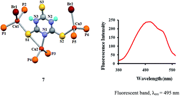
The chemistry of copper(I) with scarcely investigated heterocyclic thioamides, particularly 2,4,6-trimercaptotriazine, purine-6-thione, 2,4-dithiouracil, 2-thiouracil and pyrimidine-2-thione is described. The interaction of 2,4,6-trimercaptotriazine (tmtH3) with [Cu(CH3COO)(PPh3)2] resulted in a pair of bond isomers: [Cu(κ1N-tmtH2)(PPh3)2] (6a), [Cu(κ1N,κ1S-tmtH2)(PPh3)2] (6b); with copper(I) bromide and PPh3, it formed a trinuclear complex, [Cu3Br21N,κ1S, μ-S-tmtH2)(PPh3)6] (7) with anionic tmtH2. The 2,4-dithiouracil with copper(I) halides (CuCl, CuBr) and PPh3 yielded dinuclear complexes: [Cu2(μ-Cl)(κ1S,κ1S-dtucH)(PPh3)4] (4) and [Cu2(μ-Br)(κ1S,κ1S-dtucH)(PPh3)4] (5) with unusual eight-membered metallacyclic rings. Pyrimidine-2-thione (pymSH) is an N,S-chelated anion in a mononuclear complex, [Cu(κ1N,κ1S-pymS)(PPh3)2] (1), whereas 2-thiouracil (tucH2) with copper(I) chloride and PPh3 yielded a tetrahedral complex, [CuCl(κ1S-tucH2)(PPh3)2] (3). Purine-6-thione (purSH2) coordinated to CuI in two different modes yielding mono- and di-nuclear complexes, [Cu(κ1N,κ1S-purS)(PPh3)2]·CH3OH (2a) and [Cu21N,μ-S-purS)2(PPh3)2] (2b). The existence of bond isomers (6a and 6b), synthesis of novel dinuclear (4 and 5) and rare trinuclear (7) complexes with unusual bonding patterns are the novel features of the present study. Complexes show intense emission bands in the 380–530 nm region with λem at 490 to 495 nm.


Introduction

The coordination chemistry of heterocyclic thioamides with transition/main-group metals has been the focus of numerous studies, particularly with pyridine-2-thiones, imidazolidine-2-thiones, thiazolidine-2-thiones and a few other allied bases. These studies deal with the synthesis and structures of a variety of compounds, as well as their biochemical and other applications.1–29 Among applications, copper complexes have shown antitumor, anti-inflammatory and antimicrobial properties.2,16–18 There is keen interest in the chemistry of Cu(I)/Cu(II) with analogous N-based ligands because their complexes with 6-(2-chlorobenzylamino)purine,30 2,2′-biquinolines,31 substituted guanidines,32 phenanthroline33 and quinolin-2(1H)-one-derived Schiff bases34 have shown anticancer, antitumor, antifungal, antimicrobial and antibacterial properties.

Considering the importance of copper complexes with N/S donor ligands, it was decided to pursue copper chemistry with multifunctional heterocyclic thioamides, as shown in Scheme 1, having the propensity to bind to more than one metal ion. Coordination chemistry of copper(I) with pyrimidine-2-thione,8–13,35 purine-6-thione,36–38 2-thiouracil,39 2,4-dithiouracil40,41 and 2,4,6-trimercaptotriazine is limited.35,42,43 Herein, it is planned to react copper(I) salts with multifunctional heterocyclic thioamides (Scheme 1) with the objective of obtaining mono-, di- and polynuclear copper complexes by changing the number of donor atoms in organic thiobases. In the preparation of complexes, the reactions of pyrimidine-2-thione (pymSH, I), purine-6-thione (purSH2, II), 2,4,6-trimercaptotriazine (tmtH3, III), 2-thiouracil (tucH2, IV) and 2,4-dithiouracil (dtucH2, V) (Scheme 1) were carried out with copper(I) halides/Cu(CH3COO)(PPh3)2. The resulting isolated mono-, di- and trinuclear complexes isolated were characterized using analytical data, spectroscopy, ESI-MS spectrometry and X-ray crystallography.


image file: c4ra02748e-s1.tif
Scheme 1 Molecular structures of thiol ligands under study.

Results and discussion

Synthesis and IR spectroscopy

Scheme 2 shows a bonding pattern of the synthesized complexes. In the synthesis of complexes 1, 2 and 6, no refluxing was carried out because high temperature led to the formation of a solid which could not be crystallized, and thus the reaction mixture was kept undisturbed at room temperature. For the synthesis of complexes 3, 4, 5 and 7, a copper(I) halide was first reacted with a thio-ligand under magnetic stirring at room temperature, followed by the addition of PPh3. Slow evaporation of the resulting solution gave pale yellow (3), light orange (4, 5) or yellow (7) crystals. The reaction of pyrimidine-2-thione (pymSH) with [Cu(CH3COO)(PPh3)2] involved the removal of the N–H proton and the anionic pymS chelated to the {Cu(PPh3)2} moiety yielding the complex, [Cu(κ1N,κ1S-pymS)(PPh3)2] (1). Similarly, in the reaction of purine-6-thione (purSH2) with [Cu(CH3COO)(PPh3)2], the acetate removed one proton from an –NH– group.
image file: c4ra02748e-s2.tif
Scheme 2 A view of bonding pattern of complexes synthesized.

The anionic purSH coordinated to CuI in two different modes yielding mono- and di-nuclear complexes, [Cu(κ1N,κ1S-purS)(PPh3)2]·CH3OH (2a) and [Cu21N,μ-S-purS)2(PPh3)2] (2b) from the same reaction. The crystals of 2a and 2b were manually separated. The interaction of tmtH3 with [Cu(CH3COO)(PPh3)2] resulted in a pair of bond isomers: [Cu(κ1N-tmtH2)(PPh3)2] (6a) and [Cu(κ1N,κ1S-tmtH2)(PPh3)2] (6b) in the same unit cell (X-ray crystallography, vide infra). The reaction of tmtH3 with copper(I) bromide in the presence of two moles of PPh3 involved the removal of one –NH– hydrogen and uninegative tmtH2, yielding a trinuclear complex, [Cu3Br21N,κ1S, μ-S-tmtH2)(PPh3)6] (7). The reaction of 2-thiouracil with copper(I) chloride in the presence of two moles of PPh3 yielded light yellow crystals of a mononuclear complex, [CuCl(κ1S-tucH2)(PPh3)2] (3). The reaction of 2,4-dithiouracil with copper halides (CuCl, CuBr) in the presence of two moles of PPh3 also removed one –NH– hydrogen and reacted with two CuX units yielding yellow complexes, [Cu2(μ-Cl)(κ1S,κ1S-dtucH)(PPh3)4] (4) and [Cu2(μ-Br)(κ1S,κ1S-dtucH)(PPh3)4] (5) with unusual bonding patterns. All the complexes except 2b were soluble in organic solvents such as chloroform and dichloromethane.

The IR spectral data of complexes is given in the Experimental section and that of the ligands is provided in the ESI. The ν(N–H) band of the free pyrimidine-2-thione ligand at 3078 cm−1 disappeared in complex 1. Similarly, the diagnostic ν(C–S) band at 1187 cm−1 shifted to 1026 cm−1 in the complex, which revealed that the thio-ligand is coordinating as anion through its N and S donors. The ν(N–H) bands in complexes 2–7 occur in the region 3082–3250 cm−1, which are at different positions from those of the thio-ligands. In complex 3, the ν(O–H) band is assigned to 3447 cm−1. In complexes 27, the ν(C–S) bands are assigned in the region 844–1184 cm−1, which are generally at low-energy regions relative to the respective free thio-ligand. The ν(P–CPh) bands in the region of 1094 to 1091 cm−1 indicated the presence of PPh3 in the complexes.

Molecular structures

The crystal data of complexes are given in Table 1, and their important bond parameters are given in Table 2. Complexes 1, 2b, 3–5 and 7 crystallized in the triclinic crystal system with the space group P[1 with combining macron], whereas other complexes (2a, 6) crystallized in the monoclinic crystal system with the space groups P21/n (2a) and P21(6).
Table 1 Crystal data for complexes 1–7
  1 2a 2b 3
Empirical formula C40H33N2CuP2S C42H37N4OCuP2S C46H36N8Cu2P2S2 C40H34ClCuN2OP2S
M 699.24 771.30 953.97 751.68
T/K 296(2) 173(2) 295(2) 170(2)
Crystal system Triclinic Monoclinic Triclinic Triclinic
Space group P[1 with combining macron] P21/n P[1 with combining macron] P[1 with combining macron]
a(Å) 9.349(3) 9.0783(2) 12.6132(6) 10.8164(9)
b(Å) 11.140(4) 25.2036(6) 12.6674(6) 13.1393(10)
c(Å) 18.860(6) 16.9815(5) 17.0373(7) 14.7805(10)
α (°) 93.050(13) 88.319(3) 76.189(6)
β (°) 92.208(12) 102.171(3) 69.754(4) 81.704(6)
γ (°) 113.907(11) 65.581(4) 67.452(7)
V3) 1789.2(10) 3798.13(17) 2304.8(2) 1880.7(2)
Z 2 4 2 2
Dcalcd (g cm−3) 1.298 1.349 1.375 1.327
μ (mm−1) 0.788 0.753 2.966 0.826
F(000) 724 1600 976 776
Reflections collected 19105 39221 17070 19880
Unique reflections 7179 (Rint,0.0292) 9794 (Rint,0.0222) 9009 (Rint,0.0388) 9706 (Rint,0.0205)
Data/restraints/parameters 7179/0/416 9794/2/466 9009/0/544 9706/2/439
Reflens. With [I > 2σ(I)] 5988 8534 6944 7531
 
R Indices [I > 2σ(I)]
R1 0.0340 0.0395 0.0556 0.0433
WR2 0.0837 0.1027 0.1583 0.0960
 
R Indices (all data)
R1 0.0470 0.0481 0.0715 0.0660
WR2 0.1031 0.1095 0.1757 0.1122
Largest diff. Peak and hole 0.453 and −0.412 e Å−3 0.360 and −0.219 e Å−3 0.763 and −0.355 e Å−3 0.546 and −0.381 e Å−3

  4 5 6 7
Empirical formula C76H63ClCu2N2P4S2 C76H63BrCu2N2P4S2 C39H33N3O0.50CuP2S3 C111H92Br2Cu3N3P6S3
M 1354.81 1399.27 773.34 2100.32
T/K 173(2) 123(2) 100 (2) 173(2)
Crystal system Triclinic Triclinic Monoclinic Triclinic
Space group P[1 with combining macron] P[1 with combining macron] P21 P[1 with combining macron]
a(Å) 17.6065(5) 17.5439(3) 12.864(4) 12.8825(5)
b(Å) 18.9904(6) 18.7998(3) 16.167(5) 13.2054(5)
c(Å) 22.8262(7) 22.7125(4) 17.506(5) 33.4043(1)
α (°) 66.994(3) 67.538(2) 93.2233(3)
β (°) 69.512(3) 69.421(2) 95.271(4) 96.511(3)
γ (°) 81.169(3) 80.7420(10) 119.084(4)
V3) 6579.4(3) 6477.95(19) 3625.4(19) 4893.4(3)
Z 4 4 4 2
Dcalcd (g cm−3) 1.368 1.435 1.417 1.425
μ (mm−1) 0.893 1.483 0.898 1.675
F(000) 2800 2872 1596 2148
Reflections collected 77752 134549 42507 50419
Unique reflections 31317 (Rint,0.0475) 65025 (Rint,0.0641) 14793 (Rint,0.0473) 23304 (Rint,0.0341)
Data/restraints/parameters 31317/2/1573 65025/0/1573 14793/1/875 23304/4/1124
Reflens. With [I > 2σ(I)] 19640 37449 12435 17703
 
R Indices [I > 2σ(I)]
R1 0.0542 0.0683 0.0495 0.0679
WR2 0.1041 0.1416 0.1121 0.1345
 
R Indices (all data)
R1 0.1037 0.1342 0.0675 0.0932
WR2 0.1234 0.1722 0.1215 0.1475
Largest diff. Peak and hole 0.849 and −0.497 e Å−3 1.480 and −2.228 e Å−3 1.012 and −0.904 e Å−3 1.529 and −1.227 e Å−3


Table 2 Important bond lengths(Å) and bond angles(°)b
Mono-nuclear complexes (1, 2a, 3, 6a, 6b)
  1 2a 3 6aa 6ba
a A case of bond isomerism (6a and 6b) in the crystal lattice.b Each of the complexes, 4 and 5, has independent molecules in the asymmetric unit.
Cu–N 2.144(2) 2.130(2) 2.015(4) 2.008(4)
Cu–S 2.419(1) 2.433(1) 2.381(1) 3.144(2) (Cu2–S22) 2.879(2) (Cu1–S12)
Cu–P 2.262(1), 2.251(1) 2.262(1), 2.277(1) 2.281(1), 2.256(1) 2.249(1), 2.273(1) 2.220(1), 2.254(1)
S–C 1.711(3) 1.715(2) 1.666(2) 1.680(5), 1.680(5), 1.652(5) 1.681(5), 1.664(5), 1.663(5)
N–Cu–S 69.10(6) 88.24(5) 56.14(12) 61.59(12)
P–Cu–P 126.25(3) 126.08(2) 124.53(3) 125.88(5) 120.03(5)
Cu–S–C 78.98(9) 93.60(7) 106.18(8) 69.40(17)

Dinuclear complexes (2b, 4 and 5)
2b Molecule 1 Molecule 2   Molecule 1 Molecule 2
Cu–N 2.036(3) 2.058(3) N–Cu–S 85.85(8), 101.43(8) 87.66(9), 105.87(11)
Cu–S 2.362(1), 2.608(1) 2.345(1), 2.535(1) Cu–S–Cu, Cu–S–C 70.60(3), 89.85(13) 67.52(4), 90.88(12)
Cu–P 2.223(1) 2.215(1) S–Cu–S 109.40(3) 112.48(4)
Cu–Cu 2.880(1) 2.717(1) N–Cu–P 127.62(8) 117.64(11)
S–C 1.710(3) 1.739(3) P–Cu–S 103.35(3), 122.19(4) 110.98(5), 118.24(4)

4 Molecule 1 Molecule 2 5 Molecule 1 Molecule 2
Cu–S 2.330(1), 2.344(1) 2.316(1), 2.362(1) Cu–S 2.356(1), 2.311(1) 2.321(1), 2.337(1)
Cu–P 2.287(1), 2.283(1) 2.289(1), 2.290(1) Cu–P 2.281(1), 2.283(1) 2.265(1), 2.291(1)
Cu–Cl 2.439(1) 2.462(1) Cu–Br 2.553(1) 2.554(1)
S–C 1.693(3), 1.701(3) 1.695(3), 1.711(3) S–C 1.717(3) 1.708(3)
Cu–S–C 112.14(11), 109.33(11) 110.58(11) 106.47(12) Cu–S–C 107.42(9), 110.56(9) 112.94(9), 109.76(9)
Cu–Cl–Cu 146.92(4), (Cl) 145.94(4), (Cl) Cu–Br–Cu 139.89(2), (Br) 140.94(2), (Br)
P–Cu–S 113.20(4), 106.13(3), 114.06(3), 105.66(3) 113.66(4), 105.52(4), 114.57(3), 104.43(3) P–Cu–S 115.45(3), 104.25(3) 114.96(3) 105.86(2)
P–Cu–P 122.44(3), 121.14(3) 122.66(3), 122.36(3) P–Cu–P 122.76(3), 122.77(2) 122.35(2), 121.31(3)
S–Cu–Cl 109.23(3), 110.38(3) 106.98(3), 108.82(3) S–Cu–Br 109.73(2) 111.21(2)

Trinuclear complex (7)
Cu–N 2.080(3) S–C 1.693(4), 1.672(4), 1.631(4) N–Cu–S 62.93(9)
Cu–S 2.395(1), 2.769(1) (Cu2–S1), 2.423(1) Cu–Br 2.478(), 2.477(1) P–Cu–P 121.19(4), 124.61(5)
Cu–P 2.270(1), 2.293(1), S–Cu–Br 104.32(3), 109.13(3) Cu–S–C 72.03(14), 109.84(14), 114.30(14)


Mononuclear complexes

The pyrimidine-2-thiolate chelates to the copper atom in complex 1 through its N1, S– donor atoms with the N–Cu–S bite angle of 69.10(6)° being the shortest, whereas the P–Cu–P angle {126.25(3)°} is the largest among the various angles around the Cu atom (Fig. 1). Purine-6-thiolate also chelates to the copper atom in 2a through its N7, S– donor atoms with the N–Cu–S bite angle of 88.24(5)° being the shortest, whereas the P–Cu–P angle {126.081(19)°} is the largest among angles around the Cu atom (Fig. 2). The geometry is severely distorted from the tetrahedron in two cases; however, the Cu–N, Cu–S and Cu–P bond distances are similar. Methanol was hydrogen-bonded to N4 nitrogen with an N–O distance of 2.801 Å, which is less than the sum of the van der Waals radius of N and O, viz. 3.05 Å, indicating weak O–H⋯N interaction.44 The thiouracil did not chelate to CuI in complex 3, rather it bonded to the metal atom as a neutral ligand through its S-donor atom, whereas other donor atoms remained unbound (Fig. 3). As before, the P–Cu–P angle is the largest {124.53(3)°}, whereas other angles around the metal center fall in a narrow range, 103–109°. Complexes 6a and 6b exist as bond isomers in the same unit cell (Fig. 4). In 6a, the tmtH2 as mono anion is bonded to CuI through its one N donor atom only forming a three coordinate complex, and in 6b it is bonded to one N donor and to one S donor atom. Whereas, the Cu–N bond distances in 6a and 6b are nearly equal, although shorter than in 1 and 2a, the Cu–S distance of 2.879(2) Å in 6b is significantly longer than that in other mononuclear complexes (1, 2a, 3). In three-coordinate 6a, the Cu–S distance of 3.144(2) Å is still longer than 6b. The expected Cu–S bond distance is 2.61 Å, based on the ionic radii of Cu+ (0.91 Å) and S2− (1.70 Å), and the Cu–S distances found in 6a and 6b complex species are significantly longer and reveal a weak Cu–S bond.44 The Cu–S bonds in other complexes are similar to the literature trends.1–15
image file: c4ra02748e-f1.tif
Fig. 1 Molecular structure of [Cu(κ1N,κ 1S-pymS)(PPh3)2] 1.

image file: c4ra02748e-f2.tif
Fig. 2 Molecular structure of [Cu(κ1N,κ1S-purSH)(PPh3)2]·CH3OH 2a.

image file: c4ra02748e-f3.tif
Fig. 3 Molecular structure of [CuCl(κ1S-tucH2)(PPh3)2] (3).

image file: c4ra02748e-f4.tif
Fig. 4 Molecular structures of Cu(κ1N-tmtH2)(PPh3)2] 6a, [Cu(κ1N,κ1S-tmtH2)(PPh3)2]·H2O 6b (bond isomers).

Di- and trinuclear complexes

Complex 2a appears to lose one PPh3 forming species, {Cu(purSH)(PPh3)}, which dimerized via sulfur to yield the dinuclear complex 2b (Fig. 5). It exists as two independent entities: molecule 1 and molecule 2 in the asymmetric unit. Two molecules differ significantly in various bond lengths and bond angles, particularly pertaining to the bridging sulfur donor atoms. The N–Cu–S bite angles decrease to 85.85(8)° and 87.66(9)° vis-à-vis that 88.24(5)° of 2a. The central cores Cu2S2 that have unequal Cu–S distances form parallelograms and have Cu–S–Cu and S–Cu–S bond angles of 70.60(3)/67.52(4)° and 109.40(3)/112.48(4)° of molecules 1 and 2, respectively, and are typical of such cores observed in different dinuclear complexes.47 Compound 4, [Cu2(μ-Cl)(κ1S,κ1S-dtucH)(PPh3)4], exists as two independent molecules in the asymmetric unit with small differences in various bond parameters. In this hetero-bridged dinuclear complex, each copper is bonded to one S donor atom of dtucH anion, two P donor atoms of two PPh3 molecules, and one bridging μ-Cl (Fig. 6a and b). The binding pattern of complex [Cu2(μ-Br)(κ1S,κ1S-dtucH)(PPh3)4] (5) is similar to that of complex 4 (Fig. 7a and b). Each metal center acquires tetracoordination in a distorted tetrahedral geometry. The trinuclear complex, [Cu3Br21N,κ1S,μ-S-tmtH2)(PPh3)6] 7 has three types of Cu metal ions differently bonded (Fig. 8 and b). Here, Cu1 is bonded to one Br, two P and one bridging S donor atoms; Cu3 is bonded to one Br, two P and one S donor atoms; and finally, Cu2 is bonded to two P, one N and one bridging S donor atoms. The Cu2–S1 distance is the longest among various Cu–S distances found in complexes 1, 2, 3–5, except 6b, which is similarly bonded but has a longer distance than that observed in 7.
image file: c4ra02748e-f5.tif
Fig. 5 Molecular structure of [Cu21N,κ2S-purSH)2(PPh3)2] 2b.

image file: c4ra02748e-f6.tif
Fig. 6 (a) Molecular structure of dinuclear [Cu22Cl)(κ1S,κ1S-dtucH)(PPh3)4] (4). (b) Skeletal structure of 4 (phenyl rings omitted).

image file: c4ra02748e-f7.tif
Fig. 7 (a) Molecular structure of dinuclear [Cu22Br)(κ1S,κ1S-dtucH)(PPh3)4] (5). (b) Skeletal structure of 5 (phenyl rings omitted).

image file: c4ra02748e-f8.tif
Fig. 8 (a) Molecular structure of [Cu3Br21N,κ1S,κ2S-tmtH2)(PPh3)6] (7). (b) Skeletal view of 7(phenyl rings omitted).

Variations in C–S, Cu⋯Cu bond lengths and Cu–S–C bond angles

It is known that the C[double bond, length as m-dash]S double-bond length is 1.62 Å, whereas the C–S single-bond length is 1.81 Å.44,45 A comparison of C–S bond distances observed in different complexes (Table 2) reveal a wide variation from 1.631(4) to 1.739(3) Å. In complex 3, the thio-ligand tucH2 binds to CuI as a neutral ligand, and the C–S distance of 1.666(2) Å exhibits a weakening of this bond on coordination but there is considerable double-bond character. Complexes 1, 2, 4 and 5 with pymS(1), purSH(2) and dtucH(4, 5) uninegative anionic thio-ligands have C–S distances in the range of 1.693(3) (4, molecule 1)-1.739(3) (2b, molecule 2), suggesting significantly larger weakening of C–S bonds in the complexes. The C–S distances in three-coordinated 6a and tetracoordinated 6b are highly similar and have considerable double-bond character. Finally, in trinuclear complex 7, all three C–S distances are distinctly separate: 1.693(4) Å (bridging S), 1.672(4) Å (terminal S) and 1.631(4) Å (uncoordinated S). Regarding the Cu⋯Cu separation, the shortest distances are found in molecule 1{2.880(1) Å} and molecule 2 {2.717(1) Å}, which are comparable with 2.80 Å, which is twice the sum of the van der Waals radius of Cu.46–48 In other cases, this metal-metal separation was well above 3.0 Å and is not cited in this paper.

The Cu–S–C angle of 78.98(9) in 1 changes to 93.60(7)° in 2a, the cases in which thio-ligands are N,S-chelating. When the thio-ligand is N,S-chelating –cum-S bridging as in 2b, the Cu–S–C angles slightly contract to 89.85(13) and 90.88(12)°. The Cu–S–C angle further changes to 69.40(17)° in 6b in which there is N,S-chelation. This angle lies in the range of 106–113° in the complexes 3, 4 and 5 where a thione sulfur is terminally (unidentate) bonded. Finally, the Cu–S–C angles in trinuclear cluster 7 are 72.03, 109.84(14) and 114.30(14)° depending on whether copper metal is chelated (Cu2, N,S-chelation), bonded to bridging S (Cu1) or nonbridging S(Cu3).

A comparison with literature reports

A comparison with literature reports reveals that pyrimidine-2-thione(pymSH) bonds to CuI as a neutral thio-ligand (S-bonded or N,S-bridged) in its complexes with copper(I) halides.9–13,35 Complex 1, obtained from Cu(OAc)(PPh3)2 and pymSH in methanol-dichloroform, represents the first example in which the pymSH is chelating to CuI as the anion. Copper(II) acetate with pymSH and PPh3 in a 1[thin space (1/6-em)]:[thin space (1/6-em)]2[thin space (1/6-em)]:[thin space (1/6-em)]2 molar ratio in ethanol also formed the same product.35 Here, it is inferred that pymSH did not rupture the Cu-halogen bonds; however, with acetate as the counter anion, it deprotonated pymSH and the thio-ligand acted as the anion. This behavior is unlike that shown by purine-6-thione (purSH2), which ruptured both the Cu-halogen bonds (Cl, Br) in [Cu(κ2-N,S-purSH)(PPh3)2]·CH3OH, or bonded to the metal center as the neutral ligand in its complexes with copper(I) halides.36–38 With acetate as the counter anion, purSH2 formed mononuclear Cu(κ1N,κ1S-purS)(PPh3)2]·CH3OH 2a and dinuclear [Cu21N,μ-S-purS)2(PPh3)2] 2b complexes. Thiouracil did not rupture Cu-halogen bonds and formed the complex 3, a behavior similar to the one noticed earlier in its only other trigonal planar complex, [CuCl(κ1-S-tucH2)2].39 Dithiouracil with copper(I) halides shows dual behaviour forming mononuclear [CuBr(κ2-P,P-dppbz)(κ1-S-dtucH2)] {dppbz = 1,2-bis(diphenylphosphanyl)benzene} and polynuclear complexes [Cu(μ-S,S-dtucH2)(PPh3)X]n (X = Cl, Br, I) in which there is no Cu-halogen rupture,40,41 as well as dinuclear complexes 4 and 5 in which there is a rupture of Cu-halogen bonds {one Cu–X bond per two CuX units is ruptured}. Complexes 4 and 5 display unusual eight-membered metallacyclic rings. The reaction of Cu(OAc)(PPh3)4 with a sodium salt of tmtH3 (tmtH2Na+) only gave [Cu(κ1N-tmtH2)(PPh3)2] (6a), as reported earlier.42 This multidentate ligand has shown the formation of unusual bond isomers, [Cu(κ1N-tmtH2)(PPh3)2] (6a) and [Cu(κ1N,κ1S-tmtH2)(PPh3)2] (6b) in which the thio-ligand is differentially bonded, Cu–N (6a) and N,S-chelated (6b). A few other tmtH3 reported complexes are hexanuclear [(Cu6(PPh3)66-tmt)2]43 and polynuclear, [Cu(μ3-S,S,S-tmtH3)X]n (X = Cl, Br, I).35 Finally, tmtH3 with copper(I) bromide in the presence of PPh3 ligand loses one N–H proton and thio-ligand as anion tmtH2 binds to CuI ions in three different bonding modes, resulting in a novel trinuclear cluster [Cu3Br21N,κ1S,μ-S-tmtH2)(PPh3)6] (7). The only other trinuclear cluster of copper(I) with heterocyclic thioamides, namely [Cu3I3L13L2 ] (L1 = Ph2P–CH2–CH2-PPh2; L2 = pyridine-2-thione) was reported from this laboratory.13

NMR (1H and 31P) spectroscopy, electronic absorption and fluorescence spectroscopy

The 1H NMR spectra of complexes 17 reveal proton signals because of the thio-ring protons and phenyl protons of PPh3 (Experimental and ESI). The PPh3 protons appeared as complex multiplets, and in only a few cases, signals for o-H, m-H and p-H protons of phenyl rings could be separated. The complex 1 did not show a signal for N1H proton, which supported that the thio-ligand coordinates to CuI as the anion. The H4 and H5 protons appear at 7.93 and 6.44 ppm, respectively, and are at high fields relative to the free pymSH ligand, which is attributed to the aromatization of the pyrimidine ring in the complex. The N9H, H2 and H8 protons of complex 2a moved at a low field relative to the free purSH2 ligand. Similarly, N1,3H protons of tucH2 in complex 3 appeared as a combined signal, which is at low field, and H5 and H6 signals are marginally affected relative to a free ligand. In dtucH2 complexes 4 and 5, the expected N3H proton signals could not be identified because it was observed to be involved in H⋯X (X = Cl, Br) hydrogen bonding (X-ray crystallography, vide supra). The H5 and H6 protons move to a low field and appeared as a combined signal at 7.58 and 7.56 ppm in 4 and 5, respectively. Regarding H3tmt complexes 6 and 7, while complex 6 exhibited one signal at 9.90 ppm assigned to N3,5H protons, no NH signal could be detected in 7 probably due to broadening by quadrupolar nitrogen.

The 31P NMR spectrum of complex 1 showed one signal with a coordination shift of 2.84 ppm, suggesting that both phosphorus atoms are under identical chemical environments. Complex 2a with similar stoichiometry showed two 31P NMR signals, suggesting two types of PPh3 groups. It shows the formation of 2b in solution. The coordination shifts of 1.60 and 3.34 ppm are assigned to complexes 2a and 2b, respectively. Thiouracil and dithiouracil complexes 35 showed one signal each with Δδ values of 1.29, 1.45 and 1.07 ppm, respectively. Complex 6 whose X-ray crystallography indicated the existence of bond isomers 6a and 6b revealed equivalent chemical environments for PPh3 ligands in a solution state (Δδ, 6.10 ppm). Finally, complex 7 also showed a signal with Δδ = 2.13 ppm.

Complexes 17 show electronic absorption in the region of 259–450 nm (Fig. 9a and b). The absorption bands in the region 250–291 nm are because of π → π* transitions, whereas those in the region 334–370 nm are attributed to the metal to thio-ligand/n → π* transitions. To investigate if these copper(I) complexes display any emission properties, the complexes were excited in the region 270 to 291 nm. The complexes exhibited intense emission bands in the 380–530 nm region with λmax at 490 to 495 nm (Fig. 10).


image file: c4ra02748e-f9.tif
Fig. 9 (a) Electronic absorption spectra of complexes 13. (b) Electronic absorption spectra of complexes 47.

image file: c4ra02748e-f10.tif
Fig. 10 Fluorescence spectra of complexes 17. (It may be noted that emission shown in the region of 540–586 nm is shown by DMSO in the absence of the sample and appears at twice the wavelength of excitation at 276 nm-spectrum of DMSO placed in ESI).

ESI-mass spectral studies

The ESI-mass spectral data of most of the complexes (13, 57) have been obtained. The purpose of carrying out this study was manifold: to determine the molecular ion ([M]+), to understand the fragmentation pattern and the formation of new species, and to investigate the possibility of rupture of the Cu–P, Cu–S or C–S bonds. The C–S rupture is significant, particularly because of the presence of PPh3 in the complexes, which might assist in such cleavage. The significant species identified are listed in Table 3 and are discussed. The most significant feature of this study is the formation of the species [Cu(PPh3)2]+, which is most abundant in complexes 37 and is reasonably intense in complexes 1 and 2. This shows that Cu–P bond is the most stable, and the rupture predominantly occurs at Cu–S/N bonds. Species that could be attributed only to [Cu(thio-ligand)n]+were not observed. Nevertheless, there is a tendency to form mononuclear, dinuclear and trinuclear species. In complexes 3, 5, species containing both the thio-ligand and PPh3 components were not observed; rather, only PPh3 and halogens bonded to CuI were found to be present. In addition, there is no evidence to support the rupture of C–S bonds, and this could be attributed to the high stability of [Cu(PPh3)2]+ moiety, which favored the de-ligation of thio-ligand moiety. Mixed-ligand species containing both the thio-ligand and PPh3 components in complexes 1, 2 and 6 support that thio-ligands remain intact in the reaction system without any degradation and recombine with Cu+ in different manners, generating unusual species, as given in Table 3. Only complex 6 showed the presence of a molecular ion, [M]+ (m/z obsd. 765.0; calcd. 763.1). Interestingly, ESI-mass has very clearly supported our contention that 2b appears to have been formed from 2a via the formation of the most intense species, [Cu(purSH)(PPh3) +2H]+, which is strongly implicated in its spectrum (m/z, obsd. 477.0; calcd. 478.0). Finally, the spectrum of complex 1 has shown the formation of dinuclear and trinuclear complex species. Fig. 11 shows ESI-mass peak due to [Cu(PPh3)2]+ with an isotopic pattern found in complex 6, and Fig. 12 shows its molecular ion peak.
Table 3 ESI-mass data indicating assignment
Complex % Intensity m/z: obsd m/z: calcd. Species identified
1 20 587.1 587.1 [Cu(PPh3)2]+
100 761.0 761.0 [Cu2(pymS)(PPh3)2]+
15 935.0 935.0 [Cu3(pymS)2(PPh3)2]+
70 1023.1 1023.1 [Cu2(pymS)(PPh3)3]+
25 1197.1 1197.1 [Cu3(pymS)2(PPh3)3]+
2 100 477.0 478.0 [Cu(purS)(PPh3) + 2H]+
40 803.0 803.0 [Cu2(purS)(PPh3)2 + 2H]+
20 587.1 587.1 [Cu(PPh3)2]+
10 1065.1 1065.2 [Cu2(purS)(PPh3)3 + 2H]+
3 100 587.1 587.1 [Cu(PPh3)2]+
5 100 587.1 587.1 [Cu(PPh3)2]+
10 729.0 729.0 [Cu2Br(PPh3)2]+
15 991.0 991.0 [Cu2Br(PPh3)3]+
6 100 587.1 587.5 [Cu(PPh3)2]+
85 676.9 676.9 [Cu(tmtH2)2(PPh3)]+
25 765.0 763.1 [Cu(tmtH2)(PPh3)2]+
80 939.0 939.0 [Cu(tmtH2)2(PPh3)2]+
7 100 587.1 587.1 [Cu(PPh3)2]+


image file: c4ra02748e-f11.tif
Fig. 11 ESI-mass peak due to [Cu(PPh3)2]+ with isotopic pattern (complex 6).

image file: c4ra02748e-f12.tif
Fig. 12 ESI-mass peak due to molecular ion with isotopic pattern (complex 6).

Conclusion

The present investigation significantly contributes to copper-heterocyclic thioamide chemistry with scarcely investigated multifunctional thio-ligands, particularly purine-6-thione, thiouracils and 2,4,6-trimercaptotriazine. The formation of mono- and di-nuclear complexes (2a, 2b), display of bond isomerism by complex 6, formation of dinuclear complexes 4 and 5 with unusual bonding patterns, and the synthesis of rare trinuclear complex 7 are novel features of this study. The only other trinuclear cluster of copper(I) with heterocyclic thioamides, particularly [Cu3I3L13L2 ] (L1 = Ph2P–CH2–CH2–PPh2; L2 = pyridine-2-thione), was reported from this laboratory.13

Experimental

Materials and techniques

The precursor, particularly Cu(PPh3)2(CH3COO), was prepared by the reduction of Cu(CH3COO)2 using a four-fold excess of PPh3 in CH3OH at a reflux temperature for 3 h.49 Copper(I) halides were prepared by the reduction of CuSO4·5H2O using SO2 in the presence of stoichiometric amounts of NaCl, NaBr, and NaI in water.50 Organic ligands, namely 2,4,6-trimercaptotriazine, purine-6-thione, pyrimidine-2-thione, 2,4-dithiouracil and 2-thiouracil were procured from Sigma-Aldrich Ltd. Melting point was determined with a Gallenkamp electrically heated apparatus. IR spectra were recorded using KBr pellets on Varian 660 FT IR and Perkin Elmer FT IR Spectrometer in 4000–200 cm−1 range. 1H NMR spectra were recorded in CDCl3 using a Bruker Avance II 400 NMR spectrometer at 400 MHz and JEOL AL300 FT 1H NMR at 300 MHz using TMS as an internal reference. 31P NMR spectra were recorded in CDCl3 with Bruker Avance II 400 NMR at 161.97 MHz spectrometers with o-phosphoric acid as the external reference (δ = 29.15 ppm). The ESI-MS mass spectra was recorded on a microTOF-QII 10356 in positive mode.

Synthesis of complexes

[Cu(κ1N, κ1S-pymS)(PPh3)2] (1). To a methanolic (5 mL) solution of pymSH (0.004 g, 0.036 mmol), a solution of [Cu(PPh3)2(CH3COO)] (0.025 g, 0.039 mmol) in dichloromethane (5 mL) was slowly, and the clear solution was kept undisturbed at room temperature for a period of three to four days. The resulting pale yellow solution was allowed to evaporate slowly at room temperature. The pale yellow crystals of 1 were formed over a period of three days. Yield: 0.018 g, 72%; m.p 166–168 °C. Anal. calc. for C40H33N2CuP2S (699.24): C, 68.65; H, 4.72; N, 4.01. Found: C, 68.87; H, 5.02; N, 3.01%. Main IR peaks (KBr, cm−1): ν(C–H), 3048m, 3013w, 3001w, 2925w; ν(C–C) + ν(C–N) + δ(C–H), 1566s, 1531s, 1479s, 1434s; 1370s, 1309w, 1238m, 1215w, 1183s, 1157w; ν(P–C), 1093s; 1070w, ν(C–S), 1026m; 997m, 850w, 796w, 768w, 747s, 695s, 651w, 619w, 516s, 505m, 467w. 1H NMR (δ, ppm, J, Hz, CDCl3): 7.93 (d, 1H, J, 4.2, Hz, H4), 7.33 (m, 31H, H6, o-H, m-H and p-H, PPh3), 6.44 (t, 1H, J, 4.5, Hz, H5). 31P NMR (CDCl3, δ ppm): −2.63 ppm, Δδ(δcomplexδPPh3) = 2.84 ppm. UV-vis. data, DMSO, λmax/nm,:[10−5 M] 250–400 nm. Fluorescence data (λemmax = 494 nm, λexmax = 274 nm).
[Cu(κ1N,κ1S-purSH)(PPh3)2]·CH3OH (2a). To a methanolic (5 mL) solution of purSH (0.006 g, 0.039 mmol), a solution of [Cu(PPh3)2(CH3COO)] (0.025 g, 0.039 mmol) in dichloromethane (5 mL)was slowly added, and the clear solution was kept undisturbed at room temperature for a period of three to four days. The resulting pale yellow solution was allowed to evaporate slowly at room temperature. The pale yellow (2a) and yellow (2b) crystals were formed simultaneously over a period of three to four days, and the crystals were manually separated. [Cu(κ2-N,S-purS)(PPh3)2]·CH3OH (2a) Yield: 0.016 g, 64%; m.p above 300 °C. Anal. calc. for C42H37N4OCuP2S (771.30): C, 65.34; H, 4.80; N, 7.26. Found: C, 65.56; H, 4.91; N, 7.43%. Main IR peaks (KBr, cm−1): ν(N–H), 3080sh; ν(C–H), 3049m, 2960w, 2936w, 2816m; ν(C–C) + ν(C–N) + δ(C–H), 1592s, 1564s, 1479s, 1434s; 1374s, 1320s, 1233s, 1183w, 1129w; ν(P–C), 1094s; 1038m; ν(C–S), 942m; 853s, 793w, 748s, 696s, 646m, 603w, 516s, 431w. 1H NMR (δ, ppm, J, Hz, CDCl3): 14.45 (sb, N9H), 8.50 (s, 1H, H8), 8.22 (sb, 1H, –OH, CH3OH), 7.68 (m, 1H, H2), 7.47 (m, 12H, o-H, PPh3), 7.22 (m, 18H, m-H and p-H, PPh3), 3.49 (s, 3H, CH3OH). 31P NMR (CDCl3, δ ppm): −2.13, −3.87 ppm, Δδ(δcomplexδPPh3) = 3.34, 1.6 ppm. UV-vis. data, DMSO, λmax/nm, ε/L mol−1 cm−1: [10−5 M] 334 (1.38 × 10⁴). Fluorescence data (λemmax = 490 nm, λexmax = 266 nm).
[Cu21N,μ-S-purS)2(PPh3)2] (2b). Yield: 0.004 g, 13%; m.p above 300 °C. Anal. calc. for C46H36N8Cu2P2S2 (953.97): C, 57.86; H, 3.77; N, 11.74. Found: C, 57.80; H, 3.19; N, 11.20%. Main IR peaks (KBr, cm−1): ν(N–H), 3130sh; ν(C–H), 3051m, 2926w, 2870w, 2766w; ν(C–C) + ν(C–N) + δ(C–H), 1579s, 1477m, 1434s; 1385s, 1324m, 1230w, 1180m, ν(P–C), 1094m; 1025m, 938w; ν(C–S), 844m; 742s, 693s, 667w, 612m, 521s.
[CuCl(κ1S-tucH2)(PPh3)2] (3). To a solution of copper(I) chloride (0.025 g, 0.25 mmol) in acetonitrile, a solution of 2-thiouracil (0.032 g, 0.25 mmol) in methanol was added, followed by stirring for 1 h at room temperature. Ph3P (0.132 g, 0.50 mmol) was added to the obtained precipitates and stirred until a clear solution was obtained. The slow evaporation of the solution at room temperature formed pale yellow crystals of 3. Yield: 0.12 g, 63%, M.p. 180–182 °C. Anal. calcd for C40H34ClCuN2OP2S (751.68): C, 63.86; H, 4.52; N, 3.72; Found: C, 63.85; H, 4.34; N, 3.56%. Main IR peaks (KBr, cm−1): ν(O–H), 3447w; ν(N–H), 3130w; ν(C–H), 3048m, 3010w, 2960w; ν(C–N) + δ(C–H) 1584m, 1480s; 1432s, 1309m, ν(C–S) 1184m, 1154m; ν(P–C), 1091(s); 1027m, 998m, 923w, 851m, 743s, 694s, 517s. 1H NMR (δ, ppm, J, Hz, CDCl3): 13.58 (sb, 2H, N1,3H), 5.82 (d, 1H, J = 7.8 Hz, H5), 7.02 (d, 1H, J = 7.8 Hz, H6), 7.19–7.42 (m, 15H, Ph3P). 31P NMR (CDCl3, δ ppm): −4.18 ppm, Δδ(δcomplexδPPh3) = 1.29 ppm. UV-vis. data, DMSO, λmax/nm, ε/L mol−1 cm−1: [10−5 M] 250–400 nm. Fluorescence data (λemmax= 493 nm, λexmax= 270 nm).
[Cu2(μ-Cl)(κ1S,κ1S-dtucH)(PPh3)4] (4). To a solution of copper(I) chloride (0.025 g, 0.25 mmol) in acetonitrile (10 mL), a solution of 2,4-dithiouracil (0.036 g, 0.25 mmol) in methanol (5 mL) was added, followed by stirring for 24 h at low temperature (20 °C). Solid Ph3P (0.132 g, 0.50 mmol) was added to the obtained orange precipitates. The contents were stirred until a clear solution was obtained. Slow evaporation of solution formed light orange crystals of [Cu2(μ-Cl)(μ-S,S-dtucH)(PPh3)4] 4. Yield: 0.032 g, 19%, M.p. 178–180 °C. Anal. calcd for C76H63ClCu2N2P4S2 (1354.81): C, 67.32; H, 4.65; N, 2.07; Found: C, 67.02; H, 4.56; N, 2.18%. Main IR peaks (KBr, cm−1): ν(N–H), 3250sh; ν(C–H), 3049w, 2995w, 2927w; ν(C–N) + δ(C–H), 1543s, 1507m, 1478m; 1433m, 1384w, 1271m, 1205w; ν(C–S) 1128m; ν(P–C), 1094m; 833m, 743s, 693s, 616w, 516s. 1H NMR (δ, ppm, J, Hz, CDCl3): 7.58 (m, 2H, H5,6), 7.26 (m, 30H, o-H, m-H and p-H, PPh3). 31P NMR (CDCl3, δ ppm): −4.02 ppm, Δδ(δcomplexδPPh3) = 1.45 ppm. UV-vis. data, DMSO, λmax/nm, ε/L mol−1 cm−1: [10−5 M] 367 (7.54 × 10³), 262 (4.76 × 10⁴). Fluorescence data (λemmax = 495 nm, λexmax = 276 nm).
[Cu2(μ-Br)(κ1S,κ1S-dtucH)(PPh3)4] (5). This was prepared by the method used for the preparation of complex 4. Slow evaporation of the solution at room temperature formed light orange crystals of [Cu2(μ-Br)(μ-S,S-dtucH)(PPh3)4] 5 (Yield: 0.024 g, 20%, M.p. 181–183 °C). Anal. calcd for C76H63BrCu2N2P4S2 (1399.27): C, 65.18; H, 4.50; N, 2.00; Found: C, 65.15; H, 4.21; N, 2.14%. Main IR peaks (KBr, cm−1): ν(N–H), 3090w; ν(C–H), 3048m, 2970w, 2925m, 2854w; ν(C–N) + δ(C–H), 1541s, 1505m, 1479s; 1433s, 1384w, 1348w, 1309w, 1270m, 1204w, 1180w, 1156w; ν(C–S) 1126m; ν(P–C), 1093m; 1026w, 996w, 976w, 920w, 832m, 742s, 693s, 616w, 516s. 1H NMR (δ, ppm, J, Hz, CDCl3): 7.56 (m, 2H, H5,6), 7.37 (m, 12H, o-H, PPh3), 7.27 (m, 6H, p-H, PPh3), 7.16 (m, 12H, m-H, PPh3). 31P NMR (CDCl3, δ ppm): −4.4 ppm, Δδ(δcomplexδPPh3) = 1.07 ppm. UV-vis. data, DMSO, λmax/nm, ε/L mol−1 cm−1: [10−5 M] 370 (8.21 × 10³), 262 (5.30 × 10⁴). Fluorescence data (λemmax = 493 nm, λexmax = 273 nm).
[Cu(tmtH2)(PPh3)2]·0.5H2O (6) (bond isomers 6a,6b). To a methanolic (5 mL) solution of H3tmt (0.007 g, 0.039 mmol), a solution of [Cu(CH3COO)(PPh3)2] (0.025 g, 0.039 mmol) in dichloromethane (5 mL) was slowly added, and the clear solution was kept undisturbed at room temperature for a period of three to four days. The reaction mixture became pale yellow and was allowed to evaporate slowly at room temperature. The pale yellow-colored crystals were formed over a period of two to three days. Yield: 0.021 g, 70%; m.p above 300 °C. Anal. calc. for C39H33N3O0.50CuP2S3 (773.34): C, 60.52; H, 4.27; N, 5.43. Found: C, 59.88; H, 4.66; N, 5.49%. Main IR peaks (KBr, cm−1): ν(N–H), 3110sh; ν(C–H), 3051m, 2959w, 2910m, 2831w; ν(C–C) + ν(C–N) + δ(C–H), 1532m, 1477s; 1434s, 1396w, 1353s, 1233s, 1171m, 1154w, 1138s; ν(P–C), 1095m; 1070w, 1026w, ν(C–S), 1014m; 998w, 917w, 893m, 882m, 849w, 743s, 693s, 618w, 517s, 492w, 454s. 1H NMR (δ, ppm, J, Hz, CDCl3): 9.90 (sb, 3H, 2NH+0.5H2O), 7.36 (m, 18H, o-H and p-H, PPh3), 7.24 (m, 12H, m-H, PPh3). 31P NMR (CDCl3, δ ppm): 0.63 ppm, Δδ(δcomplexδPPh3) = 6.10 ppm. UV-vis. data, DMSO, λmax/nm, ε/L mol−1 cm−1: [10−5 M] 346 (1.41 × 10⁴), 291 (2.66 × 10⁴). Fluorescence data (λemmax = 494 nm, λexmax = 291 nm).
[Cu3Br21N,κ1S,μ-S-tmtH2)(PPh3)6] (7). To a solution of copper(I) bromide (0.025 g, 0.25 mmol) in acetonitrile (10 mL), a solution of 2,4,6-trimercaptotriazine (0.031 g, 0.25 mmol) in methanol (5 mL) was added, followed by stirring for 24 h at room temperature. Solid Ph3P (0.091 g, 0.50 mmol) was added to the obtained orange precipitates. The contents were stirred until a clear solution was obtained, and the slow evaporation of the solution at room temperature formed yellow crystals of 7, which are stable in mother liquor. Yield: 0.084 g, 68%, M.p. 200–203 °C. Anal. calcd for C111H92Br2Cu3N3P6S3 (2100.32): C, 63.42; H, 4.38; N, 2.00; Found: C, 63.47; H, 4.51; N, 1.27%. Main IR peaks (KBr, cm−1): ν(N–H), 3140w; ν(C–H), 3049m, 3010w, 2920w, 2855m; ν(C–N) + δ(C–H), 1524s, 1460s; 1433s, 1394m, 1362s, 1274w; ν(C–S), 1177s, 1126s; ν(P–C), 1093s; 1026w, 997w, 868w, 743w, 694s, 618w, 518s, 456w. 1H NMR (δ, ppm, J, Hz, CDCl3): 7.33 (m, 30H, o-H, m-H and p-H, PPh3). 31P NMR (CDCl3, δ ppm): -3.34 ppm, Δδ(δcomplexδPPh3) = 2.13 ppm. UV-vis. data, DMSO, λmax/nm, ε/L mol−1 cm−1: [10−5 M] 363 (1.41 × 10⁴), 262 (8.11 × 10⁴). Fluorescence data (λemmax = 495 nm, λexmax = 273 nm).

X-ray crystallography

Single crystals of compounds were mounted on glass fibers, and data were collected on a Bruker CCD SMART1000 (1), Xcalibur, Eos, Gemini (2a, 4, 3, 7), Xcalibur, Ruby, Gemini (2b, 5) and Bruker APEX-II CCD (1) diffractometer equipped with a graphite monochromator and Mo–Kα radiation (λ = 0.71073 Å; 6, 2a, 1, 4, 5, 3, 7) and Cu-Kα radiation (λ = 1.54184 Å; 2b). The unit-cell dimensions and intensity data were measured at 100(2) K for 6, 173(2) for 2a, 4 and 7, 295(2) for 2b, 296(2) K for 1, 123(2) for 5 and at 170(2) for 3. The data were processed with SMART (data collection, cell refinement), SAINT (data reduction) (1), CrysAlisPro (data collection, cell refinement) (2a, 2b, 4, 5, 3, 7), CrysAlisPro RED (data reduction) (2a, 4, 3), CrysAlisPro (data reduction) (2b, 5 and 7),51 Bruker APEX2 (data collection), and Bruker SAINT (cell refinement, data reduction) (1).52 The structures were solved by direct methods using the program SHELXS-97 (ref. 53) (6, 2a, 2b, 4, 5, 3, 7), SIR-92 (1)54 and refined by full-matrix least-squares techniques against F^2 using SHELXL-97.55 The structure of 6 was refined as a racemic twin with components 0.54(1), 0.46(1).56

Acknowledgements

Financial assistance from the University Grant Commission (BSR, UGC); the Council of Scientific and Industrial Research (CSIR), New Delhi; Emeritus Grant [21(0904)/12-EMR-II] to TSL; and the Department of Science and Technology (DST) ST for the X-ray diffractometer grant to the department are gratefully acknowledged.

References

  1. E. S. Raper, Coord. Chem. Rev., 1996, 153, 199–255 CrossRef CAS.
  2. E. S. Raper, Coord. Chem. Rev., 1994, 129, 91–156 CrossRef CAS.
  3. E. S. Raper, Coord. Chem. Rev., 1997, 165, 475–567 CrossRef CAS.
  4. J. A. Garcia-Vazquez, J. Romero and A. Sousa, Coord. Chem. Rev., 1999, 193, 691–745 CrossRef.
  5. P. D. Akrivos, Coord. Chem. Rev., 2001, 213, 181–210 CrossRef CAS.
  6. P. Aslanidis, P. J. Cox and A. C. Tsipis, Dalton Trans., 2010, 39, 10238–10248 CAS.
  7. M. M. Kimani, C. A. Bayse and J. L. Brumaghim, Dalton Trans., 2011, 40, 3711–3723 RSC.
  8. P. Karagiannidis, P. Aslanidis, D. P. Kessissoglou, B. Krebs and M. Dartmann, Inorg. Chim. Acta, 1989, 156, 47–56 CrossRef CAS.
  9. C. Leconite, St. Skoulika, P. Aslanidis, P. Karagiannidis and St. Papastefanou, Polyhedron, 1989, 8, 1103–1109 CrossRef.
  10. P. Aslanidis, S. K. Hadjikakau, P. Karagiannidis, M. Gdaniec and Z. Kasturkiewicz, Polyhedron, 1993, 12, 2221–2226 CrossRef CAS.
  11. D. Li, W.-J. Shi, T. Wu and S. W. Ng, Acta Crystallogr., Sect. E: Struct. Rep. Online, 2004, 60, m776–m777 CAS.
  12. T. S. Lobana, P. Kaur, A. Castineiras, P. Turner and T. W. Failes, Struct. Chem., 2008, 19, 727–733 CrossRef CAS.
  13. T. S. Lobana, R. Sharma, R. Sharma, S. Mehra, A. Castineiras and P. Turner, Inorg. Chem., 2005, 44, 1914–1921 CrossRef CAS PubMed.
  14. T. S. Lobana, R. Sultana, Geeta Hundal and R. J. Butcher, Dalton Trans., 2010, 39, 7870–7872 RSC.
  15. T. S. Lobana, R. Sultana and R. J. Butcher, Dalton Trans., 2011, 40, 11382–11384 RSC.
  16. U. Kela and R. Vijayavargiya, Biochem. J., 1981, 193, 799–803 CAS.
  17. E. Dubler and E. Gyr, Inorg. Chem., 1988, 27, 1466–1473 CrossRef CAS.
  18. M. S. Masoud, A. A. Soayed and A. F. El-Husseiny, Spectrochim. Acta, Part A, 2012, 99, 365–372 CrossRef CAS PubMed.
  19. I. Papazoglou, P. J. Cox, A. G. Papadopoulos, M. P. Sigalasc and P. Aslanidis, Dalton Trans., 2013, 42, 2755–2764 RSC.
  20. K. Paizanos, D. Charalampou, N. Kourkoumelis, D. Kalpogiannaki, L. Hadjiarapoglou, A. Spanopoulou, K. Lazarou, M. J. Manos, A. J. Tasiopoulos, M. Kubicki and S. K. Hadjikakou, Inorg. Chem., 2012, 51, 12248–12259 CrossRef CAS PubMed.
  21. G. Christofidis, P. J. Cox and P. Aslanidis, Polyhedron, 2012, 31, 502–505 CrossRef CAS PubMed.
  22. I. I. Ozturk, S. K. Hadjikakou, N. Hadjiliadis, N. Kourkoumelis, M. Kubicki, A. J. Tasiopoulos, H. Scleiman, M. M. Barsan, I. S. Butler and J. Balzarini, Inorg. Chem., 2009, 48, 2233–2245 CrossRef CAS PubMed.
  23. M. N. Xanthopoulou, S. K. Hadjikakou, N. Hadjiliadis, M. Kubicki, S. Skoulika, T. Bakas, M. Baril and I. S. Butler, Inorg. Chem., 2007, 46, 1187–1195 CrossRef CAS PubMed.
  24. I. Ozturk, S. Filimonova, S. K. Hadjikakou, N. Kourkoumelis, V. Dokorou, M. J. Manos, A. J. Tasiopoulos, M. M. Barsan, I. S. Butler, E. R. Milaeva, J. Balzarini and N. Hadjiliadis, Inorg. Chem., 2010, 49, 488–501 CrossRef CAS PubMed.
  25. P. P. Lourido, J. A. Garcia-Vazquez, J. Romero, M. S. Louro, A. Sousa and J. Zubieta, Inorg. Chim. Acta, 1998, 271, 1–8 CrossRef.
  26. R. Castro, J. Romero, J. A. Garcia-Vazquez, A. Sousa, Y. D. Chang and J. Zubieta, Inorg. Chim. Acta, 1996, 245, 119–122 CrossRef CAS.
  27. J. A. Garcia-Vazquez, J. Romero, A. S. Pedrares, M. L. Louro, A. Sousa and J. Zubieta, J. Chem. Soc., Dalton Trans., 2000, 559–567 RSC.
  28. E. W. Ainscough, E. W. Baker, A. G. Bingham, A. M. Brodie and C. A. Smith, J. Chem. Soc., Dalton Trans., 1989, 2167–2171 RSC.
  29. R. Castro, M. L. Duran, J. A. Garcia-Vazquez, J. Romero, A. Sousa, E. E. Castellano and J. Z. Schpector, J. Chem. Soc., Dalton Trans., 1992, 2559–2563 RSC.
  30. M. Malon, Z. Travnıcek, M. Marysko, R. Zboril, M. Maslan, J. Marek, K. Dolezal, J. Rolcık, V. Krystof and M. Strnad, Inorg. Chim. Acta, 2001, 323, 119–129 CrossRef CAS.
  31. R. Starosta, A. Brzuszkiewicz, A. Bykowska, U. K. Komarnicka, B. Bazanow, M. Florek, Ł. Gadzała, N. Jackulak, J. Krol and K. Marycz, Polyhedron, 2013, 50, 481–489 CrossRef CAS PubMed.
  32. G. Murtaza, M. K. Rauf, A. Badshah, M. Ebihara, M. Said, M. Gielen, D. Vos, E. Dilshad and B. Mirza, Eur. J. Med. Chem., 2012, 48, 26–35 CrossRef CAS PubMed.
  33. R. S. Kumar, S. Arunachalam, V. S. Periasamy, C. P. Preethy, A. Riyasdeen and M. A. Akbarsha, Eur. J. Med. Chem., 2008, 43, 2082–2091 CrossRef CAS PubMed.
  34. B. S. Creaven, B. Duff, D. A. Egan, K. Kavanagh, G. Rosair, V. R. Thangella and M. Walsh, Inorg. Chim. Acta, 2010, 363, 4048–4058 CrossRef CAS PubMed.
  35. (a) D. Li, W. J. Shi and L. Hou, Inorg. Chem., 2005, 44, 3907–3913 CrossRef CAS PubMed; (b) G. K. Batsala, V. Dokorou, N. Kourkoumelis, M. J. Manos, A. J. Tasiopoulos, T. Mavromoustakos, M. Simcic, S. Golic-Grdadolnik and S. K. Hadjikakoua, Inorg. Chim. Acta, 2012, 382, 146–157 CrossRef CAS PubMed.
  36. T. S. Lobana, P. J. Kaur, G. Hundal, R. J. Butcher and C. W. Liu, Z. Anorg. Allg. Chem., 2012, 2340–2346 CAS.
  37. L. Pope, M. Laing, M. R. Caira and L. R. Nassimbani, Acta Crystallogr., Sect. B: Struct. Crystallogr. Cryst. Chem., 1976, 32, 612–613 CrossRef.
  38. A. L. Shoemaker, P. Singh and D. J. Hodgson, Acta Crystallogr., Sect. B: Struct. Crystallogr. Cryst. Chem., 1976, 32, 979–980 CrossRef.
  39. G. W. Hunt, E. A. H. Griffith and E. L. Amma, Inorg. Chem., 1976, 15, 2993–2997 CrossRef CAS.
  40. P. Aslanidis, P. J. Cox, A. Kaltzoglou and A. C. Tsipis, Eur. J. Inorg. Chem., 2006, 334–344 CrossRef CAS.
  41. R. Sultana, T. S. Lobana, R. Sharma, A. Castineiras, T. Akitsu, K. Yahagi and Y. Aritake, Inorg. Chim. Acta, 2010, 363, 3432–3441 CrossRef CAS PubMed.
  42. M. F. Mahon, K. C. Molloy, M. M. Venter and I. Haiduc, Inorg. Chim. Acta, 2003, 348, 75–81 CrossRef CAS.
  43. C. K. Chan, K. K. Cheung and C. M. Che, Chem. Commun., 1996, 227–228 RSC.
  44. J. E. Huheey and R. L. Keiter, Inorganic Chemistry; Principal of Structure and Reactivity, Harper Collins College Publishers, New York, 4th edn, 1993 Search PubMed.
  45. L. Pauling, The Nature of the Chemical Bond, Cornell University Press, New York, 3rd edn, 1960 Search PubMed.
  46. T. S. Lobana, S. Paul and A. Castineriras, Polyhedron, 1997, 16, 4023–4031 CrossRef CAS.
  47. T. S. Lobana and A. Castineriras, Polyhedron, 2002, 21, 1603–1611 CrossRef CAS.
  48. T. S. Lobana, R. Sharma, E. Bermejo and A. Castineiras, Inorg. Chem., 2003, 42, 7728–7730 CrossRef CAS PubMed.
  49. M. B. Ferrari, F. Bisceglie, E. Buluggiu, G. Pelosi and P. Tarasconi, Polyhedron, 2010, 29, 2134–2141 CrossRef PubMed.
  50. G. Brauer, Handbook of Preparative Chemistry, Academic Press, New York, 2nd edn, 1965, vol. 2 Search PubMed.
  51. Oxford Diffraction, CrysAlisPro CCD and CrysAlisPro RED, Oxford Diffraction Ltd., Yarnton, England, 2009.
  52. Bruker, SMART and SAINT. Area Detector Control and Integration Software, Bruker Analytical X–ray Instruments Inc., Madison, Wisconsin, USA, 1997 Search PubMed.
  53. G. M. Sheldrick, Acta Crystallogr., Sect. A: Found. Crystallogr., 2008, 64, 112–122 CrossRef CAS PubMed.
  54. A. Altomare, G. Cascarano, C. Giacovazzo and A. Guagliardi, J. Appl. Crystallogr., 1993, 26, 343–350 CrossRef.
  55. G. M. Sheldrick, SHELXL–97, Program for the Refinement of Crystal Structures, University of Goettingen, Germany, 1997 Search PubMed.
  56. H. D. Flack, Acta Crystallogr., Sect. A: Found. Crystallogr., 1983, 39, 876–881 CrossRef.

Footnote

Electronic supplementary information (ESI) available: CCDC reference numbers 976978 (1), 976979 (2a), 976980 (2b), 976981 (3), 976982 (4), 976983 (5), 976984 (6) and 976985 (7). For ESI and crystallographic data in CIF or other electronic format see DOI: 10.1039/c4ra02748e

This journal is © The Royal Society of Chemistry 2014
Click here to see how this site uses Cookies. View our privacy policy here.