DOI:
10.1039/C4RA02660H
(Paper)
RSC Adv., 2014,
4, 27532-27540
Validation of the 2,3-dihydroxy-propionyl group in selenium speciation by chemical synthesis and LC-MS analyses†
Received
26th March 2014
, Accepted 28th May 2014
First published on 29th May 2014
Abstract
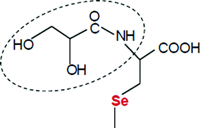
2,3-Dihydroxy-propionyl (2,3-DHP) group is a specific residue detected in selenized yeast that forms numerous stable and highly abundant Se species in several different yeast strains and fermentation batches. The conjugated form of 2,3-DHP-selenocysteine and glutathione is one of the most abundant species that is found in nearly all selenized yeast. In order to overcome the commercial unavailability of this compound, its synthesis was carried out through the active ester formation of pentachlorophenyl glycerate with selenocysteine, followed by the redox conjugation with glutathione. The optimization process of the synthesis was utilized for the production of three other Se-yeast specific compounds, namely, the conjugate of glutathione and selenocysteine, the conjugate of 2,3-DHP-selenocysteine and selenocysteine, and di-N-2,3-dihydroxy-propionyl-selenocysteine. The upstream and clean-up procedures were supported and monitored with HPLC-UV, HPLC-ICP-MS, and HPLC-ESI-QQQMS set-ups, while the identification was performed with HPLC-ESI-QTOFMS. The synthesized 2,3-DHP-selenocysteine/selenocysteine conjugates possessed fragmentation patterns identical to literature data.
Introduction
In the last decade, selenium speciation and molecule structure identification has been gravely hindered by the lack of standards. Numerous articles have been published introducing newfound Se-containing compounds, and in many cases the ESI-MS identification has been completed with fragmentation patterns and proposed molecule structures.1–3 During peptide synthesis, ESI-MSn-based structure identification counts as a routine procedure; however, there is rarely enough of the target selenium-containing components to verify the suggested molecule structure in the case of selenometabolomic studies. Fragmentation pattern may be sufficient for identification of molecules with already known S analogues; moreover, the molecular structure can also be predicted from biological pathways. However, when neither of these methods is an option, the only and ultimate way to identify molecules is through standards. Furthermore, the number of commercially available or synthetically described Se species of plant and mammal metabolism has been increasing,4–14 yet the availability of yeast specific Se species is extremely limited.15 Therefore, taking into account that almost 70 Se species have been identified from selenized yeast,1,16 and the fact that Se-yeast is the only natural (i.e., not a synthesis-based compound) and approved source for human selenium supplementation in the EU, the list of lacking (∼60) selenium standards is more than remarkable.
One of the highly abundant and commercially unavailable Se-species is the conjugate of glutathione and 2,3-dihydroxy-propionyl-selenocysteine (CAS no. 1006377-09-8; C16H27O11N4SSe+ [M + H]+, m/z 563.05568). This Se-yeast specific compound was reported first by McSheehy et al.17 and Goenega-Infante et al.,18 while its structure was tentatively identified in 2008 based on high resolution ESI-MS data.19 Since that time this compound has been detected and cited continuously from several yeast producers and strains.1,20,21 While glutathione is highly concentrated in yeast and occurs ubiquitously in eukaryotic and prokaryotic cells, the glyceroyl acid amine residue has only been previously reported either in antibiotics22 or, interestingly, in selenium-containing conjugates from yeast and, recently, from black mustard (Brassica nigra).23 Indeed, the metabolic role and origin of the 2,3-dihydroxy-propionyl (2,3-DHP; incorrectly referred to as 2,3-dihydroxy-1-oxopropyl, 2,3-DOP) group has not been elucidated yet, which is especially interesting as no sulphur analogues of any of the Se-containing species (of this group) have been found.
The ultimate goal of our study was to work out the synthesis of this compound in order to provide more reliable structure identification than standardless high resolution ESI-MSn data derived elucidation. The greatest challenge for this synthesis was the commercial unavailability of any active ester form of glycerate and the condensation/polymerization tendency of polyols. The intermediate step of the synthesis, that is, the coupling of the active ester to selenocysteine, results in two yeast-specific Se-compounds, the conjugate of 2,3-DHP-selenocysteine and selenocysteine (CAS 1246200-50-9 + 3614-08-2; C9H17O7N2Se2+ [M + H]+, m/z 424.93634),1 and di-N-2,3-DHP-selenocysteine (CAS 1357479-85-6; C12H21O10N2Se2+ [M + H]+, m/z 512.95211).21 The last step of the synthesis includes the oxidative conjugation of glutathione and a modified selenocysteine residue to form a S–Se bridge. Similar to this step, with the conjugation of glutathione and native selenocysteine, another Se-species detected in yeast (CAS no. 188609-44-1; C13H23O8N4SSe+ [M + H]+, m/z 475.03963)21 could be also synthesized and purified. The clean-up procedures were monitored with HPLC-UV, HPLC-ICP-MS, and HPLC-ESI-QQQ-MS detection, while the identification was based on HPLC-ESI-QTOFMS characterization.
To the best of our knowledge, since all the four Se species are Se-yeast specific, their availability might offer an important tool in the quantitative characterization and quality control of Se-yeast production.
Experimental
Reagents and standards
Acetonitrile (ACN; far UV HPLC grade), methanol (far UV HPLC grade) and Dowex 50WX4 cation exchange resin (200–400 mesh) were purchased from Fisher Scientific (Loughborough, Leicestershire UK). Ammonium acetate (a.r.), tris-hydroxymethyl-aminomethane (TRIS; a.r.), HCl (37 m/m %) and NaI (a.r.) were purchased from Reanal (Budapest, Hungary). Activated charcoal (4–14 mesh, granular, Norit® PK 3-5), Whatman Grade 1 filter paper, HCOOH (∼98%, puriss), dithiothreitol (DTT), 4-methylmorpholine (NMM; 98.0%), N,N′-dicyclohexylcarbodiimide (DCC; 99.0%), pentachlorophenol (PCP; 98%), selenocystine (Sec2; 97%), DL-glyceric acid hemicalcium salt hydrate (≥98%), reduced (≥98.0%) and oxidized (≥98%) glutathione stocks were purchased from the Sigma-Aldrich group (Schnelldorf, Germany). N,N-dimethylformamide (DMF; 99%) and H2O2 (a.r., 30 m/m%) were ordered from Merck (Darmstadt, Germany).
Milli-Q water (18.2 MΩ*cm, Merck-Millipore, Molsheim, France) was used throughout the experiments.
Instrumentation
Inductively coupled plasma-mass spectrometry (ICP-MS), using an Agilent 7500 cs (Agilent, Santa Clara, CA, USA) instrument, was used to monitor the isotopes of 77Se and 82Se during the chromatographic clean-up processes if applicable. The instrument was coupled to an Agilent 1200 high-performance liquid chromatography (HPLC) system. Intermediate products of syntheses were also monitored with an HPLC-ESI-MS coupling where a QTRAP 3200 triple quadrupole-linear ion trap mass spectrometer (ESI-QQQ-MS; Applied Biosystems/Sciex; Foster City, CA, USA) was used either in the Enhanced Q3 Single MS (EMS) mode for the full-scan experiments with an integration time of 1 s or in Enhanced Product Ion (EPI) mode for MS/MS analyses. The related instrumental parameters are described in the ESI.†
For the identification of selenium species, an Agilent 6530 Accurate-Mass ESI-QTOF-MS was used with an Agilent 6220 derived dual ion spray source. The instrument was coupled to an Agilent 1290 HPLC system. The operating parameters of the ESI-QTOF-MS are reported in the ESI.†
Methods
Desalting of glyceric acid
Glyceric acid hemicalcium salt was converted to the free acid form according to Berens and Scharf24 by dissolving 465 mg glyceric acid salt in 25 ml 50 V/V% methanol–water solution, and then 19.0 g Dowex 50WX4 cation exchange resin was added with stirring. After 20 minutes of incubation, the resin was removed by filtration. Then, the solution was filtered first through 2.0 g activated charcoal, followed by filtration though a filter paper, and concentrated to about 5 ml using a vacuum rotary evaporator at 26 °C, and then strained using a 0.45 μm PTFE filter. The leftover water content was removed at 55 °C using a vacuum rotary evaporator. Glyceric acid (210 mg; 2.0 mmol) was acquired that was stored at −23 °C until used.
Synthesis and clean-up of PCP-glycerate
106 mg (1.0 mmol) glyceric acid, 237 mg (1.14 mmol) DCC, and 291 mg (1.1 mmol) PCP were dissolved in 3 ml of DMF, placed in ice water bath and stirred for 24 hours. Then, another 339 mg (1.64 mmol) DCC and 412 mg (1.54 mmol) PCP in 3 ml DMF were added, and left to incubate for an additional 24 hours. The product was dried in a vacuum rotary evaporator and dissolved in a mixture of 4.5 ml ACN and 3 ml DMF. Finally, the solution was centrifuged at 4000 g for 10 min, decanted and filtered through a 0.45 μm PTFE filter.
Clean-up of PCP-glycerate was performed with fraction collection based on semi-preparative reversed-phase HPLC-UV separation and was verified with ESI-QQQ-MS. The relevant instrumental parameters can be seen in Tables 1 and 2 in the ESI.† The corresponding fractions were pooled and lyophilized, and 13.2 mg (37 μmol) of dry matter was acquired with a yield of 3%.
Coupling of PCP-glycerate and Sec2
Sec2 was solubilized in DMF according to the procedure described by Dernovics et al.25 4.0 mg PCP-glycerate (11 μmol) was dissolved in 4 ml DMF, and then the solution was placed in an ice bath under Ar with continuous stirring, to which 500 μl Sec2 solution (45 μmol) and 2 μl (18 μmol) NMM were added. To keep the pH of the solution between 7 and 8, 2 μl (18μmol) NMM was added three times at 15 minute intervals, and then the solution was incubated at room temperature for 48 hours with constant pH monitoring. The final product was lyophilized and dissolved in 4.0 ml 10 mM ammonium acetate buffer (pH = 5.5). The solution was centrifuged at 4000 g for 10 min, decanted and filtered through 0.45 μm PTFE filters. The formation of di-2,3-DHP-Sec and 2,3-DHP-Sec-Sec was monitored with analytical scale strong anion exchange (SAX) HPLC-ICP-MS set-up, while their clean-up was carried out with sequential semi-preparative SAX-HPLC-ICP-MS- and RP-HPLC-ICP-MS-based fraction collections. The relevant parameters can be seen in Table 1 in the ESI.† The column flow was split in both cases to provide an adequately low flow rate for the nebulizer of the ICP-MS. The corresponding fractions were pooled and lyophilized.
Conjugation of Sec2 with glutathione
For this step, 0.1 M TRIS buffer (pH = 8.6) was used. 3.4 mg (10 μmol) Sec2 was reduced and dissolved in 3 ml buffer containing 26.1 mg (169 μmol) DTT. 106 mg (173 μmol) oxidized glutathione dissolved in 4.0 ml buffer was added to the solution, and then 3.8 mg NaI (25 μmol; as catalyst)26 and 24 μl (160 μmol) H2O2 were added. The solution was incubated for 2 hours at room temperature.
The screening of reaction products was performed with analytical scale SAX-HPLC-ICP-MS, while the clean-up of the selenocysteine–glutathione conjugate was performed with semi-preparative SAX-HPLC-ICP-MS (Table 1 in the ESI†). The reaction solution was 1 + 3 (V/V) diluted with 10 mM ammonium acetate buffer (pH = 5.5) prior to injection. Fractions were pooled, lyophilized, dissolved in 300 μl 10% (V/V) ACN–H2O solution, and injected into a HPLC-ESI-QTOF-MS system for characterization. The relevant instrumental parameters can be seen in Tables 1 and 3 in the ESI.†
Conjugation of (2,3-DHP)-Sec-Sec and di-N-2,3-DHP-Sec with glutathione
The pooled (2,3-DHP)-Sec-Sec and di-N-2,3-DHP-Sec compounds acquired from the semi-preparative SAX-HPLC-ICP-MS clean-up were dissolved in 2.0 ml of 0.1 M TRIS buffer (pH = 8.6). First, 2.5 mg (16 μmol) DTT was added, followed by 150 mg (244 μmol) oxidized glutathione, then 100 μl 16 mg ml−1 NaI solution (11 μmol) and finally 24 μl (160 μmol) H2O2 were mixed to the solution. The solution was incubated for 2 hours at room temperature.
The clean-up and HPLC-ESI-QTOFMS characterization of the 2,3-DHP-selenocysteine–glutathione conjugate was carried out the same manner as was done with the selenocysteine–glutathione conjugate.
Results and discussion
Conjugation of Sec2 with glutathione and the characterization of Sec–glutathione
From the family of selenium-containing glutathione conjugates, Sec-glutathione is one of the least complex compounds, and it occurs in nearly all batches and strains in selenized yeast;21 however, its concentration does not exceed that of 2,3-DHP-Sec-glutathione. Apart from very low-abundance Sec2 and Sec2 species,1,27 this compound is unique in terms of containing a non-modified Sec residue conjugated through either a S–Se or a Se–Se bond.
The difficulty of the chemical synthesis of Sec-glutathione is the effective oxidative conjugation in the presence of the huge excess of DTT required for the solubilization of Sec2,28 which can be resolved by NaI-catalyzed oxidation26 and by the depletion of DTT with oxidized glutathione. As presented in Fig. 1a, the arising Sec-glutathione elutes between Sec2 and oxidized/reduced glutathione on SAX-HPLC, thus providing adequate separation for chromatographic clean-up.
 |
| Fig. 1 (a) Analytical scale SAX-HPLC-ICP-MS chromatogram of the products obtained from the oxidative conjugation of Sec2 and glutathione. Dashed line indicates the selenium signal, while the continuous line refers to sulphur. The compound eluting before reduced glutathione at 520 s was collected for further characterization with preparative scale SAX-HPLC. (b) HPLC-ESI-QTOF-MS total ion chromatogram (TIC) of the compound obtained from SAX-HPLC. The inset presents the extracted ion chromatogram (EIC) for m/z 475.0396. (c) Full scan spectrum recorded near the apex of the EIC for m/z 475.0396. The inset shows the selenium pattern of the target compound. (d) MS/MS spectrum and structure of the compound at m/z 475.0396. | |
The purified Sec-glutathione was characterized with HPLC-ESI-QTOFMS and MS/MS experiments. Fig. 1b shows the TIC and EIC of the compound, Fig. 1c shows the full scan recorded at the apex of the related EIC (C13H23O8N4SSe+ [M + H]+, m/z 475.03959, Δ = −0.08 ppm) and Fig. 1d presents the MS/MS data (see also Table 4 and the pathway of synthesis in the ESI†). Similarly to the fragmentation of the Se-containing glutathione family and Sec2 in positive ion mode,19,29 the intense fragments are caused by the loss of Gly and γ-Glu residues and by the neutral loss of NH3 and HCOOH. Also, the S–Se bond is hardly fragmented and the intact glutathione and Sec residues are in low abundance.
It is important to mention that the synthesis of Sec-glutathione was addressed both as an optimization step and as an independent method for the synthesis of a commercially unavailable compound.
Synthesis and clean-up of PCP-glycerate
The use of pentafluorophenol (PFP) might be preferred over PCP, as PFP esters react faster and the removal of PCP may be difficult.30 However, in our research, it was found that the reaction of glyceric acid with PFP did not yield any detectable amount of ester (results not shown); therefore, this step was repeated with the use of PCP.
PCP renders to the PCP-glycerate hydrophobic properties, thus providing the possibility for a reversed phase HPLC based clean-up. Fig. 2a presents the relevant HPLC-UV chromatogram, in which the compound eluting at 14.3 min was identified with ESI-MS/MS as PCP-glycerate after preparative scale fraction collection. The compound could be identified due to its unique isotopic pattern containing five chlorine atoms, and it could be characterized with the same fragmentation mechanism during both the ionization process in the ion source (Fig. 2b) and the MS/MS fragmentation (Fig. 2c), i.e., the production of pentachlorophenyl anion (m/z 351.0 [C9H4Cl5O4]− → m/z 264.8 [C6Cl5O]−). The low yield of this synthesis can be partly attributed to the polyolic structure of glyceric acid that facilitates the formation of by-products, and partly to the need for a water-containing HPLC eluent.
 |
| Fig. 2 (a) Preparative scale reversed-phase (RP) HPLC-UV chromatogram of the products obtained after the coupling of PCP and glyceric acid. The compound that eluted at 14.3 min was collected for further characterization and synthesis. (b) ESI-QQQ-MS full scan spectrum of the compound obtained from RP-HPLC. The inset presents the theoretical (left) and experimental (right) isotopic pattern of PCP-glycerate. (c) MS/MS spectrum of the compound at m/z 351.0 together with the proposed fragmentation event. | |
Coupling of PCP-glycerate to Sec2 and the characterization of the (2,3-DHP)-Sec-Sec and di-N-2,3-DHP-Sec species
Active ester coupling to Sec2 yields a mixture of non-derivatized, single- and double-derivatized species,25 thus requiring a clean-up step. As the free –NH2 groups are bound in the reaction with PCP-glycerate, the resulting species will show anionic properties even at slightly acidic pH, which enables the SAX-HPLC-based purification.
Fig. 3a presents the HPLC-ICP-MS chromatogram of the synthesized products, in which three selenium containing peaks could be observed: Sec2 elutes close to the dead volume, followed by the theoretical (2,3-DHP)-Sec-Sec and di-N-2,3-DHP-selenocysteine species, respectively. The latter two compounds were cleaned-up and characterized with HPLC-ESI-QTOFMS analyses. Fig. 3b shows the TIC and the EICs of the two compounds extracted at the theoretical m/z values.
 |
| Fig. 3 (a) Analytical scale SAX-HPLC-ICP-MS chromatogram of the products obtained from the coupling of PCP-glycerate and Sec2. The two peaks marked with the hypothetical compound structures eluting at 605 s and 1230 s were pooled for further synthesis and characterization with preparative scale SAX-HPLC. (b) HPLC-ESI-QTOF-MS-based TIC of the compounds collected from SAX-HPLC. The inset presents the EICs for m/z 424.9370 and m/z 512.9532. (c) Full scan spectrum recorded near the apex of the EIC for m/z 424.9370. The inset shows the selenium pattern of the target compound. (d) MS/MS spectrum of the compound at m/z 424.9370. (e) Full scan spectrum recorded near the apex of the EIC for m/z 512.9532. The inset shows the selenium pattern of the target compound. (f) MS/MS spectrum of the compound at m/z 512.9532. | |
Fig. 3c presents the full scan recorded at the related EIC of the m/z 424.93 compound. The accurate mass (C9H17O7N2Se2+ [M + H]+, m/z 424.93607, Δ = −0.64 ppm), isotopic distribution and MS/MS fragments (see Fig. 3d) match with those reported by Arnaudguilhem et al.1 Concerning di-N-2,3-DHP-selenocysteine, the data presented in Fig. 3e (C12H21O10N2Se2+ [M + H]+, m/z 512.95203, Δ = −0.16 ppm) are in agreement with those published by Casal et al.,21 while the MS/MS fragments have been presented here for the first time (Fig. 3f).
The suggested fragmentation pathways of the two compounds, and the pathway of synthesis are included in the ESI.† It should be highlighted that the fragmentation of both species results in the abundant appearance of the m/z 255.97 and m/z 167.95 fragments, that are also characteristic for the conjugate of 2,3-DHP-selenocysteine and glutathione (m/z 563.05).31
Taking into account the low efficiency of the 2,3-DHP coupling process, both single- and double-derivatized compounds were purified and pooled in order to increase the yield of the following conjugation step with glutathione.
Conjugation and characterization of 2,3-DHP-Sec-glutathione
Combining the optimized process of Sec-glutathione conjugation and the clean-up of 2,3-DHP-containing Sec2 species was a prerequisite to arrive at a detectable amount of 2,3-DHP-Sec-glutathione. However, this compound is slightly retained on special reversed-phase HPLC columns intended for use with eluents with low organic solvent content,18 a more robust clean-up technique with SAX-HPLC was chosen.19
The SAX HPLC-ICP-MS chromatogram of the reaction products can be seen in Fig. 4a. The first Se-containing compound, eluting at 500 s, was identical to the conjugate of Sec-glutathione that was formed in the reaction of non-derivatized Sec residues. The HPLC-ESI-QTOFMS characterization of the more intense second peak, eluting at 720 s, is presented in Fig. 4b. The targeted search for m/z 563.05568 resulted in an EIC of a single peak with the full scan shown on Fig. 4c and MS/MS fragmentation data shown on Fig. 4d. Both the MS (C16H27O11N4SSe+ [M + H]+, m/z 563.05546 [M + H]+, Δ = −0.39 ppm) and MS/MS data (see Table 4 and the pathway of synthesis in the ESI†) correspond to the previously reported information on this compound,19 which indicates that the synthesized compound matches the genuine, Se-yeast specific 2,3-DHP-Sec-glutathione conjugate.
 |
| Fig. 4 (a) Analytical scale SAX-HPLC-ICP-MS chromatogram of the products obtained from the oxidative conjugation of (2,3-DHP)-Sec-Sec and di-N-2,3-DHP-Sec with glutathione. The compound eluting at 720 s was collected for further characterization with preparative scale SAX-HPLC. (b) HPLC-ESI-QTOF-MS-based TIC of the compound collected from SAX-HPLC. The inset presents the EIC for m/z 563.0554. (c) Full scan spectrum recorded near the apex of the EIC for m/z 563.0554. The inset shows the selenium pattern of the target compound. (d) MS/MS spectrum and structure of the compound at m/z 563.0554. | |
While some (e.g., the γ-Glu specific) of the MS/MS fragments of Sec-glutathione and 2,3-DHP-Sec-glutathione are shared, the majority of the fragments are different. The most significant difference is the high abundance of the Sec residue that appears both in native (m/z 167.95) and 2,3-DHP-derivatised (m/z 255.97) forms during the fragmentation of 2,3-DHP-Sec-glutathione, but appears only as a minor fragment during the fragmentation of Sec-glutathione. This great difference in fragmentation pattern is unusual, as the two compounds share their basic structure. Indeed, the addition of the 2,3-DHP residue, which can be broken off during fragmentation at the amide bond, could stabilize the Sec residue and increase its abundance while affecting the bond strength of the S–Se bridge.17 The high fragmentation event of the S–Se bridge in positive ion mode together with the abundant appearance of the Sec residue is a unique feature of 2,3-DHP-containing glutathione derivatives and it is reported exclusively in such structures.1,31
Conclusions
Both the quality control and the quantitative characterization of selenized yeast batches require standards to monitor stability and to identify sample origin. As non-Se-yeast-specific selenium compounds (namely, selenomethionine, selenocysteine and inorganic selenium species) specified by the Commission Regulation (EC) No 1170/2009 cannot provide customized options for these purposes, the newly synthesized 2,3-DHP containing species and the conjugate of selenocysteine and glutathione may offer a viable solution. On the other hand, the more than 50 Se-species discovered during the last five years from plant and yeast samples call attention to the evident lag in the number of available standards that may be synthesized systematically with an approach similar to our method, i.e., with a grouped batch of synthesis.
Acknowledgements
The authors acknowledge the financial help of the TÁMOP 4.2.1./B-09/1/KMR-2010-0005 grant for the HPLC-ESI-QTOF-MS experiments.
References
- C. Arnaudguilhem, K. Bierla, L. Ouerdane, H. Preud'homme, A. Yiannikouris and R. Lobinski, Anal. Chim. Acta, 2012, 757, 26–38 CrossRef CAS PubMed.
- K. Bierla, J. Szpunar, A. Yiannikouris and R. Lobinski, TrAC, Trends Anal. Chem., 2012, 41, 122–132 CrossRef CAS PubMed.
- Y. Ogra and Y. Anan, J. Anal. At. Spectrom., 2009, 24, 1477–1488 RSC.
- E. Block, M. Birringer, W. Jiang, T. Nakahodo, H. J. Thompson, P. J. Toscano, H. Uzar, X. Zhang and Z. Zhu, J. Agric. Food Chem., 2001, 49, 458–470 CrossRef CAS.
- T. W. M. Fan, A. N. Lane, D. Martens and R. M. Higashi, Analyst, 1998, 123, 875–884 RSC.
- K. Wrobel, K. Wrobel, S. S. Kannamkumarath and J. A. Caruso, Anal. Bioanal. Chem., 2003, 377, 670–674 CrossRef CAS PubMed.
- Y. Ogra, T. Kitaguchi, K. Ishiwata, N. Suzuki, T. Toida and K. T. Suzuki, Metallomics, 2009, 1, 78–86 RSC.
- Y. Ogra, T. Kitaguchi, K. Ishiwata, N. Suzuki, Y. Iwashita and K. T. Suzuki, J. Anal. At. Spectrom., 2007, 22, 1390–1396 RSC.
- A. J. Matich, M. J. McKenzie, D. A. Brummell and D. D. Rowan, Phytochemistry, 2009, 70, 1098–1106 CrossRef CAS PubMed.
- Y. Kobayashi, Y. Ogra, K. Ishiwata, H. Takayama, N. Aimi and K. T. Suzuki, Proc. Natl. Acad. Sci. U. S. A., 2002, 99, 15932–15936 CrossRef CAS PubMed.
- K. Wrobel, K. Wrobel and J. A. Caruso, J. Anal. At. Spectrom., 2002, 17, 1048–1054 RSC.
- P. Braga, M. Montes-Bayon, J. Alvarez, J. M. Lopez and A. Sanz-Medel, J. Anal. At. Spectrom., 2004, 19, 1128–1133 RSC.
- P. Traar, F. Belaj and K. A. Francesconi, Aust. J. Chem., 2004, 57, 1051–1053 CrossRef CAS.
- T. Tamura, T. Oikawa, A. Ohtaka, N. Fujii, N. Esaki and K. Soda, Anal. Biochem., 1993, 208, 151–154 CrossRef CAS PubMed.
- E. Block, R. S. Glass, N. E. Jacobsen, S. Johnson, C. Kahakachchi, R. KaminskI, A. Skowronska, H. T. Boakye, J. F. Tyson and P. C. Uden, J. Agric. Food Chem., 2004, 52, 3761–3771 CrossRef CAS PubMed.
- H. Preud'Homme, J. Far, S. Gil-Casal and R. Lobinski, Metallomics, 2012, 4, 422–432 RSC.
- S. McSheehy, P. Pohl, J. Szpunar, M. Potin-Gautier and R. Lobinski, J. Anal. At. Spectrom., 2001, 16, 68–73 RSC.
- H. G. Infante, G. O'Connor, M. Rayman, R. Hearn and K. Cook, J. Anal. At. Spectrom., 2006, 21, 1256–1263 RSC.
- M. Dernovics and R. Lobinski, J. Anal. At. Spectrom., 2008, 23, 72–83 RSC.
- Y. Rao, M. McCooeye, A. Windust, E. Bramanti, A. D'Ulivo and Z. Mester, Anal. Chem., 2010, 82, 8121–8130 CrossRef CAS PubMed.
- S. G. Casal, J. Far, K. Bierla, L. Ouerdane and J. Szpunar, Metallomics, 2010, 2, 535–548 RSC.
- T. Kato, H. Hinoo, Y. Terui, J. Nishikawa, Y. Nakagawa, Y. Ikenishi and J. Shoji, J. Antibiot., 1987, 40, 139–144 CrossRef CAS.
- L. Ouerdane, F. Aureli, P. Flis, K. Bierla, H. Preud'homme, F. Cubadda and J. Szpunar, Metallomics, 2013, 5, 1294–1304 RSC.
- U. Berens and H.-D. Scharf, J. Org. Chem., 1995, 60, 5127–5134 CrossRef CAS.
- M. Dernovics, A. Vass, A. Németh and A. Magyar, Talanta, 2012, 99, 186–193 CrossRef CAS PubMed.
- M. Kirihara, Y. Asai, S. Ogawa, T. Noguchi, A. Hatano and Y. Hirai, Synthesis, 2007, 3286–3289 CrossRef CAS PubMed.
- A. Németh, J. F. García Reyes, J. Kosáry and M. Dernovics, Metallomics, 2013, 5, 1663–1673 RSC.
- J. N. Burnell, J. A. Karle and A. Shrift, J. Inorg. Biochem., 1980, 12, 343–351 CrossRef CAS.
- H. M. Crews, P. A. Clarke, D. J. Lewis, L. M. Owen, P. R. Strutt and A. Izquierdo, J. Anal. At. Spectrom., 1996, 11, 1177–1182 RSC.
- M. M. Joullié and K. M. Lassen, ARKIVOC, 2010, (viii), 189–250 CrossRef.
- M. Dernovics and R. Lobinski, Anal. Chem., 2008, 80, 3975–3984 CrossRef CAS PubMed.
Footnote |
† Electronic supplementary information (ESI) available. See DOI: 10.1039/c4ra02660h |
|
This journal is © The Royal Society of Chemistry 2014 |
Click here to see how this site uses Cookies. View our privacy policy here.