LDI-MS examination of oxygen plasma modified polymer for designing tailored implant biointerfaces

M. Gołda-Cępaa, N. Aminlashgarib, M. Hakkarainenb, K. Engvallc and A. Kotarba*a
aFaculty of Chemistry, Jagiellonian University, ul. Ingardena 3, 30-060 Krakow, Poland. E-mail: kotarba@chemia.uj.edu.pl
bDepartment of Fibre and Polymer Technology, KTH Royal Institute of Technology, SE-100 44 Stockholm, Sweden
cDepartment of Chemical Engineering and Technology, KTH Royal Institute of Technology, SE-100 44 Stockholm, Sweden

Received 26th March 2014 , Accepted 28th May 2014

First published on 9th June 2014


Abstract

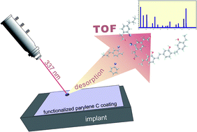
A versatile polymer coating for biomaterials was fabricated by the mild oxygen plasma treatment of Chemical Vapour Deposited (CVD) parylene C. The surface properties were tailored while the excellent protective properties of the bulk were preserved. The species, formed due to the plasma functionalisation, were fingerprinted by a novel Laser Desorption/Ionisation-Mass Spectrometry (LDI-MS) method. Improved osteosarcoma cells (line MG-63) attachment and viability on a modified surface were demonstrated.


Polymers represent a large and mostly multipurpose fraction of biomaterials.1,2 The physical and chemical bulk properties of synthetic polymers are often excellent, but they do not usually exhibit the right surface properties for specific bio-applications.3 Thus, there is a strong need for surface modification methods, which can transform these popular materials into highly valuable final products. The essential issue involves biocompatibility, which can be considered as the adjustment of the polymer surface characteristics matching the desired interactions with the tissue while concurrently maintaining the bulk properties. Parylene C is an excellent protective coating material for metal implants due to its in vivo compatibility, mechanical properties (hardness, elasticity, friction) and stability in body fluids.4–6 It is not only corrosion resistant, but it substantially decreases the release of hazardous metal ions from metal implants into the body. However, for good attachment of cells and/or drug-loaded biodegradable polymers, their surface must be adjusted via the introduction of functional groups, which will work as tangible adsorption sites. Low pressure plasmas are considered one of the universal options for such surface treatment, where the chemical structure of a top polymer layer can be tailored.7 By adjusting the plasma parameters the modification is confined to a shallow surface layer (∼10 nm) of the polymer, where energetic particles and vacuum UV radiation interact with the material. The plasma contains a large concentration of free electrons, and highly excited atomic, molecular, ionic, and radical species. Free radical intermediates react with polymer surfaces in the plasma environment, breaking covalent chemical bonds on the polymer surfaces.8 The final polymeric material is somewhat uniformly modified over the whole surface. Moreover, the amount of toxic by-products is low compared to other modification methods, which makes plasma treatment the most versatile surface tuning method.9,10 During the modification of biomedical polymers by oxygen plasma several processes occur simultaneously on the polymer surface. These include cleaning (reactions of atomic oxygen with the surface carbon giving volatile reaction products), chemical modification (formation of oxygen-containing functional groups)11 and degradation (depolymerisation, pyrolysis, crosslinking, etc.).7 Due to the multitude of elementary reactions occurring simultaneously, it is impossible in most cases to describe in detail the physical and chemical effects of plasma. Additionally, there is a lack of accurate characterisation method of the surface species formed on exposure to plasma. The classical surface sensitive techniques – such as XPS, RS, ATR-FTIR, AFM – offer general information concerning the surface topography and identification of elements, and functional groups. However, they are not specific enough for univocal identification of the formed compounds and placement of functional groups. That is the reason why a new approach for thorough surface investigation is necessary. This also makes studies on oxygen plasma treatment of polymer biomaterials at the centre of extensive and intensive research.12,13

In this communication, we present a novel application of LDI-MS (Laser Desorption/Ionisation-Mass Spectrometry) for the direct identification of functionalised compounds containing groups such as –COOH, –OH, and –C(O)H on the oxygen plasma treated parylene C surface (Fig. 1). Such a modification is crucial from the medical point of view because it leads to higher hydrophilicity,6 and provides adhesion centres and further determines the bond strength between the cells and polymer surface. As a result, the polymer material is transformed into a highly valuable finished biomimetic interface.14


image file: c4ra02656j-f1.tif
Fig. 1 Graphical representation of the used experimental methodology: parylene C coating fabrication (CVD), its oxygen plasma functionalisation; and the most important characterisation results (contact angle, LDI-MS, cells adhesion).

Parylene C (8 μm of thickness) films were prepared by CVD technique. The samples were provided by ParaTech Coating Scandinavia AB. A dimer of chloro-para-xylylene was used as a precursor and heated to 690 °C for decomposition to monomeric form. The monomers in vapor phase were polymerised at room temperature and subsequently deposited at 10−3 mbar. The thickness of the coating was controlled by the deposition time. Samples with a size 2 × 2 cm were prepared and cleaned in isopropyl alcohol and distilled water before further investigations. In order to modify the parylene C surface, the oxygen plasma treatment was carried out using a Diener Electronic Femto plasma system (Diener Electronic GmbH, Nagold, Germany). We found that the parylene C modification process strongly depends on plasma homogeneity (e.g., application of rotary drum), oxygen pressure in the chamber, power of the plasma generator, and time of the modification. Thus, these parameters were precisely adjusted to 0.2 mbar, 50 W and 60 min, respectively, as optimal for surface functionalisation while maintaining the bulk intact. For the parameters adjustment, several experiments were performed for different times of treatment from 0.1 min to 60 min. The differences in the oxygen content in parylene C coating were observed by X-ray Photoelectron Spectroscopy (XPS) and the oxygen containing surface groups were attributed, however, the exact place of oxygen insertion to the polymer backbone could not be identified.6

The general scheme illustrating the developed LDI-MS process is shown in Fig. 2. In previous work, we demonstrated that polymer nanocomposite films can be used as surfaces for the direct analysis of low molecular mass pharmaceuticals.15 Here, the thin parylene C film was placed directly on the LDI-MS plate and analysed.


image file: c4ra02656j-f2.tif
Fig. 2 Illustration of the process of LDI-MS identification of oxygen plasma modified species on the surface of parylene C coating.

The solid film surface containing the oxidised species was irradiated by the laser. The analytes were desorbed and ionised from the surface. Here, the surface functions as a matrix by absorbing the laser energy and transferring it to the isolated analyte molecules in order to desorb them. An important advantage of the developed method is that it allows direct analysis of surface species, without any sample preparation, concurrently. The surface analysis of unmodified and oxygen plasma treated parylene C was performed on a Bruker UltraFlex time-of-flight (TOF) mass spectrometer with a SCOUT-MTP ion source (Bruker Daltonics, Bremen, Germany) in reflector mode and equipped with a 337 nm nitrogen laser. The acceleration voltage was 25 kV and the reflector voltage was 26.3 kV. Each surface was placed on a modified stainless steel LDI plate. The spectrum obtained for each sample is an accumulation of 1000 shots. The mass-to-charge (m/z) ratio range was set to scan from 60 m/z to 2000 m/z. Fig. 3 illustrates the LDI-MS spectra for untreated and treated surfaces after 60 minutes of oxygen plasma modification. A table with LDI-MS interpretation and identification of the detected compounds is shown in the ESI.


image file: c4ra02656j-f3.tif
Fig. 3 LDI-MS spectra showing the chemical species on the unmodified and oxygen plasma modified parylene C surfaces.

The LDI-MS analysis revealed that insertion of oxygen to the polymer backbone took place exclusively in the linkers between the aromatic rings of the polymer chain. The characteristic m/z peaks collected in table (see ESI) prove that the rings remained untouched during the plasma treatment, being in line with the difference in chemical reactivity between stable six-carbon benzene core and alkane linkers. This is important for keeping the crystalline structure of parylene C, which prevents extensive penetration of the oxygen active species into the bulk during plasma treatment. Additional confirmation of stability of aromatic rings with chlorine substituents is provided by characteristic splitting of the main line of 2 m/z due to the presence of the natural abundance of 35Cl and 37Cl isotopes, as can be seen on the grey inset in Fig. 3.

Cell adhesion to solid surfaces is always the result of non-specific interactions mediated by biopolymer adlayers. In general, surface-associated physical and chemical cues control cells' proliferation and differentiation.16 Therefore, tuning the surface properties of biomaterials can be used as a powerful tool to control the cells adhesion and accelerate the integration of biomaterials with host tissues.17 In this context for the evaluation of cells adhesion (MG-63), sterilised parylene C disks, as a model polymer coating, were used. In the experiments, both unmodified and oxygen plasma modified samples were investigated. Cells were seeded in 24-well plates (BD Falcon) at a density of 15 × 104 per well, grown in Dulbecco's modified essential medium (MEM), supplemented with 2 mM glutamine and 10% fetal bovine serum (FBS), purchased from Gibco (Life Technologies). The tests were performed for 24 hours and a sample without parylene C film was used as a control. After the test, films were investigated using an Olympus IX51 optic microscope. The microscopic observations clearly show superior cell attachment to the modified parylene C compared to the unmodified one (Fig. 1). For the modified surface, the cells were evenly spread, displaying a closely packed congruent structure. For reference, with regard to the unmodified parylene C film, the cells were separated from each other and the surface coverage was uneven. The different affinity of the cells to the investigated parylene C surfaces was reflected in their viability. In order to quantify the effect of plasma treatment, standard MTT assay procedure was applied.18 For these tests, cells were grown in the same way as for the adhesion test, and the absorbance was measured using a microplate reader (Infinite 200 M PRO NanoQuant, Tecan, Switzerland). The cell viability tests for each polymer surface were completed in at least three independent experiments. The data from assays were normalised, defining control cells viability as 100%. Statistical comparisons were made by a standard one way ANOVA with Dunnett's multiple comparison tests.

As shown in our previous studies, easily vacuum-deposited parylene C exhibits excellent mechanical properties, biocompatibility, and forms continuous thin and inert films on metallic substrates.6 Long-term electrochemical (EIS) tests in Hanks' solution, with and without H2O2, revealed that the parylene C layer can be successfully used for corrosion protection of implant surface. The coating was also proved to have excellent wear resistant properties. The optimal thickness of the coating (from the investigated 2–20 μm) was determined to be 8 μm which limited the wear debris formation efficiently.5 Such coatings demonstrate sufficient elastomeric properties, essential in sustaining strains during surgical implantation and long-term usage in the body. Moreover, the 8 μm layer of parylene C coating reduced the corrosion process, and as a consequence the hazardous heavy metal ions release is lowered by more than one order of magnitude.19 However, the material is strongly hydrophobic and not permeable for water, so its interface is not tuned for interactions with cells or suitable for further functionalisation (e.g., an attachment of additional, biodegradable layers or drug molecules). Therefore, the surface should be tailored with suitable adsorption sites to provide anchors for bioactive molecules. As described above, the oxygen containing groups can be successfully used for such purposes.

The insertion of oxygen into the parylene C surface can lead to the formation of adsorption sites. Drug molecules usually contain polar groups, typically: [double bond, length as m-dash]O, –OH, –COOH, –NH–, which results in considerable dipole moments, for example ibuprofen reaches 2.1 D, diclofenac 0.9 D,20 ketoprofen 2.12 D,21 and tend to attach to so-called hydrophilic surfaces. That is the reason why the oxygen containing functional groups may be utilised in therapeutic applications. This is also related to the adhesion of biodegradable drug-loaded polymer layers, which can be applied in regenerative medicine. After the implantation surgery the most common complications are prolonged inflammatory response and massive infection,22 thus the material must be resistant to H2O219 and biodegradation. Due to its crystalline structure parylene C is hydrogen peroxide resistant, and the plasma treatment subtly affects only the surface properties. The treatment also provides possibilities for kinetically controlled, in-site delivery of anti-inflammatory medication or antibiotics directly from the implants surface.

The second beneficial effect of the oxygen plasma treatment on parylene C consists in a substantial increase in its surface-free energy from 40 mJ m−2 for unmodified to 70 mJ m−2 for modified surfaces, as revealed by contact angle measurements with the use of Owens–Wendt method.23 Higher surface-free energy is a good descriptor of surface cells' adhesion and their viability.24

In summary, we demonstrate that the mild oxygen plasma technology provides a direct route for facile engineering of polymeric biointerface. The developed LDI-MS method provided a direct way to analyse the oxygen containing functional groups and species formed on the parylene C surface, as well as their placement in the polymer chain. Thus, LDI-MS was shown to be a versatile and powerful tool for direct characterisation of modified surfaces. The developed method also has huge potential more generally for the analysis of modified surfaces, understanding and optimisation of surface modification processes. It could also be further used for characterisation of all kinds of surface degradation reactions.

It should be emphasised that the modification is confined to the very top polymer layer, at the same time, keeping the excellent physical and chemical bulk properties invariant. The oxygen plasma modified parylene C implant coating extends beyond the anti-corrosive applications related to the suppression of heavy metal ions release, and stimulates the adhesion and growth of osteoblast cells. The development of the next-generation biosurfaces with controlled in-site drug delivery for regenerative medicine applications is highlighted.

Acknowledgements

This work was sponsored by the Polish National Science Centre decision number DEC-2012/05/N/ST8/03321.

Notes and references

  1. D. Li, Q. Zheng, Y. Wang and H. Chen, Polym. Chem., 2014, 5, 14 RSC.
  2. L. G. Griffith, Acta Mater., 2000, 48, 263 CrossRef CAS.
  3. P. Roach, D. Eglin, K. Rohde and C. C. Perry, J. Mater. Sci.: Mater. Med., 2007, 18, 1263 CrossRef CAS PubMed.
  4. M. Cieślik, M. Kot, W. Reczyński, K. Engvall, W. Rakowski and A. Kotarba, Mater. Sci. Eng., C, 2012, 32, 31 CrossRef PubMed.
  5. M. Cieślik, S. Zimowski, M. Gołda, K. Engvall, J. Pan, W. Rakowski and A. Kotarba, Mater. Sci. Eng., C, 2012, 32, 2431 CrossRef PubMed.
  6. M. Gołda, M. Brzychczy-Włoch, M. Faryna, K. Engvall and A. Kotarba, Mater. Sci. Eng., C, 2013, 33, 4221 CrossRef PubMed.
  7. J. Friedrich, The Plasma Chemistry of Polymer Surfaces, Wiley-VCH, 2012 Search PubMed.
  8. Polymer Surfaces and Interfaces, ed. M. Stamm, Springer-Verlag, Berlin and Heidelberg, 2008 Search PubMed.
  9. V. Sunkara, D. K. Park and Y. K. Cho, RSC Adv., 2012, 35, 9066–9070 RSC.
  10. J. S. Meena, M. C. Chu, Y. C. Chang, H. C. You, R. Singh, P. T. Liu, H. P. D. Shieh, F. C. Chang and F. H. Ko, J. Mater. Chem. C, 2013, 1, 6613 RSC.
  11. X. Guo, J. Jian, L. Lin, H. Zhua and S. Zhua, Analyst, 2013, 138, 5265 RSC.
  12. E. Fortunati, S. Mattioli, L. Visai, M. Imbriani, J. L. G. Fierro, J. M. Kenny and I. Armentano, Biomacromolecules, 2013, 14, 626 CrossRef CAS PubMed.
  13. T. Jacobs, R. Morent, N. De Geyter, P. Dubruel and C. Leys, Plasma Chem. Plasma Process., 2012, 32, 1039 CrossRef CAS.
  14. J. M. Goddard and J. H. Hotchkiss, Prog. Polym. Sci., 2007, 32, 698 CrossRef CAS PubMed.
  15. N. Aminlashgari, M. Shariatgorji, L. L. Ilag and M. Hakkarainen, Anal. Methods, 2011, 3, 192 RSC.
  16. R. A. Gittens, T. McLachlan, R. Olivares-Navarrete, Y. Cai, S. Berner, R. Tannenbaum, Z. Schwartz, K. H. Sandhage and B. D. Boyan, Biomaterials, 2011, 32, 3395 CrossRef CAS PubMed.
  17. M. Y. Tsai, C. Y. Lin, C. H. Huang, J. A. Gu, S. T. Huang, J. Yu and H. Y. Chen, Chem. Commun., 2012, 48, 10969 RSC.
  18. K. Kyzioł, Ł. Kaczmarek, G. Brzezinka and A. Kyzioł, Chem. Eng. J., 2014, 240, 516 CrossRef PubMed.
  19. M. Cieślik, K. Engvall, J. Pan and A. Kotarba, Corros. Sci., 2011, 53, 296 CrossRef PubMed.
  20. I. Vergili, J. Environ. Manage., 2013, 127, 177 CrossRef CAS PubMed.
  21. M. L. Vueba, M. E. Pina, F. Veiga, J. J. Sousa and L. A. E. Batista de Carvalho, Int. J. Pharm., 2006, 307, 56 CrossRef CAS PubMed.
  22. D. Campoccia, L. Montanaro and C. R. Arciola, Biomaterials, 2013, 34, 8018 CrossRef CAS PubMed.
  23. M. Kobayashi, Y. Terayama, H. Yamaguchi, M. Terada, D. Murakami, K. Ishihara and A. Takahara, Langmuir, 2012, 28, 7212 CrossRef CAS PubMed.
  24. L. Ponsonnet, K. Reybier, N. Jaffrezic, V. Comte, C. Lagneau, M. Lissac and C. Martelet, Mater. Sci. Eng., C, 2003, 23, 551 CrossRef.

Footnote

Electronic supplementary information (ESI) available: Table S1. LDI-MS interpretation of the detected oxygen containing species on parylene C surface. See DOI: 10.1039/c4ra02656j

This journal is © The Royal Society of Chemistry 2014
Click here to see how this site uses Cookies. View our privacy policy here.