DOI:
10.1039/C4RA02404D
(Paper)
RSC Adv., 2014,
4, 17347-17353
A molecular level understanding of interaction between FTY720 (Fingolimod hydrochloride) and DMPC multilamellar vesicles†
Received
19th March 2014
, Accepted 31st March 2014
First published on 1st April 2014
Abstract
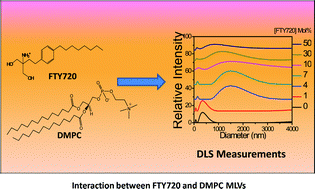
This work focuses on the molecular level understanding of interaction between FTY720 (Fingolimod hydrochloride) and dimyristoylphosphatidylcholine (DMPC) multilamellar vesicles (MLVs) as a drug molecule carrier by investigating the structural changes, solubilisation effect and thermotropic phase behaviour. Three different techniques like dynamic light scattering (DLS), differential scanning calorimetry (DSC) and fluorescent molecular probe based techniques were used to study the effect of FTY720 on DMPC MLVs. DLS studies showed that FTY720 induces a marked change in the DMPC MLVs' structure and solubilises the lipid membrane with the increase in concentration of FTY720. DSC and temperature dependent fluorescence intensity studies of 1-naphthol showed that FTY720 broadens and shifts the phase transition temperature of the DMPC MLVs to a lower temperature. This is in contrast to the effect of the sphingosine which is a structural analogous to FTY720. Fluorescence anisotropy study of 1,6-diphenyl-1,3,5-hexatriene (DPH) also revealed the above phenomena. Fluorescence lifetime decay analysis of 1-naphthol suggested that incorporation of FTY720 causes a redistribution of 1-naphthol molecules between the core and interfacial regions of the membrane.
Introduction
Liposomal delivery systems for drugs and cosmetics are becoming increasingly popular in recent years.1 Generally the lipid bilayer membranes of multilamellar vesicles (MLVs) are considered as the preferred media for hydrophobic and amphiphilic drug carriers due to their high encapsulation efficiency.2–5 A molecular level understanding of the incorporation of a drug molecule of interest into the lipid bilayer membrane, and thereby the drug induced changes in the physical properties of the membrane like membrane organisation, thermotropic phase transition etc., are important in this respect.
FTY720 (Fingolimod hydrochloride) is an important amphipathic drug molecule (Fig. 1), which has recently got FDA approval as an oral drug molecule for multiple sclerosis.6,7 FTY720 is suggested to be involved in the reduction of lymphocyte migration to central nervous system through blood barrier of brain by interacting with CD4+T cell, CD8+T cell and B cells.8–10 It also acts as an immunosuppressive drug molecule for organ transplantation.10 FTY720 has a close structural homology with sphingosine (Fig. SI1†), which is a metabolite of cell membrane. It is known in the literature that sphingosine affects the lipid bilayer membranes properties.11–16 Sphingosine has been found to increase the phase transition temperature of zwitterionic phosphatidylcholine (PC) membrane. At higher concentrations, it solubilises and broadens the transition temperature of lipid bilayer membranes.13,14,16 The close structural similarity of sphingosine with FTY720 prompts the investigation towards the effects of FTY720 on lipid bilayer membrane. In a recent work,17 we studied the aqueous phase aggregation of FTY720 drug and its effect on DMPC small unilamellar vesicles (SUVs). FTY720 in water showed an efficient micellar aggregation with critical micellar concentration (CMC) at ∼75 μM with an aggregation number 42 ± 3. The interaction of FTY720 with DMPC SUVs was observed to prevent partitioning of small molecules into the membrane in both solid gel (SG) and liquid crystalline (LC) phases. Temperature dependent fluorescence intensity and fluorescence anisotropy measurements have shown that above CMC of FTY720 the SG to LC main phase transition temperature (TM) of lipid bilayer membrane decreases from 23 °C to 21 °C in aqueous medium without affecting the intactness of DMPC SUVs.17
 |
| Fig. 1 Molecular structure of FTY720 (2-amino-2-(4-octylphenyl) ethylpropane-1,3-diol hydrochloride). | |
In view of the importance of MLVs as carriers of amphipathic drugs, a study of the structural changes, solubilisation effect, fluidity and thermotropic phase behaviour of DMPC MLVs incorporating FTY720 was carried out. Fluorescence molecular probes for lipid bilayer membranes like 1,6-diphenyl-1,3,5-hexatriene (DPH) (fluorescence anisotropy probe) and 1-naphthol (excited state proton transfer probe) were used for a molecular level understanding of the membrane organization. In addition to these studies, DLS (dynamic light scattering) and DSC (differential scanning calorimetry) measurements were carried out.
Experimental procedure
Materials
1-Naphthol purchased from SRL, India was sublimed and used after checking its purity. Dimyristoylphosphatidylcholine (DMPC) and 1,6-diphenyl-1,3,5-hexatriene (DPH) were purchased from Sigma Chemical Co. (Bangalore, India). The requisite sample of FTY720 was prepared according to the synthetic scheme disclosed earlier.17,18 All the solvents used were of spectroscopic grade. Triple-distilled water, prepared using alkaline permanganate solution, was also used for the experiments.
Methods
Dynamic light scattering (DLS) analysis was carried out with Malvern Zetasizer nano series, with a path length of 1 cm. The wavelength of the laser used was 632.8 nm and the scattering angle was kept as 90 °C. Differential Scanning Calorimetry (DSC) measurements were performed by using TA-DSC Q-200 instrument. Fluorescence emission and fluorescence anisotropy measurements were performed using Fluoromax-4 fluorescence spectrophotometer. The fluorescence lifetime measurements were carried out using Horiba Jobin Yvon TCSPC lifetime instrument. 295 nm and 370 nm nano-LEDs were used as light source for the experiments with fluorescent probes 1-naphthol and DPH respectively. The pulse repetition rate was set to 1 MHz, and the pulse widths were ∼800 ps for 295 nm LED, and ∼1.1 ns for 370 nm LED. The detector response time is less than 1 ns. The instrument response function was collected using a scatterer (Ludox AS40 colloidal silica). The decay data were analyzed using IBH software. A value of χ2, in-between 0.99–1.22 and symmetrical distribution of residual was considered as a good fit. The average fluorescence lifetime (τavg) values were calculated by the following equation19
where τi is the individual lifetime with corresponding amplitude αi.
Liposome preparation
Multilamellar vesicles (MLVs) were prepared by a solvent evaporation method.20 DMPC lipid was dissolved in chloroform–methanol 2
:
1 (v/v) at the desired molar ratio. The solution was evaporated to dryness. The solvent was removed by using a rotary evaporator and residual solvent if any was removed by leaving the round bottomed flask in vacuum for one hour. Multilamellar vesicles (MLVs) were prepared by adding the appropriate volume of triple-distilled water, to the lipid film with vigorous vortexing and then warming at 40–45 °C, to yield a final lipid concentration of 0.4 mM. FTY720 was incorporated by adding a required amount to the lipid solution in methanol–chloroform (1
:
2) mixture, before preparation of vesicle. The required mol% of FTY720 was calculated with respect to the lipid concentration taken. The final concentration of DMPC MLVs for DLS and fluorescence measurements was 0.4 mM. For DSC measurements, the final concentration was fixed 2 mg mL−1. The concentrations of the fluorescent probe were maintained at 4 μM to get a desired lipid to probe ratio of 100.
Results and discussions
Differential scanning calorimetry (DSC)
DSC was used to study the thermotropic phase behaviour of DMPC MLVs in presence and absence of FTY720. DSC has been a useful technique for the study of thermotropic phase transition in lipid bilayer membranes.21,22 Fig. 2 shows the DSC thermogram plots of the DMPC MLVs in presence and absence of FTY720. A shift of the main transition temperature to a lower temperature is observed with increase in mol% of FTY720. The TM was found to decrease from 22.6 °C to 21.5 °C at 10 mol% of FTY720 and 19.6 °C at 30 mol% of FTY720. A decrease in height and marginal increase in peak half width (°C) were observed from thermograms plot of DSC (Fig. 2). Table 1 gives the changes in TM and the peak half width (°C) for DMPC MLVs in the presence and absence of FTY720.
 |
| Fig. 2 Endothermic plot for differential scanning calorimetry analysis of DMPC MLVs in absence and in presence of FTY720. ([DMPC] = 2 mg mL−1). | |
Table 1 Changes in main phase transition and peak half width of the DMPC MLVs in the presence and absence of the FTY720
mol% (FTY720) |
TM (°C) |
Peak half width (°C) |
0 |
22.6 |
1.03 |
10 |
21.5 |
1.08 |
30 |
19.6 |
1.24 |
DSC analysis indicates that FTY720 significantly alters the structural organization and phase transition temperature of DMPC MLVs. Earlier studies suggested that the sphingosine, having a close structural proximity with FTY720, increases the phase transition temperature of DMPC MLVs with increase in mol% and at higher concentration the intactness of membrane is lost.14,15 Possible reasons for such behaviour on lipid bilayer membranes are (1) electrostatic interaction between the partially protonated amino group of sphingosine and the lipid phosphate (2) hydrogen bonding between sphingosine hydroxyls and the lipid phosphate, which rigidify the membrane and stabilize the SG phase.14,15 However FTY720 shows a reverse effect on thermotropic phase behaviour of DMPC MLVs by decreasing the TM of vesicle. The presence of bulky benzene ring in FTY720 causes a disturbance in the acyl chain packing and destabilizes the SG phase of DMPC MLVs, which could be a possible reason for the decrease of the TM of vesicles.
Dynamic light scattering (DLS) studies
Dynamic light scattering analysis has been a very reliable technique for the determination of structural changes and solubilisation of lipid vesicles induced by surfactants and drugs molecule.23–26 Fig. 3 shows the variations in size distributions curves for the DMPC MLVs with or without different mol% of FTY720. There are two peaks observed in the size distribution curve. The large intense curve at longer diameter region is expected to be due to the formation of pure DMPC MLVs in absence of FTY720 or mixed FTY720-DMPC vesicle in presence of FTY720 (Fig. SI2†). The less intense peak at smaller diameter region could be due to the formation of smaller complex aggregate system like mixed micelles and small unilamellar mixed vesicle (Fig. SI3†). It is known that surfactants or drug molecules interact with lipid bilayer membranes to form small complex aggregates and mixed vesicle systems.23–26 The solubilisation of lipid vesicles by surfactants or drugs molecules is known to proceed through four steps: (1) mixed vesicle formation, (2) formation of both mixed vesicles and mixed bilayer sheets, (3) transformation of mixed bilayers to mixed micelles, (4) solubilisation of lipid in the surfactants micelles.27 The DLS analysis (Fig. 3) shows the similar behaviour from DMPC MLVs incorporating FTY720.
 |
| Fig. 3 DLS plot for distribution of hydrodynamic diameter of DMPC MLVs in SG phase with increase in mol% FTY720. ([DMPC] = 0.4 mM). | |
Fig. 4 shows the plot for the variation in mean diameter of DMPC MLVs with or without FTY720. The mean diameter of DMPC MLVs increases with an increase in concentration of FTY720. A maximum is observed at the 10 mol% of FTY720 and then it decreases. The variation in mean diameter (Fig. 3 and 4), indicates that up to 10 mol%, FY720 combined with DMPC lipid forms mixed saturated FTY720-DMPC MLVs with sizes larger than pure DMPC vesicles. On the other hand, the smaller complex aggregates start increasing their size distribution within the saturated vesicle (Fig. SI3†). Around 50 mol%, FTY720 solubilises DMPC MLVs significantly, which leads to decrease in the mean diameter of vesicles and increases the size distribution of small complex aggregates.
 |
| Fig. 4 DLS plot for the variation of mean diameter of DMPC MLVs with the increase in mol% of FTY720. ([DMPC] = 0.4 mM). (Error = ±5%.) | |
Fluorescence studies
Fluorescence intensity of 1-naphthol. 1-Naphthol is an excited state proton transfer fluorescent molecular probe. In water, ultrafast deprotonation of 1-naphthol leads to predominant anionic form emission. Incorporation of 1-naphthol in lipid bilayer membranes gives both anionic (NpO−*) and neutral (NpOH*) form of emission, due to the slowing down of proton transfer process.27 Neutral form intensity (NpOH*) of 1-naphthol in lipid bilayer membranes has been informative towards membrane related changes induced by additives.29,30Fig. 5 shows the changes in NpOH* intensity in DMPC MLVs with the variation in mol% FTY720 in both SG and LC phase. The NpOH* intensity plot (Fig. 5 and SI4†) shows the decrease in fluorescence intensity with increase in mol% of FTY720 in both SG and LC phase of DMPC MLVs. Sujatha et al. proposed a model to explain the decrease in NpOH* intensity of 1-naphthol in SG and LC phase of lipid bilayer membranes.28 According to the model, neutral form (NpOH*) emission appeared from water inaccessible inner core site of membrane. Thus any modification in DMPC MLVs properties is efficiently reflected in the change of NpOH* intensity. The decrease in NpOH* emission (Fig. 5) indicates that the FTY720 efficiently alter the membrane properties both in SG and LC phase.
 |
| Fig. 5 Plots of variation in NpOH* fluorescence intensity with increase in mol% FTY720 at 13 °C and 35 °C in DMPC MLVs. (λex = 290 nm), [DMPC] = 0.4 mM, [1-naphthol] = 4 μM. (Error = ±5%.) | |
Phase transition temperature of DMPC MLVs using 1-naphthol fluorescence intensity
The neutral form intensity of 1-naphthol has been found to be significantly sensitive towards the main phase transition of lipid bilayer membranes.28–31 Fig. 6 shows the variation of NpOH* intensity of 1-naphthol in DMPC MLVs with variation in temperature and increase in mol% of FTY720.
 |
| Fig. 6 Plot of NpOH* fluorescence intensity to the changes induced by different mol% of FTY720 with temperature in DMPC MLVs (λex = 290 nm, λem = 370 nm). [DMPC = 0.4 mM], [1-naphthol = 4 μM]. (Error = ±5%.) | |
The plot of the neutral form fluorescence intensity (Fig. 6) with FTY720 mol% shows a gradual decrease of TM from 23 °C to 21 °C at above 7 mol%. At higher concentrations above 10%, the intensity response of NpOH* gets widened. It is to be noted that the response of 1-naphthol fluorescence is generally broader than that of DSC measurements. This could explain the small mismatch of the phase transition data obtained by the two methods. At 50 mol% FTY720, the curve is rather too broad. As discussed earlier in the context of DLS data, at this high concentration of FTY720, membrane solubilization could be a possibility that can explain this observation.
DLS analysis suggested that the FTY720 solubilises the DMPC MLVs to form small complex aggregates and mixed vesicle systems. Thus the broadening of phase transition behaviour with the disappearance of phase transition temperature (Fig. 6) is due to the solubilisation of DMPC MLVs by FTY720. The decrease in phase transition temperature of DMPC MLVs suggests the alteration of acyl chain packing in hydrophobic core region.17,31
Fluorescence lifetime study of 1-naphthol
The time resolved fluorescence decay dynamic of 1-naphthol in lipid bilayer membrane has been a useful tool to study the membrane properties.28,31 NpOH* fluorescence decay as well as NpO−* fluorescence decay in lipid bilayer membranes are biexponential in nature, each having a longer and a shorter component. According to the model proposed by Sujatha et al.,28 the NpOH* forms longer lifetime component which is originated from water inaccessible membrane core region and shorter lifetime component (NpOH*) is originated from water accessible membrane interface region. For NpO−*, the longer lifetime component (NpO−*) is originated from membrane interface region and shorter lifetime component (NpO−*) is originated from unpartition 1-naphthol present in aqueous medium.31 The amplitude of each lifetime component corresponds to the population corresponding to that lifetime.
Table 2 gives the time resolved fluorescence data of NpOH* in SG and LC phases of DMPC MLVs with increasing mol% FTY720. The amplitude of NpOH* shorter lifetime component increases with decrease in the corresponding longer lifetime component without any change in lifetimes of either component in both SG and LC phases. This is in accordance with the 1-naphthol population model of Sujatha et al.28,30 This is also supported by the decrease of fluorescence intensity of neutral form in both SG and LC phase DMPC vesicle (Fig. 5). It is to be noted that in our recent studies on SUV-FTY720 interactions,17 an increase in the amplitude of shorter lifetime component of NpO−*, originating from bulk water medium was observed, implying the prevention of 1-naphthol partitioning to the membrane. In the present case, however, the amplitude of shorter lifetime component of NpO−* remain constant (Table SI1†). This observation further corroborates with the redistribution model. The longer lifetime component of NpO−* decreases with increase in mol% FTY720 in both SG and LC phase of DMPC vesicle (Table SI1†), indicating a possible increase in the fluidity of bilayer membrane due to FTY720 incorporation. The shorter lifetime component of NpOH*, which is associated with 1-naphthol present at the interface, does not show any noticeable change possibly implying negligible changes in water permeability. It is seen that in presence of 50 mol% FTY720 in the vesicle suspension medium, the longer lifetime of NpOH* both in LC and SG phase increases. A likely explanation of this observation could be due to the possibility of membrane solubilization and mixed surfactant aggregate formation, as mentioned in the context of DLS (Fig. 3) and phase transition studies (Fig. 6).
Table 2 Variation in fluorescence lifetimes and amplitudes of NpOH* form with increase in mol% FTY720 for SG (13 °C) and LC phase (35 °C) of DMPC MLVs. (λex = 295 nm, λem = 360 nm), [DMPC] = 0.4 mM, [1-naphthol] = 4 μM (error = ±5%)
[FTY 720] (mol%), Em = 360, 13 °C (SG phase) |
τs (ns) (α1) |
τl (ns) (α2) |
τavg (ns) |
[FTY 720] (mol%), Em = 360, 35 °C (LC phase) |
τs (ns) (α1) |
τl (ns) (α2) |
τavg (ns) |
0 |
2.52 (0.48) |
7.48 (0.52) |
6.30 |
0 |
2.76 (0.66) |
6.95 (0.34) |
5.12 |
1 |
2.77 (0.48) |
7.36 (0.52) |
6.12 |
1 |
2.91 (0.65) |
6.80 (0.35) |
5.06 |
4 |
2.76 (0.48) |
7.29 (0.52) |
6.22 |
4 |
2.89 (0.67) |
6.68 (0.33) |
4.90 |
7 |
2.89 (0.50) |
7.54 (0.50) |
6.25 |
7 |
2.88 (0.67) |
6.66 (0.33) |
4.89 |
10 |
2.74 (0.51) |
7.53 (0.49) |
6.21 |
10 |
2.81 (0.68) |
6.76 (0.33) |
4.90 |
30 |
2.46 (0.53) |
7.67 (0.47) |
6.28 |
30 |
2.54 (0.68) |
6.80 (0.32) |
4.91 |
50 |
2.56 (0.55) |
8.24 (0.45) |
6.67 |
50 |
2.45 (0.72) |
7.75 (0.28) |
5.37 |
Fluorescence study of DPH in DMPC MLV
1,6-Diphenyl-1,3,5-hexatriene (DPH) is one of the sensitive and widely used fluorescence anisotropy probes for lipid bilayer membranes.29,32–35 The fluorescent molecular probe DPH is insoluble in water and shows very weak fluorescence emission. Micellar aggregation of FTY720 in water solubilises DPH thereby showing an increase in the fluorescence emission intensity. The observed fluorescence anisotropy of DPH in these aggregates are also reasonably high (Fig. SI5 and 6†).17 Fig. 7 shows the emission intensity of DPH in SG phase of DMPC vesicle with increase in mol% of FTY720. The fluorescence intensity and fluorescence anisotropy of DPH are much higher in DMPC MLVs as compared to that of the 50 mol% of FTY720 in water (Fig. 7, SI5 and 6†), implying increasing solubilization of DPH in liposomic suspensions and highly restricted rotational motion of DPH in membrane environments. Thus any change in fluorescence intensity and fluorescence anisotropy of DPH is due to the change in MLVs properties. In SG phase of DMPC MLVs, the fluorescence intensity and anisotropy of DPH appears to decrease with increase in mol% of FTY720 (Fig. 7 and 8), indicating possible expulsion of DPH from hydrocarbon core of membrane to a mixed surfactant environment.
 |
| Fig. 7 Fluorescence intensity plot of DPH in DMPC MLVs in SG phase with increase in mol% of FTY720. (λex = 370 nm, λem = 428 nm). [DMPC] = 0.4 mM, [DPH] = 4 μM. | |
 |
| Fig. 8 3D Plots of the variation of fluorescence anisotropy of DPH in DMPC MLV with increase in mol% of FTY720 and temperature. ([DMPC] = 0.4 mM) (λex = 370 nm, λem = 428 nm), [DPH] = 4 μM. (Error = ± 5%.) | |
Fig. 8 shows the temperature dependent variation of fluorescence anisotropy of DPH in DMPC MLVs with increase in mol% of FTY720. In SG phase, a significant decrease in fluorescence anisotropy with increase mol% of FTY720 is observed, indicating that FTY720 efficiently fluidizes the DMPC MLVs when it is in more compact or rigid phase. DPH anisotropy plot shows a decrease of the main phase transition temperature of DMPC vesicle (23 °C) with increase in FTY720 mol% (Fig. 8). This decrease in TM of DMPC MLVs with increase in mol% of FTY720 shows good agreement with the results obtained from DSC analysis and fluorescence intensity measurements of 1-naphthol. The decrease in fluorescence anisotropy in SG and LC phase and phase transition temperature of DMPC MLVs with increase in mol% FTY720 shows the perturbation of lipid packing of hydrocarbon core region and significant increase in fluidity of membrane with solubilisation of membrane. At higher concentration FTY720, above ca. 30% (Fig. 8), the integrity of DMPC MLVs membrane appears to be significantly reduced.
Conclusions
FTY720 interaction with DMPC MLVs results a significant perturbation and fluidization of the acyl chain packing of lipid bilayer membrane resulting in the lowering of the main phase transition temperature. This is in contrast to the effect of the sphingosine which is a structural analogue of FTY720, that increases the phase transition temperature. At higher concentrations above 30 mol%, integrity of the lipid membrane is significantly reduced, which is evident from the differential scanning calorimetry measurements (DSC), neutral form fluorescence intensity of 1-naphthol, fluorescence anisotropy measurements of DPH and fluorescence lifetime measurements of 1-naphthol.
Acknowledgements
The authors thank Dr E. Prasad, Department of Chemistry, IIT Madras, for his kind help in carrying out DLS measurements. This research was supported by a research project funded by the Department of Science and Technology, Govt. of India. J.S. thanks IIT Madras his research fellowships.
References
- M. A. Theresa and R. C. Pieter, Adv. Drug Delivery Rev., 2013, 65, 36 CrossRef PubMed.
- A. Akbarzadeh, R. Rezaei-Sadabady, S. Davaran, S. Woo Joo, N. Zarghami, Y. Hanifehpour, M. Samiei, M. Kouhi and K. Nejati-Koshki, Nanoscale Res. Lett., 2013, 8, 102 CrossRef PubMed.
- G. Lopez-Berestein and R. Perez-Soler, Anticarcinogenesis and Radiation Protection, Springer, USA, 1988, pp. 441–450 Search PubMed.
- A. S. Ulrich, Biosci. Rep., 2002, 22, 129 CrossRef CAS.
- A. Laouini, C. Jaafar-Maalej, I. Limayem-Blouza, S. Sfar, C. Charcosset and H. Fessi, J. Colloid Sci. Biotechnol., 2012, 1, 147 CrossRef PubMed.
- L. Kappos, N. Engl. J. Med., 2010, 362, 387 CrossRef CAS PubMed.
- C. R. Hopkins, ACS Chem. Neurosci., 2011, 2, 116 CrossRef CAS PubMed.
- J. Chun and H. Hartung, Clin. Neuropharmacol., 2010, 33, 91 CrossRef CAS PubMed.
- V. Brinkmann, Br. J. Pharmacol., 2009, 158, 1173 CrossRef CAS PubMed.
- K. Budde, R. L. Schmouder, R. Brunkhorst, B. Nashan, P. W. Lucker, T. Mayer, S. Choudhury, A. Skerjanec, G. Kraus and H. H. Neumayer, J. Am. Soc. Nephrol., 2002, 13, 1073 CAS.
- F. Contreras, J. Sot, A. Alonso and F. M. Goni, Biophys. J., 2006, 90, 4085 CrossRef CAS PubMed.
- M. Garcia-Pacios, M. I. Collado, J. V. Busto, J. Sot, A. Alonso, J. L. R. Arrondo and F. M. Goni, Biophys. J., 2009, 97, 1398 CrossRef CAS PubMed.
- F. Lopez-Garcia, J. Villalain and J. C. Gomez-Fernandez, Biochim. Biophys. Acta, 1994, 1194, 281 CrossRef CAS.
- F. M. Goni and A. Alonso, Biochim. Biophys. Acta, 2006, 1758, 1902 CrossRef CAS PubMed.
- A. Koiv, P. Mustonen and P. K. J. Kinnunen, Chem. Phys. Lipids, 1993, 66, 123 CrossRef CAS.
- L. Siskind, S. Fluss, M. Bui and M. Colombini, J. Bioenerg. Biomembr., 2005, 37, 4 Search PubMed.
- J. Swain, M. Mohapatra, S. Borkar, I. S. Aidhen and A. K. Mishra, Phys. Chem. Chem. Phys., 2013, 15, 17962 RSC.
- B. Sivaraman, A. Senthilmurugan and I. A. Singh, Synlett, 2007, 18, 2841 Search PubMed.
- J. R. Lakowicz, Principles of Fluorescence Spectroscopy, Kluwer Academic, Plenum Publishers, New York, 1999 Search PubMed.
- R. R. C. New, Liposomes, A Practical Approach, OUP, New York, 1991 Search PubMed.
- S. M. Ohline, M. L. Campbell, M. T. Turnbull and S. J. Kohler, J. Chem. Educ., 2001, 78, 1251 CrossRef CAS.
- J. M. Sturtevant, Annu. Rev. Phys. Chem., 1987, 38, 466 CrossRef.
- O. Lopez, A. de la Maza, L. Codercha, C. Lopez-Iglesias, E. Wehrli and J. L. Parra, FEBS Lett., 1998, 426, 314 CrossRef CAS.
- O. Lopez, M. Cocera, R. Pons, N. Azemar and A. D. L. Maza, Langmuir, 1998, 14, 4671 CrossRef CAS.
- F. R. Hallett, J. Watton and P. Krygsman, Biophys. J., 1991, 59, 357 CrossRef CAS.
- A. de la Maza and J. L. Parra, Biochem. J., 1994, 303, 907 CAS.
- U. Subuddhi and A. K. Mishra, J. Chem. Sci., 2007, 119, 169 CrossRef CAS PubMed.
- J. Sujatha and A. K. Mishra, Langmuir, 1998, 14, 2256 CrossRef CAS.
- N. Pappayee and A. K. Mishra, Photochem. Photobiol., 2001, 73, 573 CrossRef CAS.
- J. Sujatha and A. K. Mishra, J. Photochem. Photobiol., A, 1997, 104, 173 CrossRef CAS.
- M. Mohapatra and A. K. Mishra, J. Phys. Chem. B, 2010, 114, 14934 CrossRef CAS PubMed.
- A. Jonas and W. R. Mason, Biochemistry, 1981, 20, 3801 CrossRef CAS.
- C. Nunes, G. Brezesinski, D. Lopes, J. Lima, S. Reis and M. Lucio, J. Phys. Chem. B, 2011, 115(12), 615 Search PubMed.
- S. Merino, J. Vazquez, O. Domenech, M. Berlanga, M. Vinas, M. T. Montero and B. J. Hernandez, Langmuir, 2002, 18, 3288 CrossRef CAS.
- Y. Onuki, M. Morishita, Y. Chiba, S. Tokiwa and K. Takayama, Chem. Pharm. Bull., 2006, 54, 68 CrossRef CAS.
Footnote |
† Electronic supplementary information (ESI) available: Molecular structure of sphingosine, DLS histogram plot for DMPC MLVs in presence of different mol% of FTY720, fluorescence intensity of 1-naphthol in water with increase in concentration of FTY720 at 13 °C and 35 °C. Temperature dependence fluorescence lifetime data of NpO−* in DMPC liposome. Response of the fluorescence intensity of DPH in water with increase in concentration of FTY720. Fluorescence anisotropy plot of DPH in DMPC MLVs and in water with increase in mol% of FTY720. See DOI: 10.1039/c4ra02404d |
|
This journal is © The Royal Society of Chemistry 2014 |
Click here to see how this site uses Cookies. View our privacy policy here.