Liwen Hua,
Zongqian Hub,
Chang Liua,
Zhijing Yua,
Xiangyu Caoa,
Yang Hana and
Shuqiang Jiao*a
aState Key Laboratory of Advanced Metallurgy, University of Science and Technology Beijing, Beijing, 100083, P.R. China. E-mail: sjiao@ustb.edu.cn; Fax: +86-10-62334204; Tel: +86-10-62334204
bBeijing Institute of Radiation Medicine, Beijing, 100850, P.R. China
First published on 15th May 2014
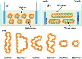
Electrodeposition of ZnO in a novel electrolyte, namely reverse micelles containing zinc nitrate and dissolved oxygen, has been successfully performed. By simply altering the applied current densities or using different substrates, various morphologies are obtained, from 2D ultrathin nanosheets to 3D hierarchical flower-like morphologies, which are quite different from those obtained in conventional electrolytes. Importantly, the behaviors of the soft colloidal templates have a great influence on the morphologies of the ZnO nanostructures. It can be inferred that the versatile deformation, coalescence and rearrangement of the soft colloidal templates also contribute to the specific surface morphologies. The soft colloidal templates are inclined to coalesce and assemble into larger templates on ITO, whereas deformation into different shapes is observed on carbon paper. In this way, an overall new idea to control the shapes and sizes of the template via electrochemistry is proposed.
Traditionally, for the soft-colloidal template synthesis of target materials, two different micelles or microemulsion droplets containing two types of reaction precursors are prepared.19–25 The reaction is enabled when the two different droplets fluctuate and collide with each other, and subsequent treatments comprise the separation of products, which is fairly complicated. The aim of this study is therefore to extend this type of electrolyte to an electrolyte in which the soft template is directly adhered to an electrode, offering simple and precise control of the dimensions of the as-synthesized structures.26 Our previous work involved the electrochemical synthesis of a conducting polymer and a transition metal oxide in water-in-oil microemulsion, namely reverse micelles and their application in supercapacitors.27–29 The results indicated that a dispersed soft colloidal template in this kind of novel electrolyte solution contributed to the uniform size distribution of the above materials. Meanwhile, it is clear that in addition to the soft template, the current density or potential applied to the electrode and the composition of solutions also influence the size, shape and composition of the materials. Therefore, further research is needed on the growth mechanism of materials in this kind of electrolyte.
ZnO, a typical II–VI compound semiconducting material with a wide band gap of 3.37 eV and a large exciton binding energy, has attracted much attention as a result of its potential applications in various fields such as UV detector, electro-optical switches, chemical sensors, photo-catalysts, and dye-sensitized solar cells.30–35 To date, a series of works concerning cathodic electrodeposition in aqueous solution or organic electrolyte have been reported and a diversity of ZnO nanostructures, including nanorods, nanowires, nanotubes, nanosheets, nanobelts, nanoplates and nanocombs have been obtained.36–43 It has been found that overpotential, temperature and composition of the electrolyte have significant influence on the morphology and composition of the nanostructure.44–46 However, to the best of our knowledge, no work on the electrodeposition of ZnO and its mechanism in this unconventional electrolyte has been reported. As stated above, it is of great significance to schematically evaluate the morphologies and compositions of the ZnO precursors obtained via electrochemical assemblies in this novel electrolyte.
In this contribution, a simple method is demonstrated for obtaining ZnO nanostructures with different morphologies (from 2D ultrathin nanosheet to 3D nanorods) by changing current densities, without tuning the ratio of surfactant in the reverse micelle. A comprehensive investigation is carried out on the effect of preparatory conditions on the morphologies and compositions of zinc oxides, including the current densities and substrates. It is the electric field at the interface of electrode/electrolyte that mainly controls the nucleation, growth and behaviors of soft templates and afterwards influences the morphologies. A probable mechanism is then proposed, indicating a new method for controlling the shapes and sizes of the soft template. This work not only lays a good foundation in fundamental electrochemistry and materials science but also shows diverse prospects and potentially valuable applications.
To investigate the effect of substrates on the surface morphologies, carbon paper was employed for electrochemical assembly of ZnO architectures for comparison. The FESEM and the corresponding TEM images of ZnO electrodeposited onto carbon paper substrate are shown in Fig. 2. The morphologies of the ZnO obtained on carbon paper are quite different from those on ITO substrates. Fig. 2a shows a thinner nanosheet covering the substrate at 1 mA cm−2, which resembles graphene. The corresponding TEM shows the obtaining of an ultrathin nanosheet. With increase of current density, the ultrathin nanosheet turns into nanoplates assembled into different shapes. The sizes of the hierarchical aggregates are much smaller than those assembled on ITO substrates, which may be because of the fast nucleation rates in all directions. This phenomenon can be ascribed to deformation rather than coalescence of the soft templates at the interface of carbon paper/solution.
The mechanism of ZnO deposition by electrochemical methods in both aqueous and organic solutions has been widely studied. This process is based on electroreduction of dissolved oxygen and/or NO3− ions to generate OH− ions at the surface of a working electrode. The generated OH− reacts with Zn2+ ions instantaneously to form zinc hydroxide, which is then dehydrated to produce the more stable zinc oxide. The general scheme for the electrodeposition of ZnO from aqueous zinc nitrate solution is thought to be as follows:47–49
NO3− + H2O + 2e → NO2− + 2OH− (Eθ = −0.240 V vs. SCE) | (1) |
NO3− + 6H2O + 8e → NH3 + 9OH− (Eθ = −0.373 V vs. SCE) | (2) |
O2 + 2H2O + 4e → 4OH− (Eθ = 0.151 V vs. SCE) | (3) |
Zn2+ + 2OH− → Zn(OH)2 → ZnO + H2O. | (4) |
However, it has been found that the potential for the reduction of nitrate shifts positively in the presence of Zn2+, as Zn2+ ion can act as a catalyst in reduction of nitrate. Moreover, the reduction potential of Zn2+ ion to its corresponding metal also shifts positively. So it can be inferred that the electrodeposition of ZnO in the present system is expected to proceed as follows:48
Zn2+ + NO3− + 2e → NO2− + ZnO (Eθ = 0.246 V vs. SCE) | (5) |
5Zn2+ + NO3− + 2H2O + 8e → ZnO + NH4+ (Eθ = 0.213 V vs. SCE). | (6) |
The probable mechanism for the behaviors of soft templates under an electric field is therefore proposed as in Scheme 1.
![]() | ||
Scheme 1 The behaviors of the soft colloidal templates at the interface of ITO/solution, (a) with no current being applied; (b) with current being applied; (c) with different current densities. |
It can be observed that the water phases constrained by the soft colloidal membrane are evenly dispersed in the continuous oil phase, as shown in Scheme 1a. When a current is applied, the droplets fluctuate and collide under the effect of the electric field, and the anions and cations inside the cores transfer between the adjacent polar aggregates. Thus, the deformation and coalescence of the soft template lead to formation of ion channels (Scheme 1b).50, 51, 53 The reactions at the interface of electrode/reverse micelle comprise three steps: first, the water phases electromigrate to the electrode; second, the anions and cations inside the cores diffuse to the surface of the electrode through the interface membrane; third, nucleation is enabled by the redox reaction of relevant ions. This process is remarkably different from that in the traditional electrolyte as the transporting behaviors at the interface membrane are more complicated and the interactive thermodynamic and kinetic properties are elusive. So it can be inferred that the nucleation of ZnO is controlled by the electric field at the interface of electrode/solution, while the growth is governed by the soft colloidal template. It is known that the ultrahigh field intensity near the electrode causes deformation, coalescence and rearrangement of the soft colloidal templates at the interface, as shown in Scheme 1c.52, 53 That is to say, it is the electric field at the interface of electrode/solution that finally influences nucleation and growth of ZnO. As shown in Scheme 1c, at different current densities, the soft colloidal template fluctuates, collides and self-assembles into different shapes. So the mechanism can be summarized: at 1 mA cm−2 constant current, the growth of ZnO has a height advantage on nucleation rate which leads to the large nanoplate morphology, and the rearrangement of soft colloidal templates at the surface of electrode shown in Scheme 1c decides the continuous plane. With increasing current density, the growth rates decrease while the nucleation rates increase. So the morphologies within the soft template are quite different, from nanoplates to octahedral nanostructures and nanorods. Reassembly of the soft colloidal templates occurs because of the destruction of balance caused by the electric field. The dispersed droplets tend to deform and coalesce under the effect of the electric field and will separate when a critical current density is reached. So soft templates with different shapes can be presented. However, the nucleation, growth and behaviors of soft colloidal templates at the carbon substrate are very different. The nanoplates tend to aggregate into hierarchical morphologies with different shapes and sizes. This is because of the faster nucleation rate resulting in formation of smaller particles and because the template tends to deform rather than coalesce.
Cyclic voltammograms (CVs) and potential–time curves are demonstrated for further illustration of the different morphologies obtained on ITO and carbon paper substrate. Fig. 3a shows the CVs of the ITO and carbon paper in the W/O microemulsion without Zn2+ ions; the polarization current for ITO substrates is higher than that of carbon paper, which indicates a faster depolarization ability. The nucleation and growth rates for ITO substrate are higher. With Zn2+ ions added in the solution, the CVs are clearly different. As shown in Fig. 3b, the reduction of nitrate becomes easier and the potential shifts positively in the presence of Zn2+, which is in accordance with the above-stated mechanism. Potential–time curves obtained with the constant current density of 1 mA cm−2 are shown in Fig. 3c (without Zn2+) and Fig. 3d (with Zn2+). The polarization potential for the ITO substrate is lower than that of carbon paper, see Fig. 3c. In the presence of Zn2+, the reduction potential of nitrate shifted positively.
The structure and crystal parameters of ZnO assembled onto carbon paper with different current densities are characterized by XRD spectra, shown in Fig. 4. The sharp diffraction peaks (Fig. 4a) are assigned to carbon substrates. The other weak diffraction patterns can be indexed as hexagonal wurtzite structures (hexagonal phase, space group P63mc) according to JCPDS card (no. 36-1451), which can be clearly observed in Fig. 4b. The four major peaks arising from the (100), (002), (101) and (102) planes of hexagonal structure indicate that they are polycrystalline. It is noted that the relative peak intensities for the ZnO film are different from the standard power sample, indicating differences in their crystallographic orientation. Clearly, there is preferential growth orientation on the (002) plane. To analyze the detailed change of crystal orientation, orientation indices of the three major peaks were calculated for each XRD pattern using the following method. An intensity factor (IFhkl) is defined as the ratio of the peak intensity for the diffraction peak of interest, relative to the sum of the intensities of the diffraction peaks.47,48 For example, the calculation of the (100) plane is presented as follows:
![]() | (7) |
![]() | ||
Fig. 4 X-ray diffraction patterns of the as-prepared ZnO films on carbon paper with different current densities. (a) 1 mA cm−2; (b) 5 mA cm−2; (c) 10 mA cm−2; (d) 15 mA cm−2. |
The I(hkl) refers to the diffraction peak intensity in XRD. The standard intensity factors IFS(hkl) were obtained from the data of a standard powder sample (JCPDS). Then the intensity factor (IFhkl) was compared with standard intensity factors (IFS(hkl)) using the defined formula:
![]() | (8) |
Any crystal faces for which these values exceed 1 are preferentially oriented. The specific values can be seen in Table 1. It is found that all the ZnO films on carbon paper substrates with different current densities are oriented with its (100) and (002) planes. But the (100) preferential orientation is less significant compared with the preferential growth on the (002) plane. The OIS(101) value becomes significantly smaller than 1. The results clearly demonstrate the preferential growth parallel rather than perpendicular to the substrate, which is consistent with the flower-like morphology observed for these films in Fig. 2.
Sample | hkl | I(hkl) | IF(hkl) | OIS |
---|---|---|---|---|
Standard card | (100) | 57 | 0.284 | |
(002) | 44 | 0.219 | ||
(101) | 100 | 0.498 | ||
a | (100) | 77.78 | 0.297 | 1.046 |
(002) | 84.03 | 0.321 | 1.466 | |
(101) | 100 | 0.382 | 0.767 | |
b | (100) | 89 | 0.317 | 1.116 |
(002) | 92.1 | 0.327 | 1.493 | |
(101) | 100 | 0.356 | 0.715 | |
c | (100) | 97.54 | 0.335 | 1.180 |
(002) | 93.44 | 0.321 | 1.466 | |
(101) | 100 | 0.344 | 0.691 | |
d | (100) | 83.4 | 0.302 | 1.063 |
(002) | 92.89 | 0.336 | 1.534 | |
(101) | 100 | 0.362 | 0.727 |
To gain further insights into the structural features and crystallography of the nanosheet of the hierarchical aggregates, TEM and HRTEM techniques were employed to investigate the sample under the condition of 8 mA cm−2. The flower-like morphology is composed of many ultrathin sheets, see Fig. 5. In the high-magnification image of the ultrathin deposit, a uniform lattice image can be observed with inter-space calculated to be 2.6 Å over the entire sheet, corresponding to the inter-space of the (002) plane. The results again confirmed the preferential growth of ZnO with the (002) plane on carbon paper substrates.
The surface compositions of the ZnO architectures were analyzed by X-ray photoelectron spectroscopy (XPS). A wide survey spectrum with the applied current density of 8 mA cm−2 is shown in Fig. 6a, with the observed peaks corresponding to the elements Zn, O and C.54,55 The Zn 2p spectrum in Fig. 6b shows a doublet whose binding energies are 1021.4 and 1044.4 eV, attributed to Zn 2p3/2 and Zn 2p1/2, respectively, indicating the presence of a single Zn2+ divalent state, corresponding to both Zn(OH)2 and ZnO. The spin–orbit splitting of 23.0 eV (±0.1 eV) is consistent with the literature value of 22.97 eV.52 Fig. 6c shows the N 1s narrow scan spectrum: the two peaks refer to ammonium (NH4+) and nitrate (NO3−) with binding energies of 399.8 and 406.7 eV, respectively. The result confirms the proposed mechanism. The O 1s core-level spectrum of ZnO is fitted with three Gaussian peaks. The lowest binding energy of 530.17 eV is assigned to the O2− ions in Zn–O bonds of the hexagonal wurtzite structure of ZnO. The other two peaks located at 531.38 and 532.70 eV are related to the absorbed OH− and NO3− on the surface with the ZnO. So it is concluded that the probable reaction at the interface of electrode/solution is eqn (6).
![]() | ||
Fig. 6 XPS spectra of the ZnO film electrochemical assembled at 8 mA cm−2: (a) a wide survey, (b) Zn 2p, (c) N 1s, (d) O 1s. |
This journal is © The Royal Society of Chemistry 2014 |