Nanostructured CuO/reduced graphene oxide composite for hybrid supercapacitors

Kamatchi Kamaraj Purushothaman*a, Balakrishanan Saravanakumarb, Inbamani Manohara Babuc, Balasubramanian Sethuramana and Gopalan Muralidharanc
aDepartment of Physics, TRP Engineering College (SRM group), Irungalur, Tamilnadu, India. E-mail: purushoth_gri@yahoo.co.in; Tel: +91 431 2908050
bFaculty of Physics, Mahalingam College of Engineering and Technology, Pollachi, Tamilnadu, India
cDepartment of Physics, Gandhigram Rural University, Gandhigram-624302, Tamilnadu, India

Received 11th March 2014 , Accepted 6th May 2014

First published on 8th May 2014


Abstract

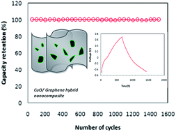
To address the issues such as low ionic conductivity, poor electrode kinetics and cyclic stability, the strategy of combining carbon-based materials with transition metal oxide (TMO) is adopted. In this article, the preparation of CuO/reduced graphene oxide (RGO) nanocomposite electrodes by a simple, low cost hydrothermal method is described. This hybrid nanocomposite exhibits a high specific capacitance of 326 F g−1 at a current density of 0.5 A g−1. It shows a high energy density of 65.7 W h kg−1 at a power density of 302 W kg−1. Further, this material does not exhibit any measureable degradation in electrochemical performance, even after 1500 cycles. Symmetric hybrid capacitors exhibit a specific capacitance of 97 F g−1 at 0.2 A g−1 with a power density of 72 W kg−1. These superior electrochemical features demonstrate that the CuO/RGO hybrid nanocomposite is a promising material for next-generation supercapacitor systems.


Introduction

Supercapacitors (SCs) have attracted significant attention from researchers owing to their special characteristics such as rapid charge–discharge, long cycling life, environmental friendliness, safety and high power density. Further, they complement other energy storage systems such as batteries and fuel cells.1 Generally, a supercapacitor is composed of three prime components namely electrodes, a separator and an electrolyte. Among these, innovative electrodes are mainly responsible for better overall performance of the SCs. Huge efforts have been devoted worldwide by researchers in this direction to fabricate better supercapacitor electrodes. Many types of material have been tested and studied as electrodes for SCs, including carbon-based materials, conducting polymers, transition metal oxides (TMOs) and hybrid composites.2 Because of their ability to exist in different oxidation states, long cycle life and ease of preparation, TMOs are considered as candidates for supercapacitor electrodes. TMOs such as RuO2, NiO, MnO2, Co3O4 and V2O5 have been studied and found to be suitable as electrodes for supercapacitor systems.3–7

Cupric oxide (CuO) is a well-known metal oxide due to its special features such as non-toxicity, abundance, low cost and ease of fabrication in the form of nano dimensions, which have led to its use in Li-ion batteries and supercapacitor electrodes.8–10 However, its low electrical conductivity and destruction of the structure during ion intercalation/deintercalation have hampered the electrochemical performance of CuO. These shortcomings are addressed by two strategies. First, the fabrication of CuO in the form of nanostructures is expected to provide facile pathways for electrolyte ion penetration. Second is blending carbon-based materials with CuO to make CuO hybrid composites. The presence of carbon-based materials (CNT, Graphene, mesoporous carbon etc.) in the composite greatly improves conductivity and electrochemical performance.

In this point of view, graphene has recently drawn great attention of researchers from many groups, since its discovery.11 Primarily, it possess tightly connected flat single layers of sp2-hybridized carbon atoms into a honeycomb lattice.12,13 Because of its hallmark features such as a 2D structure, ultra-high specific surface area, high mechanical strength and elevated conductivity have led to its application in bio- and chemical sensors,14,15 field-effect transistors,16 energy storage devices,17,18 and bio medicine,19 etc.

In this sense, the preparation of a composite containing CuO and carbon-based materials such as graphene is expected to yield SCs with an improved electrochemical nature. Earlier, the CuO/graphene nanocomposites were prepared and studied for Li-ion battery applications. Wang et al. fabricated graphene-enwrapped CuO with a high rate capacity and good cycling performance.20 Mai et al. reported the preparation of a CuO/graphene composite with improved efficiency as anode materials for Li-ion batteries.21 Rai et al. showed a microwave-assisted synthesis of a CuO/RGO nanocomposite to yield good electrochemical performance.22 However, the use of a CuO/RGO composite as a supercapacitor electrode is the least explored. This work is probably one of the few reports available on this composite or on the nature of CuO as an electrode material.

In this article, we reported the fabrication of a CuO/RGO hybrid nanocomposite by a low cost hydrothermal method and studied its electrochemical performance to explore its application as a supercapacitor electrode. We utilized RGO as a conductivity booster with CuO nanostructures. Structural and electrochemical investigations demonstrate the suitability and electrochemical features of the CuO/RGO hybrid nanocomposite as a better electrode material for a supercapacitor. The CuO/RGO is denoted as “CuG hybrid nanocomposite” in further discussions.

Results and discussion

The morphology of the CuG hybrid nanocomposite was identified using scanning electron microscope (SEM) and scanning transmission electron microscope (STEM) studies. Fig. 1a–c shows the SEM images of the CuG hybrid nanocomposite, which consists of nanosized particles. Further, STEM images (Fig. 2) confirm the presence of graphene sheets stacked one over the other. CuO nanoparticles appear to be covered with graphene sheets. Scheme 1 shows the schematic of the fabrication of the CuG hybrid nanocomposite. Fig. 3 shows the EDS spectrum with elemental mapping of the CuG composite, which confirms the presence of copper (64.4 wt%), oxygen (24.4 wt%) and carbon (9.92 wt%).
image file: c4ra02107j-f1.tif
Fig. 1 (a–c). SEM images of the CuG hybrid nanocomposite.

image file: c4ra02107j-f2.tif
Fig. 2 STEM images of the CuG hybrid nanocomposite.

image file: c4ra02107j-s1.tif
Scheme 1 Schematic of the fabrication of the CuG hybrid nanocomposite.

image file: c4ra02107j-f3.tif
Fig. 3 EDS spectrum and elemental mapping images of the CuG hybrid nanocomposite.

In order to confirm the structure, phase and purity of the materials, X-ray diffraction patterns (XRD) were recorded for the CuG hybrid nanocomposites (Fig. 4). The diffraction peaks coincide well with the standard pattern of CuO (JCPDS card no. 05-0661). Further, it shows monoclinic symmetry with a C2/C space group and possess lattice constant values of a = 0.4684 nm, b = 0.3425 nm and c = 0.5129 nm: β = 99.47°. No impurity peaks could be detected in the diffraction pattern; this is indicative of the phase purity of CuO.


image file: c4ra02107j-f4.tif
Fig. 4 (a) XRD pattern of the CuG hybrid nanocomposite (b) FT Raman spectrum of the CuG hybrid nanocomposite.

FT Raman spectroscopy is an effective analytical method to identify the structure and defects in composite materials. The FT Raman spectrum of the CuG hybrid nanocomposite (Fig. 4b) shows D and G bands at 1335 and 1593 cm−1, respectively. This band corresponds to the characteristic peaks of graphene. The G peak represents sp2 carbon vibrations, and the D band represents sp3 carbon in the graphitic lattice.23,24 The intensity of the D and G bands is less because of the low loading of graphene in CuO. CuO belongs to space group C62h with Raman active optical phonons (Ag + 2Bg). The well-resolved bands at 294, 615 and 1087 cm−1 confirm the presence of CuO.25,26

The electrochemical performance of the CuG hybrid nanocomposite is primarily investigated by cyclic voltammetry measurements (CV) at a scan rate of 1 mV s−1 within a potential range of −0.55 to 0.65 V (shown in Fig. 5a). The presence of anodic and cathodic peaks in the CV curve are proof of the fact that the origin of capacitance in the present nanocomposite is mainly from the surface redox reactions. The CuG hybrid nanocomposite exhibited a specific capacitance of 318 F g−1 at a scan rate of 1 mV s−1. Fig. 5b displays the CV profiles of the nanocomposite at different scan rates. It is indicative of good kinetic reversibility of the CuG hybrid nanocomposite as an electrode material. The variation of the specific capacitance with the scan rate is displayed in Fig. S1. At higher scan rates, the electrolyte ions are not provided with enough time to access the interior parts of the electrode, and hence exhibit reduced charge storage characteristics, i.e., a lower specific capacitance. The electrolyte ion utilizes a maximum area of the electrode at lower scan rates and finds enough time to percolate into the material, resulting in superior values of specific capacitance.27


image file: c4ra02107j-f5.tif
Fig. 5 (a) CV curve of the CuG hybrid nanocomposite at a scan rate of 1 mV s−1 (b) CV curve at different scan rates (c) charge–discharge profile of the CuG hybrid nanocomposite at 500 mA g−1(d) charge–discharge profile of the CuG hybrid nanocomposite at different current densities.

To understand the charge storage mechanism and rate capacity of the CuG hybrid nanocomposite, galvanostatic charge discharge (GCD) measurements were performed. A typical charge–discharge profile for the nanocomposite prepared in this work from −0.5 to 0.7 V at a constant current of 0.5 A g−1 is displayed in Fig. 5c. The near symmetric nature of the charge and discharge curve shows the improved capacitive nature and charge–discharge reversibility. Further, the specific capacitance value reaches up to 326 F g−1 at 0.5 A g−1. This capacitance is higher than the values reported by other researchers.23,28 To the best of our knowledge, this is one of the best capacitance values ever reported for a CuG hybrid nanocomposite. This superior enhancement in the specific capacitance is attributed to the presence of RGO with CuO. RGO acts as a conductivity enhancer and provides better pathways for ion intercalation. Fig. 5d shows the charge discharge profiles at different current densities. At higher current rates, electrolyte ion movement was not properly synchronized with the current rates.29 Because of this high current density, specific capacitance is significantly reduced. The variation of specific capacitance with current density is shown in Fig. S2.

Energy density and power density are two of the most important parameters of an electrode material to decide its suitability for commercial usage. The Ragone plot for the electrodes prepared using the nanocomposite is shown in Fig. S3. CuG hybrid nanocomposite electrodes exhibit a maximum energy density of 65.7 W h kg−1 and power density of 302 W kg−1 at 0.5 A g−1 and a maximum power density of 2.4 kW kg−1 and energy density of 20.8 W h kg−1 at 4 A g−1. The retention of capacity after long-term cycling of charge–discharge is one of the key factors to evaluate the supercapacitor electrodes for practical applications. Reduced graphene oxide (RGO) shows 100% stability, and copper oxide (CuO) exhibits 74% up to 1500 cycles at 5 A g−1 (Fig. S4). The CuG hybrid nanocomposite does not show any measureable degradation after 1500 continuous cycles of charge–discharge at 5 A g−1 (Fig. 6a). The presence of RGO with CuO seems to greatly enhance the stability of the nanocomposite. The excellent cyclic stability exhibited by the CuO/RGO hybrid nanocomposite reveals that it is possible to use the nanocomposite in practical energy storage systems.


image file: c4ra02107j-f6.tif
Fig. 6 (a) Cyclic stability of the CuG hybrid nanocomposite measured at 5 Ag−1. (b and c) The Nyquist plot of the CuG hybrid nanocomposite and equivalent circuit model.

Better understanding of the fast ion diffusion in the CuG hybrid nanocomposite can be achieved by performing EIS measurements. They were made in the frequency range of 0.01 Hz to 100 kHz. The Nyquist profiles of the CuG hybrid nanocomposite is shown in Fig. 6b. The inset of Fig. 6b represents the high-frequency region of the Nyquist plot. Fig. 6c shows the equivalent circuit diagram to which the impedance data has been fitted to. It contains six components: solution resistance (Rs), charge transfer resistance (Rct), leakage resistance (RL), double layer capacitance (Cdl), mass capacitance (CL) and the Warburg element (W). These elements have their usual meaning. The semicircle intercept on the real axis (Z′) at higher frequencies represents the charge transfer resistance (Rct). The Rct value is observed to be 2.47 Ω for the CuG hybrid nanocomposite. This low value of charge transfer resistance helps in faster charge–discharge possibilities, which is an essential feature of an energy storage device where the generation of energy is fast and for a short period, and in some cases, the load may require instantaneous delivery of large power.

To analyze the capacitive features of the CuG hybrid nanocomposite electrode in a complete cell set up, a symmetric supercapacitor was fabricated. The schematic illustration of the fabrication process of the symmetric supercapacitor is shown in Fig. 7. This is composed of the CuG hybrid nanocomposite as the positive and negative electrodes, polypropylene (Celgard) as a separator and 0.5 M K2SO4 used as the electrolyte. Fig. 8 shows the CV curves of the CuG hybrid nanocomposite-based symmetric supercapacitor device at different scan rates (2 to 50 mV s−1) in a potential window of 1.2 V (0 to 1.2 V). The estimated specific capacitance from the CV measurements are 64, 58, 49, 39, 27 and 18 F g−1 at scan rates of 2, 3, 5, 10, 25 and 50 mV s−1, respectively. Further, the GCD curves with various current densities are shown in Fig. 9. The symmetric supercapacitor shows the triangular charge–discharge curves, confirming the linear relationship between potential and time. Further symmetric charge and discharge curves validate the fact that the material has superior electrochemical reversibility. Specific capacitance values derived from the GCD measurements are 97, 71, 55 and 36 F g−1 at current densities of 0.2, 0.5, 1 and 2 Ag−1, respectively, and the estimated energy density is 19 W h kg−1 at a power density of 72 W kg−1. The mixing of CuO and graphene has led to a new type of a hybrid nanocomposite as an electrode material for high-performance supercapacitors.


image file: c4ra02107j-f7.tif
Fig. 7 Schematic representation of the fabrication of the CuO/RGO-based symmetric supercapacitor.

image file: c4ra02107j-f8.tif
Fig. 8 CV curves of the symmetric supercapacitor at different scan rates.

image file: c4ra02107j-f9.tif
Fig. 9 GCD curves of the symmetric supercapacitor at different current densities.

Conclusion

A nanostructured CuG hybrid composite material has been synthesized by a simple and inexpensive hydrothermal method. Electrochemical investigations reveal that the electrodes can offer high specific capacitance (326 F g−1 at a current density of 0.5 A g−1), high energy density (65.7 W h kg−1) and large power density (302 W kg−1). The material did not show any loss of specific capacity after 1500 cycles of charge–discharge. The symmetric supercapacitor fabricated using the CuG hybrid nanocomposite material as electrodes exhibited a high specific capacitance of 97 F g−1 at 0.2 A g−1. These enhanced electrochemical features of the CuG hybrid composite is attributed to the synergy between the CuO and RGO in the storage of charge. These results permit us to state with confidence that a new class of nanocomposites with high electrochemical performance has been identified.

Experimental details

Analytical grade graphite powder, NaNO3, KMnO4, hydrazine hydrate (N2H4), Cu (NO3)2·3H2O and disodium citrate (Na2HC6H5O7) were purchased from Sigma Aldrich. H2SO4, H2O2 (30%), HCl and NaOH were procured from SD Fine Chemicals Ltd., India. All chemicals and reagents were used as supplied without further purification.

Synthesis of RGO

The synthesis of reduced graphene was done with the help of a modified Hummers method. The synthesis process consists of two stages, namely the synthesis of graphene oxide and reduction. Initially, 100 ml of H2SO4 was taken in a flask. First, a mixture containing 3 g of graphite powder and 1.5 g of NaNO3 was poured into the flask and stirred whilst keeping inside an ice bath. To avoid overheating, 8 g of KMnO4 was added slowly and stirred at room temperature for 2 h. The colour of the suspension becomes bright brown. 90 ml of double distilled water (DDW) was then added to the suspension. The addition of the DDW makes the solution change to yellow in colour. The suspension was again stirred for 12 h at 98 °C, after this, 30% H2O2 (30 ml) was added to the mixture. After a thorough wash with 5% HCl and DDW, the suspension was centrifuged and dried. It produced a black-coloured graphene oxide powder. Second, the mixture of 30 ml DDW and graphene oxide (100 mg) was ultrasonicated for 1 h to get a homogenous suspension of graphene oxide. The suspension was kept at 100 °C, and 3 ml of hydrazine hydrate was mixed with the suspension. Further, the suspension was maintained at 98 °C for 24 h. With the help of filtration, black-coloured reduced graphene oxide powders were obtained from the suspension. The as-prepared powders were washed several times with DDW to remove excess hydrazine. Then the suspension was centrifuged and dried to get the final product.

Synthesis of CuO/RGO

Initially, 100 mg of graphene was dispersed in 50 ml DDW and ultrasonicated for 1 h to get a homogeneous dispersion. Second, copper nitrate (0.906 g) was dissolved in 20 ml of DDW, and 10 wt% of disodium citrate was dissolved in 5 ml DDW. Both solutions were slowly added to the graphene solution, and it was stirred for 1 h. Further, a 2 M NaOH (10 ml) solution was prepared and added dropwise until the pH value reached 10. The complete solution was kept under vigorous magnetic stirring for 2 h at room temperature, and finally, a dark brown colloidal suspension was formed. The colloidal suspension was transferred to a 100 ml Teflon lined stainless steel autoclave and kept at 180 °C for 12 h. The resulting precipitate obtained was washed thoroughly using DDW. The washed precipitate was dried at 80 °C for 12 h. Finally, a brown-coloured byproduct was obtained, and the same product was annealed at 300 °C for 2 h in air.

Material characterization

X-ray diffraction analysis was performed from 10° to 80° using a PANalytical XPERT-PRO X-ray diffractometer with CuKα radiation to determine the phase and purity of the samples. FT-Raman spectra have been recorded using a FEKI-Japan STR-500 mm focal length laser Raman spectrometer. A scanning electron microscope with EDS (JEOL JSM 3690 scanning electron microscope) was used to investigate the morphology of the samples. Cyclic voltammetry (CV), galvanostatic charge−discharge (GCD), and electrochemical impedance spectroscopy (EIS) analyses were performed using a CHI 660 D electrochemical workstation (CH Instruments).

Electrode preparation and evaluation

Electrochemical analysis of the CuG nanocomposite samples was tested using a three-electrode cell set up by employing CuG as the working electrode, Ag/AgCl as the reference electrode and platinum wire as the counter electrode. The electrode material was prepared by mixing 85 wt% sample, 10 wt% activated carbon (Sigma-Aldrich), and 5 wt% polytetrafluoroethylene (Sigma-Aldrich) with ethanol. The mixture was coated onto a nickel foam (1 cm2) surface. Following this, the electrode was dried at 80 °C for 5 h. 0.5 M K2SO4 was used as the electrolyte solution at atmospheric temperature. Based on the mass of the active material (1 mg) in the electrode, specific capacitances and energy densities were calculated for the three electrode and two electrode systems.

The specific capacitance (C, F g−1) values from CV measurements were obtained using the following equation,

image file: c4ra02107j-t1.tif
where I (mA) is the average current obtained by integrating CV, Δν (V) is the potential window and m (mg cm−2) is the mass of the active material.

Specific capacitance values were calculated from the galvanostatic discharge curves according to the following equation,

image file: c4ra02107j-t2.tif
where I (ma) is the discharge current, Δt (s) the time needed for discharge, Δν (V) the potential range and m (mg cm−2) is the mass of the active material.

Energy and power densities were estimated from the following equations,

image file: c4ra02107j-t3.tif

image file: c4ra02107j-t4.tif
where C (F g−1) is the discharge specific capacitance, Δv (V) is the potential, E (W h kg−1) is the energy density, P (W kg−1) is the power density and t (s) the discharge time.

References

  1. P. Simon and Y. Gogotsi, Nat. Mater., 2008, 7, 845 CrossRef CAS PubMed.
  2. M. Toupin, T. Brousse and D. Bélanger, Chem. Mater., 2004, 16, 3184–3190 CrossRef CAS.
  3. M. T. Brumbach, T. M. Alam, P. G. Kotula, B. B. McKenzie and B. C. Bunker, ACS Appl. Mater. Interfaces, 2010, 2, 778–787 CAS.
  4. K. K. Purushothaman, I. Manohara Babu, B. Sethuraman and G. Muralidharan, ACS Appl. Mater. Interfaces, 2013, 5(21), 10767–10773 CAS.
  5. Y. Huang, Y. Li, Z. Hu, G. Wei, J. Guo and J. Liu, J. Mater. Chem. A, 2013, 1, 9809–9813 CAS.
  6. D. Wang, Q. Wang and T. Wang, Inorg. Chem., 2011, 50, 6482–6492 CrossRef CAS PubMed.
  7. B. Saravanakumar, K. K. Purushothaman and G. Muralidharan, ACS Appl. Mater. Interfaces, 2012, 4, 4484–4490 CAS.
  8. R. Wu, X. Qian, F. Yu, H. Liu, K. Zhou, J. Wei and Y. Huang, J. Mater. Chem. A, 2013, 1, 11126–11129 CAS.
  9. S. Gao, S. Yang, J. Shu, S. Zhang, Z. Li and K. Jiang, J. Phys. Chem. C, 2008, 112(49), 19324–19328 CAS.
  10. G. S. Gund, D. P. Dubal, D. S. Dhawale, S. S. Shinde and C. D. Lokhande, RSC Adv., 2013, 3, 24099–24107 RSC.
  11. K. S. Novoselov, A. K. Geim, S. V. Morozov, D. Jiang, Y. Zhang, S. V. Dubonos, I. V. Grigorieva and A. A. Firsov, Science, 2004, 306, 666–669 CrossRef CAS PubMed.
  12. X. C. Dong, H. Xu, X. W. Wang, Y. X. Huang, M. B. Chan-Park, H. Zhang, L. H. Wang, W. Huang and P. Chen, ACS Nano, 2012, 6(4), 3206–3213 CrossRef CAS PubMed.
  13. C. Zhu, S. Guo, Y. Fang, L. Han, E. Wang and S. Dong, Nano Res., 2011, 4(7), 648–657 CrossRef CAS PubMed.
  14. J. D. Fowler, M. J. Alle, V. C. Tung, Y. Yang, R. B. Kaner and B. H. Weiller, ACS Nano, 2009, 3, 301–306 CrossRef CAS PubMed.
  15. Y. X. Liu, X. C. Dong and P. Chen, Chem. Soc. Rev., 2012, 41, 2283–2307 RSC.
  16. F. Schwierz, Nat. Nanotechnol., 2010, 5, 487–496 CrossRef CAS PubMed.
  17. F. Zhang, T. Zhang, X. Yang, L. Zhang, K. Leng, Y. Huang and Y. Chen, Energy Environ. Sci., 2013, 6, 1623–1632 CAS.
  18. C. Liu, Z. Yu, D. Neff, A. Zhamu and B. Z. Jang, Nano Lett., 2010, 10, 4863–4868 CrossRef CAS PubMed.
  19. h. Shen, L. Zhang, M. Liu and Z. Zhang, Theranostics, 2012, 2(3), 283–294 CrossRef CAS PubMed.
  20. b. Wang, X. L. Wu, C. Y. Shu, Y. G. Guo and C. R. Wang, J. Mater. Chem., 2010, 20, 10661–10664 RSC.
  21. Y. J. Mai, X. L. Wang, J. Y. Xiang, Y. Q. Qiao, D. Zhang, C. D. Gu and J. P. Tu, Electrochim. Acta, 2011, 56, 2306–2311 CrossRef CAS PubMed.
  22. A. K. Rai, L. T. Anh, J. Gim, V. Mathew, J. Kang, P. J. Paul, N. K. Singh, J. Song and J. Kim, J. Power Sources, 2013, 244, 435–441 CrossRef CAS PubMed.
  23. D. W. Kim, K. Y. Rhee and S. J. Park, J. Alloys Compd., 2012, 530, 6–10 CrossRef CAS PubMed.
  24. Y. Qi, J. R. Eskelsen, U. Mazur and K. W. Hipps, Langmuir, 2012, 28, 3489–93 CrossRef CAS PubMed.
  25. A. Pendashteh, M. F. Mousavia and M. S. Rahmanifar, Electrochim. Acta, 2013, 88, 347–357 CrossRef CAS PubMed.
  26. J. S. Shaikh, R. C. Pawar, R. S. Devan, Y. R. Ma, P. P. Salvi, S. S. Kolekar and P. S. Patil, Electrochim. Acta, 2011, 56, 2127–2134 CrossRef CAS PubMed.
  27. G. Wee, H. Z. Soh, Y. L. Cheah, S. G. Mhaisalkar and M. Srinivasan, J. Mater. Chem., 2010, 20, 6720–6725 RSC.
  28. Y. Liu, H. Huang and X. Peng, Electrochim. Acta, 2013, 104, 289–294 CrossRef CAS PubMed.
  29. L. Wang, H. Ji, S. Wang, L. Kong, X. Jiang and G. Yang, Nanoscale, 2013, 5, 3793–3799 RSC.

Footnote

Electronic supplementary information (ESI) available: The variation of specific capacitance with various scan rates from the CV measurements (Fig. S1), the variation of specific capacitance with different current densities from the GCD measurements (Fig. S2) and the Ragone plot (Fig. S3). See DOI: 10.1039/c4ra02107j

This journal is © The Royal Society of Chemistry 2014
Click here to see how this site uses Cookies. View our privacy policy here.