Enhancing photocatalytic activity by tuning the ratio of hexagonal and orthorhombic phase Nb2O5 hollow fibers

Ruyi Shao, Zhenzhu Cao*, Yang Xiao, Hongjie Dong, Weiyan He, Yanfang Gao and Jinrong Liu*
College of Chemical Engineering, Inner Mongolia University of Technology, Hohhot 010051, China. E-mail: czz03@163.com; Fax: +86 471 6503298; Fax: +86 579 8228226; Tel: +86 471 6575722 Tel: +86 579 8228226

Received 9th March 2014 , Accepted 5th June 2014

First published on 5th June 2014


Abstract

Cotton was used as template for the fabrication of a biomorphic Nb2O5 photocatalyst with hexagonal and orthorhombic structures. The as-prepared Nb2O5 was characterized through X-ray diffraction (XRD), scanning electron microscopy (SEM), specific surface area and UV-Vis diffused reflectance spectra measurements. The photocatalytic activity of the Nb2O5 fibers was evaluated through degradation of methylene blue (MB) in aqueous solution exposed to UV light. SEM results show that biomorphic Nb2O5 fibers are both hollow and have porous walls. The architectures allow a more accessible surface for the photocatalytic decomposition of dyes molecules. The photocatalytic activity of physically mixed fibers with 75% H-Nb2O5 and 25% O-Nb2O5 phases is superior to those of pure H- and O-Nb2O5 phases, and other samples with different H- and O-Nb2O5 ratios. Such superior photocatalytic activity is mainly due to an optimal balance between the adsorption of MB and the absorption of UV light. Moreover, the large size of the Nb2O5 hollow fibers allows them to be easily separated from the solution.

Introduction

Fujishima and Honda1 reported on the photoelectrochemical splitting of water on n-TiO2 electrodes. Subsequently, semiconductor-based photocatalysis has been extensively studied for potential environmental remediation and as a new energy source. In general, the photocatalytic reactions of semiconductors involve the formation of electron–hole pairs derived from interband excitation under light irradiation. Thus, a charge migrates from electron–hole pairs to the adsorbed reactants on the semiconductor surface, resulting in a redox reaction with the surrounding media.2 Numerous oxide semiconductors, such as TiO2,1,3 ZnO,4 Bi2O3 (ref. 5) and Ta2O5,6 have been used as photocatalysts in organic pollutant degradation and water-splitting reactions.

Among oxide semiconductors, niobium oxide (Nb2O5) is a promising n-type transition metal oxide7 with numerous significant photocatalytic activities, including selective photooxidation of amines,8,9 photocatalytic hydrogen production10 and photodegradation of harmful organic contaminants.11–13 Studies have shown that niobium oxide is a novel photocatalyst that can be used for the degradation of organic pollutants.11–14 Prado and coworkers11 found that Nb2O5 has sufficiently high photocatalytic activity for the decomposition of indigo carmine dye. Zhao et al.12 reported that Nb2O5 nanorod exhibits higher photocatalytic activity than nanospheres for the decomposition of methylene blue. Recently, Qi and coworkers14 prepared Nb2O5 nanofibers with hexagonal (H) and orthorhombic (O) structures by electrospun method and characterized its photocatalytic activity. Results showed that the H-Nb2O5 nanofibers exhibit much higher activity than O-Nb2O5 during decomposing methyl orange (MO). Doping with metal ions,15,16 nonmetal ions,17,18 and mixed metal oxides19 has been conducted to enhance the photocatalytic activity of Nb2O5. The coexistence of anatase/rutile phases in commercial TiO2 (Degussa P25) contributes to its excellent photocatalytic activity.2,20 Nb2O5 exists in many polymorphic forms, such as H-Nb2O5 (hexagonal), O-Nb2O5 (orthorhombic) and M-Nb2O5 (monoclinic).7 However, information is lacking regarding the effect of H- and O-Nb2O5 ratio on photocatalytic degradation of organic dyes.

To improve photocatalytic activity, the preparation of the nanometer catalyst2 has been intensively studied. Reusability of a nanosized photocatalyst is limited because of the difficulty of separating the photocatalyst from the suspension.21 Micrometer hollow fibers have been developed to overcome this drawback.22,23 Cotton fibers are stable, low cost, abundant, and reproducible natural products that have a hierarchically built anatomy.22 Micrometer hollow fibers prepared using cotton as templates have large surface areas and strong organic adsorption capacities, which are favorable characteristics for photocatalytic reactions.24 In the present study, micrometer Nb2O5 hollow fibers were obtained by calcining cotton fibers infiltrated by niobium oxalate solution for the first time. The photocatalytic properties of Nb2O5 hollow fibers with different ratios of H- to O-Nb2O5 phases were evaluated by photodegradation of methylene blue solution under UV irradiation. The physical mixed-phase Nb2O5 hollow fibers comprising a H- and O-Nb2O5 ratio of 75/25 w/w are more highly efficient than pure H-Nb2O5, O-Nb2O5 and other mixing phases.

Experimental

Synthesis

Dried and loose medical absorbent cotton (Fig. S1) were immersed in niobium oxalate solution (3 wt%, with water as solvent) for 24 h. The cotton fibers were calcined at either 500 °C or 700 °C in air for 6 h to remove cotton templates. Biomorphic Nb2O5 fibers with H- and O-Nb2O5 phases were obtained and subsequently physically mixed. In the mixing process, the Nb2O5 fibers were dispersed in ethanol, ground for 30 min, and dried under 60 °C for 2 h.

Characterization

Phases of the prepared biomorphic Nb2O5 fibers were identified through X-ray diffraction (XRD) with Cu Kα radiation (Bruker D8 Advance, Germany). The microstructures of the biomorphic Nb2O5 were investigated through scanning electron microscopy (SEM, S-3400, Hitachi, Ltd., Japan). The band gap was derived from the UV-vis diffused reflectance spectra (UV3150, Shimadzu, Japan). The specific surface area of samples were determined according to Brunauer–Emmett–Teller (BET) method on liquid nitrogen adsorption at 77 K by using equipment (ASAP 2010, Micromeritics, USA).

Photocatalysis measurement

The photocatalytic activities of biomorphic Nb2O5 fibers were evaluated by degrading 15 mg L−1 methylene blue (MB) aqueous solution under UV light (mercury lamp, 500 W; maximum emission = 365 nm). Prior to UV illumination, the samples (0.2 g) were dispersed in 200 mL MB aqueous solution and magnetically stirred in darkness for 50 min until adsorption/desorption equilibrium was reached. During irradiation, approximately 10 mL of MB degradation solution was continually extracted at 10 min intervals. Retrieved samples were centrifuged and subsequently examined using a UV-vis spectrometer (UVT6, Purkinje General, China). Absorption measurements were taken at 663 nm. The value of C/C0 was calculated according to the calibration curve of concentration and absorption.25 The photocatalytic activity of physically mixed H-Nb2O5/O-Nb2O5 phase fibers was evaluated under the abovementioned conditions for comparison.

Results and discussion

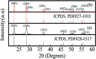
XRD patterns of cotton template-derived Nb2O5 catalysts calcined at 500 and 700 °C for 6 h are shown in Fig. 1. All the original components of cotton have been removed. As shown in Fig. 1a, the sample calcined at 500 °C was considered as the hexagonal Nb2O5 crystal (H-Nb2O5). Splitting of the peaks at 2θ = 28.4°, 36.7°, 50° was observed when calcination temperature was increased to 700 °C (Fig. 1b). Such splitting indicates transformation of Nb2O5 crystals from H-Nb2O5 to O-Nb2O5, which is consistent with the previous report.13 The calculated lattice constants of H-Nb2O5 were a = b = 0.3619 nm, c = 0.3929 nm and those of O-Nb2O5 were a = 0.164 nm, b = 2.9337 nm, c = 0.3931 nm. These constants were in good agreement with values form standard cards of H-Nb2O5 (JCPDS, PDF#28-0317, a = b = 0.3607 nm, c = 0.3925 nm) and O-Nb2O5 (JCPDS, PDF# 27-1003, a = 0.168 nm, b = 2.9312 nm, c = 0.3936 nm.). This result further confirms phase purity of the as-prepared samples. The XRD peaks of samples progressively narrowed and strengthened with the increase in annealing temperature, indicating the growth of the Nb2O5 crystallite.26 To elucidate crystallite growth, the crystallite size of the samples was estimated using the Debye–Scherrer equation from line broadening of the (001) diffraction peak.27 Results testified the crystallite growth of the Nb2O5 nanoparticles (Table 1).
image file: c4ra02038c-f1.tif
Fig. 1 XRD patterns of synthesized biomorphic Nb2O5 hollow fibers (a) calcined at 500 °C and (b) 700 °C.
Table 1 Structural properties of calcined biomorphic Nb2O5 hollow fibers
Sample Calcination temperature (°C) Crystallite size (nm) BET surface area (m2 g−1) Average pore size (nm)
H-Nb2O5 500 18 32.8 14.1
O-Nb2O5 700 35 23.0 19.6


SEM images of the biomorphic Nb2O5 fibers calcined at 500 and 700 °C are shown in Fig. 2. As shown in Fig. 2a and c, Nb2O5 has the same morphology as that of the cotton templates in Fig. S1 and retains the original fibrous morphology. The cotton fibers are crucial in the formation of biomorphic Nb2O5 hollow fibers. The niobium oxalate solution covers the outer surface of the cotton fibers during the infiltration process. Crystalline biomorphic Nb2O5 hollow fibers are obtained after the removal of the cotton templates during the calcination process.22,23 The length of each biomorphic Nb2O5 fiber is approximately 70 μm. Magnified SEM images in Fig. 2b and d demonstrate that biomorphic Nb2O5 fibers are hollow with inner diameters in the range of 3 μm to 10 μm. Each hollow fiber has a wall thickness of approximately 500 nm. Pores in the wall are the results of the collapse of cotton and gases released from the decomposition of cotton during the calcination (Fig. 2e and f). This special morphology provides more accessible active sites and easier pathways for mass transferring.28 Clearly, biomorphic Nb2O5 hollow fibers with the large dimension could be very easily recovered from the treated suspension by a less energy-consumed filtration process. The characteristics of micrometer Nb2O5 hollow fibers make it an ideal photocatalysis.


image file: c4ra02038c-f2.tif
Fig. 2 SEM images of biomorphic Nb2O5 fibers, (a and b) calcined at 500 °C, (c and d) calcined at 700 °C, and (e and f) single biomorphic Nb2O5 hollow fibers with porous walls calcined at 700 °C.

The optical absorption properties of the biomorphic Nb2O5 fibers were investigated through UV-vis diffused reflectance spectra. As shown in Fig. 3, the absorption cut-off wavelength of the H-Nb2O5 and O-Nb2O5 samples are approximately 400 nm, suggesting that biomorphic Nb2O5 fibers can be used as potential UV light-absorbing materials. Close inspection found that the absorption edge of O-Nb2O5 shows a slight red shift, suggesting that the O-Nb2O5 could absorb longer wavelength light. Meanwhile, O-Nb2O5 exhibits weak UV-light absorption in the wavelength shorter than 330 nm. The difference is possibly due to the quantum-size effect and the effects of the crystalline phase.29,30 The optical band gap of the as-synthesized biomorphic Nb2O5 fibers can be estimated by the Kubelka–Munk function.31 The band gaps (Fig. 3 inset) are approximately 3.05 and 3.19 eV for H- and O-Nb2O5, respectively, and these values are in agreement with those in the literature.29


image file: c4ra02038c-f3.tif
Fig. 3 UV-vis diffuse reflectance spectra (the inset is a plot of the modified Kubelka–Munk function vs. energy of exciting light).

The photocatalytic activity of the biomorphic Nb2O5 hollow fibers was evaluated by degradation of methylene blue. As shown in Fig. 4, biomorphic O-Nb2O5 showed low degradation rate (35%) after 50 min of irradiation, whereas biomorphic H-Nb2O5 showed significant activity (about 96%). Similar results of methylene blue decomposition over Nb2O5 nanorods under the same conditions are reported.12 Many factors could contribute to such a notable increase in photocatalytic activity. Chai et al.32 demonstrated that the surface of H-Nb2O5 is covered with more Lewis acid sites than that of O-Nb2O5. MB is readily immobilized on the Lewis acid sites of H-Nb2O5 surface; photoinduced electrons and holes can directly attack these sites.12,13,33 The surface area of a catalyst greatly affect its activity.24 The larger BET surface area of H-Nb2O5 hollow fibers (32.8 m2 g−1) than that of O-Nb2O5 hollow fibers (23 m2 g−1) is also beneficial for photodecomposition of MB. Besides, Qi and coworkers proposed that the stronger oxidation property and the lower recombination rate of the e/h+ pairs of H-Nb2O5 leads to its higher degradation efficiency.14 In contrast to the discrete nanoparticles, micrometer Nb2O5 hollow fibers could be very easily filtered from the treated solution by a common filter paper. The pore in the wall not only allows more active sites available for photocatalysis but also provides much more pathways for mass transferring during the photocatalytic process.28Therefore, the easily recyclable feature and the high photocatalytic efficiency made micrometer Nb2O5 hollow fibers act as low-cost and feasible photocatalyst for the large-scale applications.


image file: c4ra02038c-f4.tif
Fig. 4 Photocatalytic activities of biomorphic H- and O-Nb2O5 hollow fibers.

The overall heterogeneous photocatalysis involves three key steps as follows: (1) adsorption of reactant on the surface, (2) chemical reaction on the catalyst surface under UV irradiation, and (3) desorption of final products on the catalyst surface.34 The adsorption and photodegradation rate of MB on biomorphic Nb2O5 hollow fibers with different ratios of H- and O-Nb2O5 mixed phases are shown in Fig. 5. The adsorption rate of the sample monotonously increases with decreasing O-Nb2O5 content, while the rate of photodegradation shows a maximum value. The biomorphic hollow fiber mixture of 25% O-Nb2O5 and 75% H-Nb2O5 (Fig. 5 and S2) exhibits the highest activity (83%) after 20 min irradiation among all the samples. This activity, even higher than that of previously reported Nb2O5 nanorods (about 75%),12 can interpreted by the following synergistic effect. The O-Nb2O5 with a narrower optical band gap efficiently absorbs UV light with longer wavelength, and this absorption benefits the formation of the photo-electron and hole pair, as shown in Fig. 3. However, O-Nb2O5 is covered with a low number of Lewis acid sites, consequently lowering the absorption and degradation of MB. Although the immobilization of MB molecules on the surface acid sites of the H-Nb2O5 is facilitated by the high number of Lewis acid sites, a wide optical gap suppresses the absorption of UV light. However, stronger oxidation property and the lower recombination rate of the e/h+ pairs of H-Nb2O5 benefits degradation of MO.14 When excess O-Nb2O5 coexists with H-Nb2O5, which shields the Lewis acid sites of the H-Nb2O5 and reduce the absorption of MB molecules (Fig. 5).35,36 At the same time, the oxidation ability of the mixture is decreased due to the decrease of H-Nb2O5 content. Consequently, the optimal balance of MB adsorption, the use of UV light and the oxide ability of the Nb2O5 with different structure results in the significantly enhanced photocatalytic activity of the mixture with optimal ratio of H- to O-Nb2O5 (75/25).


image file: c4ra02038c-f5.tif
Fig. 5 Dependence of adsorption and photodegradation on O/H-Nb2O5 value in the suspending solution.

Conclusions

Micrometer bimophic H- and O-Nb2O5 hollow fibers with the easily recyclable feature were successfully prepared using cotton fibers as bio-templates. The hollow structure and the porous wall of Nb2O5 fibers provide more accessible surface areas, allowing them act as a high-efficiency photocatalyst. Besides, the ratio of phases H- to O-Nb2O5 greatly affects the physicochemical properties and photocatalytic activity of the biomorphic Nb2O5 hollow fibers by balance the adsorption of MB, the capture of UV light and the oxidation property. The biomorphic Nb2O5 hollow fibers comprising a H- and O-Nb2O5 ratio of 75/25 w/w show enhanced photocatalytic activity. These findings may lead to further research on the photocatalytic performance of Nb2O5 with different structures.

Acknowledgements

This study was supported by the Program for the National Natural Science Foundation of China (Grant no. 51102130) and the Program for Key Laboratory of Inorganic Function Material and Device, Chinese Academy of Sciences (Grant no. KLIFMD-2011-01).

Notes and references

  1. A. Fujishima and K. Honda, Nature, 1972, 238, 37–38 CrossRef CAS.
  2. M. R. Hoffmann, S. T. Martin, W. Choi and D. W. Bahnemann, Chem. Rev., 1995, 95, 69–96 CrossRef CAS.
  3. C. Wang, L. Yin, L. Zhang, Y. Qi, N. Lun and N. Liu, Langmuir, 2010, 26, 12841–12848 CrossRef CAS PubMed.
  4. P. Zhu, J. Zhang, Z. Wu and Z. Zhang, Cryst. Growth Des., 2008, 8, 3148–3153 CAS.
  5. A. Hameed, T. Montini, V. Gombac and P. Fornasiero, J. Am. Chem. Soc., 2008, 130, 9658–9659 CrossRef CAS PubMed.
  6. J. Duan, W. Shi, L. Xu, G. Mou, Q. Xin and J. Guan, Chem. Commun., 2012, 48, 7301–7303 RSC.
  7. I. Nowak and M. Ziolek, Chem. Rev., 1999, 99, 3603–3624 CrossRef CAS PubMed.
  8. A. Esteves, L. C. A. Oliveira, T. C. Ramalho, M. Goncalves, A. S. Anastacio and H. W. P. Carvalho, Catal. Commun., 2008, 10, 330–332 CrossRef CAS PubMed.
  9. S. Furukawa, Y. Ohno, T. Shishido, K. Teramura and T. Tanaka, J. Phys. Chem. C, 2012, 117, 442–450 Search PubMed.
  10. X. Chen, T. Yu, X. Fan, H. Zhang, Z. Li, J. Ye and Z. Zou, Appl. Surf. Sci., 2007, 253, 8500–8506 CrossRef CAS PubMed.
  11. A. G. S. Prado, L. B. Bolzon, C. P. Pedroso, A. O. Moura and L. L. Costa, Appl. Catal., B, 2008, 82, 219–224 CrossRef CAS PubMed.
  12. Y. Zhao, C. Eley, J. Hu, J. S. Foord, L. Ye, H. He and S. C. Tsang, Angew. Chem., Int. Ed., 2012, 51, 3846–3849 CrossRef CAS PubMed.
  13. W. Li, R. Gao, M. Chen, S. Zhou and L. Wu, J. Colloid Interface Sci., 2013, 411, 220–229 CrossRef CAS PubMed.
  14. S. Qi, R. Zuo, Y. Liu and Y. Wang, Mater. Res. Bull., 2013, 48, 1213–1217 CrossRef CAS PubMed.
  15. S. Furukawa, D. Tsukio, T. Shishido, K. Teramura and T. Tanaka, J. Phys. Chem. C, 2012, 116, 12181–12186 CAS.
  16. S. Furukawa, A. Tamura, T. Shishido, K. Teramura and T. Tanaka, Appl. Catal., B, 2011, 110, 216–220 CrossRef CAS PubMed.
  17. X. Wang, G. Chen, C. Zhou, Y. Yu and G. Wang, Eur. J. Inorg. Chem., 2012, 2012, 1742–1749 CrossRef CAS.
  18. Z. Wang, J. Hou, C. Yang, S. Jiao, K. Huang and H. Zhu, Phys. Chem. Chem. Phys., 2013, 15, 3249–3255 RSC.
  19. Y.-H. Pai and S.-Y. Fang, J. Power Sources, 2013, 230, 321–326 CrossRef CAS PubMed.
  20. T. Ohno, K. Sarukawa, K. Tokieda and M. Matsumura, J. Catal., 2001, 203, 82–86 CrossRef CAS.
  21. B. Su, K. Wang, N. Dong, H. Mu, Z. Lei, Y. Tong and J. Bai, J. Mater. Process. Technol., 2009, 209, 4088–4092 CrossRef CAS PubMed.
  22. P. Song, Q. Wang, Z. Zhang and Z. Yang, Sens. Actuators, B, 2010, 147, 248–254 CrossRef CAS PubMed.
  23. D. I. Choi, H. Lee, D. J. Lee, K.-W. Nam, J.-S. Kim, R. A. Huggins, J.-K. Park and J. W. Choi, J. Mater. Chem. A, 2013, 1, 5320–5325 CAS.
  24. T. Zhao, Z. Liu, K. Nakata, S. Nishimoto, T. Murakami, Y. Zhao, L. Jiang and A. Fujishima, J. Mater. Chem., 2010, 20, 5095–5099 RSC.
  25. G. T. Wei, C. Y. Fan, L. Y. Zhang, R. C. Ye, T. Y. Wei and Z. F. Tong, Catal. Commun., 2012, 17, 184–188 CrossRef CAS PubMed.
  26. T. Sreethawong, S. Ngamsinlapasathian and S. Yoshikawa, J. Mol. Catal. A: Chem., 2013, 374–375, 94–101 CrossRef CAS PubMed.
  27. B. D. Cullity and S. R. Stock, Elements of X-ray Diffraction, Prentice hall Upper Saddle River, NJ, 2001 Search PubMed.
  28. Q. Zhang and L. Gao, J. Eur. Ceram. Soc., 2006, 26, 1535–1545 CrossRef CAS PubMed.
  29. H. Y. Lin, H. C. Yang and W. L. Wang, Catal. Today, 2011, 174, 106–113 CrossRef CAS PubMed.
  30. R. Brayner and F. Bozon-Verduraz, Phys. Chem. Chem. Phys., 2003, 5, 1457–1466 RSC.
  31. P. Kubelka, J. Opt. Soc. Am., 1948, 38, 448 CrossRef CAS.
  32. S. H. Chai, H. P. Wang, Y. Liang and B. Q. Xu, J. Catal., 2007, 250, 342–349 CrossRef CAS PubMed.
  33. A. S. Weber, A. M. Grady and R. T. Koodali, Catal. Sci. Technol., 2012, 2, 683–693 CAS.
  34. K. Pirkanniemi and M. Sillanpää, Chemosphere, 2002, 48, 1047–1060 CrossRef CAS.
  35. I. M. Arabatzis, T. Stergiopoulos, D. Andreeva, S. Kitova, S. G. Neophytides and P. Falaras, J. Catal., 2003, 220, 127–135 CrossRef CAS.
  36. A. R. Studart, U. T. Gonzenbach, E. Tervoort and L. J. Gauckler, J. Am. Ceram. Soc., 2006, 89, 1771–1789 CrossRef CAS PubMed.

Footnote

Electronic supplementary information (ESI) available. See DOI: 10.1039/c4ra02038c

This journal is © The Royal Society of Chemistry 2014
Click here to see how this site uses Cookies. View our privacy policy here.