From graphite oxide to nitrogen and sulfur co-doped few-layered graphene by a green reduction route via Chinese medicinal herbs

Bin Fengab, Jian Xie*ab, Chen Dongb, Shichao Zhangc, Gaoshao Caob and Xinbing Zhaoab
aState Key Laboratory of Silicon Materials, Department of Materials Science and Engineering, Zhejiang University, Hangzhou 310027, China. E-mail: xiejian1977@zju.edu.cn; Fax: +86-571-87951451; Tel: +86-571-87952181
bKey Laboratory of Advanced Materials and Applications for Batteries of Zhejiang Province, China
cSchool of Materials Science and Engineering, Beijing University of Aeronautics and Astronautics, Beijing 100191, China

Received 7th March 2014 , Accepted 5th April 2014

First published on 7th April 2014


Abstract

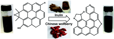
In this work, we developed a green and facile approach to prepare graphene by the reduction of graphite oxide (GO) using two Chinese medicinal herbs: inulin and Chinese wolfberry. The reduced products were systematically characterized by UV-visible absorption spectroscopy, X-ray diffraction, X-ray photoelectron spectroscopy, Raman spectroscopy, Fourier transform infrared spectroscopy, scanning electron microscopy and transmission electron microscopy. These results provided convincing evidence of the removal of oxygen-containing groups from GO to form few-layered graphene after the reduction reaction. Simultaneously, nitrogen and sulfur co-doping were achieved under a relatively low temperature (90 °C). The reduction mechanism of GO and N, S bonding configurations in graphene were also proposed.

1. Introduction

Graphene, a flat monolayer of sp2-bonded carbon atoms arranged in a tightly packed honeycomb two-dimensional (2D) lattice, has received considerable attention due to its high electronic conductivity,1 large specific surface area2 and huge mechanical strength3 since first discovered by Novoselov et al.4 These unique features offer promising applications of graphene in various fields such as supercapacitors,5 nanoelectronics,6 sensors,7 catalysis,8 solar cells,9 Li–air batteries10 and so on. Therefore, the synthesis of graphene has become one of the hot topics in scientific research in recent years.11–14

Diverse procedures have been explored for synthesizing graphene, which are mainly classified into three categories. Large-area graphene sheets can be prepared by chemical vapor deposition (CVD) on metal substrate15 or epitaxial growth on single-crystal SiC.16 The second approach is to directly exfoliate graphite through sonication,17 intercalation18 or liquid-phase method.19 The disadvantage of the direct exfoliation method is its low yield, which is not suitable for large-scale production. Chemical reduction of graphite oxide (GO)20 is another way to make graphene. This method has received a special interest because it holds a great promise for producing graphene on industrial scale. In addition, inexpensiveness, ease of functionalization and the ability to adapt it for synthesis of graphene-based hybrids make chemical reduction of GO an attractive way to prepare graphene.

Despite the advantages of chemical reduction of GO, the commonly used reducing agents, sodium borohydride, hydrazine, dimethylhydrazine, etc., however, are highly toxic and explosive in nature, which makes it difficult to handle the hazardous waste generated by the reduction reaction and could even have detrimental effect on applications of graphene if some toxic chemicals are remained. Therefore, it is necessary to utilize an eco-friendly system for deoxygenation of GO. Actually, some efforts have been made to develop green reductants based on natural products for reducing GO instead of toxic chemicals, such as tea solution,21 wild carrot root,22 reducing sugar,23 aqueous phytoextracts,24 wild plant extract,25 and protein.26 These attempts indicate that natural products show potential applications in reducing GO and preparing graphene and graphene-based hybrids.

In addition to pristine graphene, chemical modified graphene, for example nitrogen doped graphene (N-graphene) and sulfur doped graphene (S-graphene) also attracts an increasing interest because both theoretical27,28 and experimental29,30 studies have shown that chemical doping of graphene with foreign atoms such as nitrogen and sulfur can tailor its electronic property and chemical reactivity, as well as give rise to new functions. Typically, two strategies have been utilized to prepare N-graphene: direct synthesis and post treatment. CVD is a direct and effective technique to produce N-graphene.31 However, the catalyst used in CVD synthesis is expensive and the yield is low. Heating graphene in the presence of NH3 gas32 or other nitrogen-containing chemicals29,33 provides another way to produce N-graphene. However, post-treatment needs high temperature which may leads to several negative effects on N-graphene such as increased defect density in lattice, polycrystalline nature, and low device performance. Therefore, some alternative ways like solvothermal34 or hydrothermal35 route have been developed for preparing N-graphene. As to S-graphene, the incorporation of sulfur into graphene relied on the reaction between sulfur source and graphene/GO at high temperature (usually higher than 500 °C) so far.30,36 High-temperature heating will also bring unfavorable effect on graphene. Few reports, however, are available about nitrogen and sulfur co-doping of graphene.37–39 In this sense, a low-temperature synthesis route should be explored for preparing nitrogen and sulfur modified graphene.

In this work, we demonstrate a novel green and facile strategy to produce nitrogen and sulfur modified few-layered graphene by reducing GO using two Chinese medicinal herbs: Chinese wolfberry and inulin, which are commonly seen in daily life in China. Our approach exhibits several advantages on preparing graphene: (1) it is totally environmentally friendly because only eco-friendly reducing agents are used; (2) the synthesis procedure is safe and facile; (3) the reduction of GO and nitrogen and sulfur co-doping can be achieved simultaneously under a relatively low temperature (90 °C).

2. Experimental

2.1 Preparation of inulin or Chinese wolfberry solution

In a typical procedure, 4 g of inulin or Chinese wolfberry powder was added into 200 mL of deionized (DI) water and boiled at 100 °C for 40 min. The resulting suspension was filtered and centrifugalized to obtain inulin or Chinese wolfberry solution.

2.2 Reduction of GO by inulin or Chinese wolfberry solution

GO (50 mg), synthesized by the modified Hummer's method,40 was ultrasonically dispersed in 100 mL of DI water for 5 h to get exfoliated graphene oxide using an ultrasonic bath. Subsequently, 100 mL of inulin or Chinese wolfberry solution was added to the above solution followed by refluxing at 90 °C for 24 h. The resultant product was separated by centrifugation, washed with DI water and dried at 40 °C under vacuum overnight. The final products were named IGN and CWGN, corresponding to graphene converted by inulin and Chinese wolfberry, respectively. For comparison, a control sample (TGO) was prepared using the similar route without using any reductant.

2.3 Materials characterization

UV-visible spectra were recorded on Mapama UV-1800 spectrometer. Crystalline structures of the products were characterized by X-ray diffraction (XRD) on a Rigaku D/Max-2550pc powder diffractometer equipped with Cu Kα radiation (λ = 1.54 Å). Morphologies of the products were observed by field-emission scanning electron microscopy (SEM) on a FEI-sirion microscope, transmission electron microscopy (TEM) and high-resolution TEM (HRTEM) on a JEM 2100F microscope. X-ray photoelectron spectroscopy (XPS) measurements were performed on a KRATOS AXIS ULTRA-DLD spectrometer with a monochromatic Al Kα radiation ( = 1486.6 eV). Raman spectra were recorded on a Jobin-Yvon Labor Raman HR-800 Raman system by exciting a 514.5 nm Ar+ laser. Fourier transform infrared (FTIR) spectroscopy measurements were performed on a Bruker Vector 22 Fourier infrared spectrometer. The powder sample was mixed uniformly with KBr at a weight ratio of 1[thin space (1/6-em)]:[thin space (1/6-em)]100, and pressed into a pellet before FTIR measurements.

3. Results and discussion

Fig. 1 shows the photograph of ultrasonically dispersed aqueous solution of GO, TGO, CWGN and IGN. The color of GO solution was brownish yellow, while the color of CWGN and IGN solution was black, suggesting the restoration of electronic conjugation after reduction.21 It is noted that the color of TGO was also brownish yellow, which means GO could not be reduced spontaneously if there was no reducing agent when refluxed at 90 °C.
image file: c4ra01985g-f1.tif
Fig. 1 Digital images of (a) GO, (b) TGO, (c) CWGN and (d) IGN solution.

To study the disturbed and rearranged π electronic conjugation of GO, TGO, CWGN and IGN, UV-visible absorption spectra were measured as shown in Fig. 2. GO shows a peak at 232 nm which is attributed to the π–π* transition of the aromatic C–C bonds and an absorbance shoulder at 302 nm is recognized to be the n–π* transition of the C[double bond, length as m-dash]O bonds. As to CWGN and IGN, the peaks at 232 nm were red shifted to ∼270 nm and the shoulder in GO disappears, indicating the electronic conjugation has been restored. TGO shows the same absorbance peaks as GO, which further confirmed that GO could not be reduced spontaneously at 90 °C in the absence of reductant.


image file: c4ra01985g-f2.tif
Fig. 2 UV-visible spectra of GO, TGO, CWGN and IGN.

Fig. 3 gives the XRD patterns of GO, TGO, CWGN and IGN. GO and TGO show a diffraction peak at 2θ = 11°, corresponding to a large d-spacing (002 planes) of 8.1 Å. This is because GO is rich in oxygen-containing groups, mainly carboxyl, epoxy and hydroxyl groups.20 By contrast, CWGN and IGN have a peak centered at 2θ = 22–24°, while the peak at 2θ = 11° disappears, indicating the reduction of GO during which the oxygen-containing groups are removed. A small peak related to (001) plane of graphene is also observed at 2θ = 43° in these samples.


image file: c4ra01985g-f3.tif
Fig. 3 XRD patterns of GO, TGO, CWGN and IGN.

XPS was used to check the reduction state of GO. As shown in Fig. 4a, both CWGN and IGN exhibit enhanced C/O atomic ratio compared to GO as well as visible peaks corresponding to N1s and S2p. The C/O atomic ratios of in GO, CWGN and IGN are 0.6, 1.49, and 1.33, respectively based on the XPS analysis. Fig. 4b presents the C1s XPS of GO, CWGN and IGN. The peaks are fitted into four peaks, corresponding to carbon atoms in different functional groups: non-oxygenated carbon (C1: C–C, 285.6 eV, and C[double bond, length as m-dash]C, 284.8 eV), carbon in C–O group (C2: epoxy or hydroxyl, 286.3 eV), carbonyl carbon (C3: C[double bond, length as m-dash]O, 287.6 eV) and carboxyl carbon (C4: O–C[double bond, length as m-dash]O, 289.0 eV).41 Clearly, the peak intensity of the C–O, C[double bond, length as m-dash]O and O–C[double bond, length as m-dash]O groups shows a remarkable decrease in CWGN and IGN after the reactions, indicative of a sufficient reduction of GO into graphene. Note that the final products still contain residual epoxy and/or hydroxyl groups, in consistence with the theoretical calculation that these groups are difficult to be removed when located at the edges of the GO.42 The nitrogen bonding configurations in CWGN and IGN are investigated by N1s XPS (Fig. 4c). It is deconvoluted to three peaks that are assigned to pyridinic N (N1: ∼398.7 eV), pyrrolic N (N2: ∼400.1 eV), and graphitic N (N3: ∼401.8 eV).34,36 As depicted in Fig. 4e, pyridinic N bonds with two C atoms with one p-electron localized in π conjugated system and pyrrolic N with a pair of p-electrons. Graphitic N represents nitrogen atoms that substitute carbon atoms within the hexagonal ring in graphene. The nitrogen contents of CWGN and IGN are 2.8% and 2.9%, respectively. Fig. 4d gives the S2p XPS of CWGN and IGN, which characterize the sulfur bonding configurations of Chinese wolfberry and inulin converted graphene. All the S2p peaks can be resolved into three peaks at 163.9, 165.1 and 168.5 eV, respectively. The former two peaks are related to 2p3/2 (S1) and 2p1/2 (S2) of thiophene S owing to their spin–orbit coupling.36,37 The other peak arises from the oxidized sulfur (S3), as indicated in Fig. 4e. The sulfur contents of CWGN and IGN are 0.26% and 0.11%, respectively.


image file: c4ra01985g-f4.tif
Fig. 4 (a) Survey and (b) C1s XPS of GO, CWGN and IGN, (c) N1s and (d) S2p XPS of CWGN and IGN, and (e) bonding configurations of (I) nitrogen and (II) sulfur atoms in CWGN and IGN.

Raman and FTIR spectra of GO, CWGN and IGN are measured to further check the reduction of GO as seen in Fig. 5. In Raman spectra of Fig. 5a, all the samples show two bands at about 1320–1350 and 1580–1600 cm−1, corresponding to the disordered (D) and graphitic (G) bands of carbon-based materials.43 The D-to-G peak intensity ratios of GO, CWGN and IGN are calculated to be 1.05, 1.18 and 1.36, respectively. Compared with GO, both CWGN and IGN exhibit an increased D/G intensity ratio, caused by a reduction of the average size of the sp2 domains and an increased number of these domains.20 Another important point is that the position of G peak of CWGN (1587 cm−1) and IGN (1584 cm−1) is lower than that of GO (1599 cm−1). The downshift of the G peak from GO to CWGN and IGN can be attributed to the restoration of the conjugated structure as well as electron-doping of N and S atoms, which is consistent with other reports.29,34,44 In addition, the G peak shows an asymmetric character. In fact, it consists of two overlapped peaks, namely, G and D′ peaks. The D′ peak is a defect peak due to intra-valley scattering.43 The D′ peak of graphene was also observed in other work.45 The peaks at around 2660–2710 cm−1 are related to the second-order two phonon mode 2D band.46 Both CWGN and IGN exhibit a broad 2D peak, indicating the few-layered feature of graphene.46 In FTIR of Fig. 5b, the strong absorption band at 1725 cm−1 is due to the C[double bond, length as m-dash]O stretching, the band at 1622 cm−1 is due to aromatic C[double bond, length as m-dash]C, and the bands at 1398, 1224, and 1069 cm−1 correspond to carboxyl C–O, epoxy C–O and alkoxy C–O, respectively.47 Of note is the obvious decrease of the peak intensity related to C[double bond, length as m-dash]O and C–O bands after the reactions, which suggests the remarkable reduction of GO, agreeing well with the XPS and Raman results. In addition, peaks at 1565 cm−1 for sp2 C[double bond, length as m-dash]N bonds47 and peaks at 1373 and 1311 cm−1 for O[double bond, length as m-dash]S[double bond, length as m-dash]O asymmetric stretching bonds48 can also be observed.


image file: c4ra01985g-f5.tif
Fig. 5 (a) Raman and (b) FTIR spectra of GO, CWGN and IGN.

The morphologies of GO, CWGN and IGN are shown in Fig. 6. GO shows a sheet-like structure with a lateral size of tens of microns as seen in Fig. 6a. After reduction, CWGN and IGN show a much smaller size (several microns) compared with GO as shown in Fig. 6b and c. Wrinkles are observed in SEM images of CWGN and IGN. Corrugation is one of the intrinsic natures of graphene, in order to make its 2D structure become thermodynamically stable. Fig. 6d and e show TEM images of CWGN and IGN, which indicates that WGN and IGN are very thin. To better understand the structure of graphene, the folded areas of the sheets are observed by HRTEM (Fig. 6f and g). HRTEM reveals the few-layered nature of graphene (below 10 layers). Energy dispersive X-ray spectroscopy (EDS) mapping is carried out to investigate N and S distribution in CWGN and IGN (Fig. 7). The EDS result indicates that N and S are uniformly distributed in both samples.


image file: c4ra01985g-f6.tif
Fig. 6 SEM images of (a) GO, (b) CWGN and (c) IGN, TEM images of (d) CWGN and (e) IGN, and HRTEM images of (f) CWGN and (g) IGN.

image file: c4ra01985g-f7.tif
Fig. 7 Dark-field TEM images and EDS mapping of (a) CWGN and (b) IGN.

Inulin is composed mainly of fructose units, and typically has a terminal glucose, as well as additives such as amino acids, vitamins and so on. Chinese wolfberry contains sugars and proteins. It also has abundant amino acids and vitamins. Here we take typical chemical components in the two medicinal herbs, namely fructose (C6H12O6), vitamin B4 (C5H5N5) and methionine (C5H11NO2S), to explain the reduction reactions. The proposed mechanisms for reduction of GO and nitrogen and sulfur doping are shown in Fig. 8. GO contains some oxygen-containing groups, epoxy, hydroxyl, etc. For epoxy, it could be opened by oxygen anion of fructose by reacting through a SN2 mechanism24 and forms an intermediate. This is followed by another SN2 nucleophilic attack with release of H2O, during which the intermediate undergoes a thermal elimination to become graphene.49 The removal of hydroxyl groups in GO experiences a similar process. The doping of N and S occurs at the same time during the reduction reactions as indicated in Fig. 8.


image file: c4ra01985g-f8.tif
Fig. 8 Proposed reaction mechanism for GO reduction and N, S co-doping of graphene.

4. Conclusions

We demonstrate in this work that two Chinese medicinal herbs: Chinese wolfberry and inulin could be used as effective reducing agents for the reduction of GO as well as doping sources for the preparation of nitrogen and sulfur co-doped graphene. These reductants based on natural products are totally environmentally friendly for both preparation and use, in contrast to the noxious reductants based on industrial products. This work opens up a green, facile route to the preparation of elemental modified graphene.

Acknowledgements

This work was supported by National Basic Research Program of China (2013CB934001), the National Natural Science Foundation of China (no. 51101139), Key Science and Technology Innovation Team of Zhejiang Province under Grant Number 2010R50013, and Program for Innovative Research Team in University of Ministry of Education of China (IRT13037).

References

  1. S. Stankovich, D. A. Dikin, G. H. B. Dommett, K. M. Kohlhaas, E. J. Zimney, E. A. Stach, R. D. Piner, S. T. Nguyen and R. S. Ruoff, Nature, 2006, 442, 282–286 CrossRef CAS PubMed.
  2. M. D. Stoller, S. Park, Y. Zhu, J. An and R. S. Ruoff, Nano Lett., 2008, 8, 3498–3502 CrossRef CAS PubMed.
  3. C. Lee, X. Wei, J. W. Kysar and J. Hone, Science, 2008, 321, 385–388 CrossRef CAS PubMed.
  4. K. S. Novoselov, A. K. Geim, S. V. Morozov, D. Jiang, Y. Zhang, S. V. Dubonos, I. V. Grigorieva and A. A. Firsov, Science, 2004, 306, 666–669 CrossRef CAS PubMed.
  5. Y. W. Zhu, S. Murali, M. D. Stoller, K. J. Ganesh, W. W. Cai, P. J. Ferreira, A. Pirkle, R. M. Wallace, K. A. Cychosz, M. Thommes, D. Su, E. A. Stach and R. S. Ruoff, Science, 2011, 332, 1537–1541 CrossRef CAS PubMed.
  6. J. R. Williams, L. DiCarlo and C. M. Marcus, Science, 2007, 317, 638–641 CrossRef CAS PubMed.
  7. F. Schedin, A. K. Geim, S. V. Morozov, E. W. Hill, P. Blake, M. I. Katsnelson and K. S. Novoselov, Nat. Mater., 2007, 6, 652–655 CrossRef CAS PubMed.
  8. J. H. Shen, Y. H. Zhu, X. L. Yang and C. Z. Li, Chem. Commun., 2012, 48, 3686–3699 RSC.
  9. N. L. Yang, J. Zhai, D. Wang, Y. S. Chen and L. Jiang, ACS Nano, 2010, 4, 887–894 CrossRef CAS PubMed.
  10. J. Xiao, D. H. Mei, X. L. Li, W. Xu, D. Y. Wang, G. L. Graff, W. D. Bennett, Z. M. Nie, L. V. Saraf, I. A. Aksay, J. Liu and J. G. Zhang, Nano Lett., 2011, 11, 5071–5078 CrossRef CAS PubMed.
  11. D. Li, M. B. Muller, S. Gilje, R. B. Kaner and G. G. Wallace, Nat. Nanotechnol., 2008, 3, 101–105 CrossRef CAS PubMed.
  12. V. C. Tung, M. J. Allen, Y. Yang and R. B. Kaner, Nat. Nanotechnol., 2009, 4, 25–29 CrossRef CAS PubMed.
  13. S. Pei and H. M. Cheng, Carbon, 2012, 50, 3210–3228 CrossRef CAS PubMed.
  14. I. Y. Jeon, H. J. Choi, S. M. Jung, J. M. Seo, M. J. Kim, L. M. Dai and J. B. Baek, J. Am. Chem. Soc., 2013, 135, 1386–1393 CrossRef CAS PubMed.
  15. X. S. Li, W. W. Cai, J. H. An, S. Kim, J. Nah, D. X. Yang, R. Piner, A. Velamakanni, I. Jung, E. Tutuc, S. K. Banerjee, L. Colombo and R. S. Ruoff, Science, 2009, 324, 1312–1314 CrossRef CAS PubMed.
  16. K. V. Emtsev, A. Bostwick, K. Horn, J. Jobst, G. L. Kellogg, L. Ley, J. L. McChesney, T. Ohta, S. A. Reshanov, J. Rohrl, E. Rotenberg, A. K. Schmid, D. Waldmann, H. B. Weber and T. Seyller, Nat. Mater., 2009, 8, 203–207 CrossRef CAS PubMed.
  17. M. Choucair, P. Thordarson and J. A. Stride, Nat. Nanotechnol., 2009, 4, 30–33 CrossRef CAS PubMed.
  18. J. Kwon, S. H. Lee, K. H. Park, D. H. Seo, J. Lee, B. S. Kong, K. Kang and S. Jeon, Small, 2011, 7, 864–868 CrossRef CAS PubMed.
  19. Y. Hernandez, V. Nicolosi, M. Lotya, F. M. Blighe, Z. Y. Sun, S. De, I. T. McGovern, B. Holland, M. Byrne, Y. K. Gun'ko, J. J. Boland, P. Niraj, G. Duesberg, S. Krishnamurthy, R. Goodhue, J. Hutchison, V. Scardaci, A. C. Ferrari and J. N. Coleman, Nat. Nanotechnol., 2008, 3, 563–568 CrossRef CAS PubMed.
  20. S. Stankovich, D. A. Dikin, R. D. Piner, K. A. Kohlhaas, A. Kleinhammes, Y. Jia, Y. Wu, S. T. Nguyen and R. S. Ruoff, Carbon, 2007, 45, 1558–1565 CrossRef CAS PubMed.
  21. Y. Wang, Z. X. Shi and J. Yin, ACS Appl. Mater. Interfaces, 2011, 3, 1127–1133 CAS.
  22. T. Kuila, S. Bose, P. Khanra, A. K. Mishra, N. H. Kim and J. H. Lee, Carbon, 2012, 50, 914–921 CrossRef CAS PubMed.
  23. C. Z. Zhu, S. J. Guo, Y. X. Fang and S. J. Dong, ACS Nano, 2010, 4, 2429–2437 CrossRef CAS PubMed.
  24. S. Thakur and N. Karak, Carbon, 2012, 50, 5331–5339 CrossRef CAS PubMed.
  25. D. Mhamane, W. Ramadan, M. Fawzy, A. Rana, M. Dubey, C. Rode, B. Lefez, B. Hannoyer and S. Ogale, Green Chem., 2011, 13, 1990–1996 RSC.
  26. F. Lu, S. H. Zhang, H. J. Gao, H. Jia and L. Q. Zheng, ACS Appl. Mater. Interfaces, 2012, 4, 3278–3284 CAS.
  27. P. A. Denis, R. Faccio and A. W. Mombru, ChemPhysChem, 2009, 10, 715–722 CrossRef CAS PubMed.
  28. B. Uchoa and A. H. Castro Neto, Phys. Rev. Lett., 2007, 98, 146801 CrossRef.
  29. Z. Y. Lin, G. Waller, Y. Liu, M. L. Liu and C. P. Wong, Adv. Energy Mater., 2012, 2, 884–888 CrossRef CAS.
  30. Z. Yang, Z. Yao, G. F. Li, G. Y. Fang, H. G. Nie, Z. Liu, X. M. Zhou, X. Chen and S. M. Huang, ACS Nano, 2012, 6, 205–211 CrossRef CAS PubMed.
  31. Y. Z. Xue, B. Wu, L. Jiang, Y. L. Guo, L. P. Huang, J. Y. Chen, J. H. Tan, D. C. Geng, B. R. Luo, W. P. Hu, G. Yu and Y. Q. Liu, J. Am. Chem. Soc., 2012, 134, 11060–11063 CrossRef CAS PubMed.
  32. T. N. Huan, T. V. Khai, Y. Kang, K. B. Shim and H. Chung, J. Mater. Chem., 2012, 22, 14756–14762 RSC.
  33. Z. H. Sheng, L. Shao, J. J. Chen, W. J. Bao, F. B. Wang and X. H. Xia, ACS Nano, 2011, 5, 4350–4358 CrossRef CAS PubMed.
  34. D. H. Deng, X. L. Pan, L. A. Yu, Y. Cui, Y. P. Jiang, J. Qi, W. X. Li, Q. A. Fu, X. C. Ma, Q. K. Xue, G. Q. Sun and X. H. Bao, Chem. Mater., 2011, 23, 1188–1193 CrossRef CAS.
  35. L. Sun, L. Wang, C. G. Tian, T. X. Tan, Y. Xie, K. Y. Shi, M. T. Li and H. G. Fu, RSC Adv., 2012, 2, 4498–4506 RSC.
  36. S. Yang, L. Zhi, K. Tang, X. Feng, J. Maier and K. Müllen, Adv. Funct. Mater., 2012, 22, 3634–3640 CrossRef CAS.
  37. J. X. Xu, G. F. Dong, C. H. Jin, M. H. Huang and L. H. Guan, ChemSusChem, 2013, 6, 493–499 CrossRef CAS PubMed.
  38. Z. Liu, H. G. Nie, Z. Yang, J. Zhang, Z. P. Jin, Y. Q. Lu, Z. B. Xiao and S. M. Huang, Nanoscale, 2013, 5, 3283–3288 RSC.
  39. J. Liang, Y. Jiao, M. Jaroniec and S. Z. Qiao, Angew. Chem., Int. Ed., 2012, 51, 11496–11500 CrossRef CAS PubMed.
  40. W. S. Hummers and R. E. Offeman, J. Am. Chem. Soc., 1958, 80, 1339 CrossRef CAS.
  41. H. J. Shin, K. K. Kim, A. Benayad, S. M. Yoon, H. K. Park, I. S. Jung, M. H. Jin, H. K. Jeong, J. M. Kim, J. Y. Choi and Y. H. Lee, Adv. Funct. Mater., 2009, 19, 1987–1992 CrossRef CAS.
  42. X. Gao, J. Jang and S. Nagase, J. Phys. Chem. C, 2009, 114, 832–842 Search PubMed.
  43. A. C. Ferrari, Solid State Commun., 2007, 143, 47–57 CrossRef CAS PubMed.
  44. P. P. Guo, F. Xiao, Q. Liu, H. F. Liu, Y. L. Guo, J. R. Gong, S. Wang and Y. Q. Liu, Sci. Rep., 2013, 3, 3499 Search PubMed.
  45. D. Chen, W. Wei, R. Wang, J. Zhu and L. Guo, New J. Chem., 2012, 36, 1589–1595 RSC.
  46. A. C. Ferrari, J. C. Meyer, V. Scardaci, C. Casiraghi, M. Lazzeri, F. Mauri, S. Piscanec, D. Jiang, K. S. Novoselov, S. Roth and A. K. Geim, Phys. Rev. Lett., 2006, 97, 187401 CrossRef CAS.
  47. L. F. Lai, H. P. Yang, L. Wang, B. K. Teh, J. Q. Zhong, H. Chou, L. W. Chen, W. Chen, Z. X. Shen, R. S. Ruoff and J. Y. Lin, ACS Nano, 2012, 6, 5941–5951 CrossRef CAS PubMed.
  48. H. L. Poh, P. Šimek, Z. Sofer and M. Pumera, ACS Nano, 2013, 7, 5262–5272 CrossRef CAS PubMed.
  49. C. Z. Zhu, S. J. Guo, Y. X. Fang and S. J. Dong, ACS Nano, 2010, 4, 2429–2437 CrossRef CAS PubMed.

This journal is © The Royal Society of Chemistry 2014
Click here to see how this site uses Cookies. View our privacy policy here.