DOI:
10.1039/C4RA01598C
(Paper)
RSC Adv., 2014,
4, 19077-19085
Coumarin–hemicyanine conjugates as novel reaction-based sensors for cyanide detection: convenient synthesis and ICT mechanism†
Received
24th February 2014
, Accepted 8th April 2014
First published on 14th April 2014
Abstract
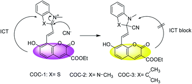
Using intramolecular charge transfer (ICT) as a signaling mechanism, a series of hybrid coumarin–hemicyanine compounds were synthesized as chemosensors for cyanide detection by taking advantage of cyanide's strong affinity toward the polarized CN bond of the hemicyanine group. Structure identification of the compounds was confirmed by 1H NMR, 13C NMR, 1H-1H COSY, HSQC, IR, and HRMS spectroscopy. Multiple sensory signals are available and can be used for both qualitative monitoring and quantitative determination of cyanide, including high-contrast visual color change, fluorescence quenching and enhancement.
1. Introduction
Anion recognition is an area of growing interest in supra-molecular chemistry due to its important role in a wide range of environmental, clinical, chemical, and biological applications.1–3 Among various anions, the cyanide anion is one of the most extensively studied, as it is widely used in synthetic fibers,4 resins,5 herbicides6 and the gold-extraction process.7 However, it is an extremely toxic anion and can affect many functions in the human body, including the vascular,8 visual,9 central nervous,10 cardiac,11 endocrine12 and metabolic systems.13 There is therefore considerable interest in developing effective detection methods for cyanide anion.
Some cyanide chemosensors have been developed using cyanide complexes including Zn(II)–porphyrin,14 Co(II)–coumarinylsalen,15 Cu(II) complex,16–18 boronic acid derivatives19 and CdSe quantum dots20 by exploiting the strong co-ordination ability of cyanide. However, these sensors only provided an indirect strategy to detect cyanide anions. In contrast to the indirect approach, we here consider whether direct detection of the cyanide anion could be achieved by regulating the acceptor moiety of the sensor molecule to affect the ICT efficiency and induce naked-eye color changes. After a careful review of the literature,21–26 we found that chemodosimetric sensors based on the special nucleophilicity of cyanide were promising owing to their characteristic features such as analyte-specific response, direct detection of cyanide anion and little competition from the aqueous medium. Specific nucleophilic reactions between the sensor molecule and cyanide accompanied by some spectroscopic changes have recently attracted the attention of many scientists as potential sensing systems, i.e., the dicyano-vinyl group,27 cyanohydrin reactions,28–31 chromogenic oxazines31–33, pyrylium or acridinium compounds,34–37 and squaraine, croconium, or triarylmethane dyes.38–40 However, hemicyanine compounds have seldom been used to detect cyanide in aqueous solution.41,42
Coumarin–hemicyanine hybrids are often exploited as fluorophores for chemosensors due to their relatively large Stokes shift and long emission wavelength.43–46 Moreover, the emission of the coumarin chromophore can be recovered due to the inhibition of π-conjugation between the hemicyanine and coumarin induced by certain chemical reactions.43,45 Hybrid coumarin–hemicyanine derivatives are therefore valuable platforms for the construction of fluorescence sensors. Herein, we report a new kind of ratiometric fluorescent sensor for cyanide detection in aqueous solution, featuring advantages such as easy implementation, large emission shift, good ratiometric response, as well as high selectivity based on the cyanide-induced ICT blocking of a hybrid coumarin–hemicyanine dye.
2. Experiment
2.1. General
All solvents and reagents were purchased from commercial sources and used as received. Absorption spectra were recorded on a UV-1700 spectrophotometer. Fluorescent spectra were recorded on a Hitachi F-4500 fluorescence spectrophotometer. NMR spectra were collected on a Varian INOVA-400 spectrometer 400 MHz for 1H NMR and 100 MHz for 13C NMR. Mass spectra were obtained with an UltiMate 3000 mass spectrophotometer. FT-IR spectra were obtained in KBr pellets on a Bruker EQUIOX-55 FT-IR spectrometer.
2.2. Synthesis
The sensors COC-1, COC-2 and COC-3 were conveniently synthesized via the condensation of ethyl-8-formoxyl-7-hydroxycoumarin-3-carboxylate (1) with 2,3-dimethylbenzothiazolium iodide (2), 1,2,3-trimethylbenzimidazolium iodide (3) and 1-methyl-2,3,3-trimethyl-3H-indolium in ethanol (4) (Scheme 1).
|
| Scheme 1 Synthetic procedure of the sensors. | |
2.2.1. Synthesis of COC-1. Ethyl-8-formoxyl-7-hydroxycoumarin-3-carboxylate (1)47 was synthesized according to our previous work. 2,3-Dimethylbenzothiazolium iodide (2) (96 mg, 0.33 mmol) was treated with compound 1 (86 mg, 0.33 mmol) in anhydrous ethanol (20 mL) and piperidine (3 drops). The reaction was then refluxed for 2 h. After cooling the solid was collected, washed with anhydrous ethanol, then dried, giving a purple solid (135 mg, yield: 76%). 1H NMR (400 MHz, DMSO-d6-CF3COOD): δ 1.29 (t, J = 8.0 Hz, 3H), 4.26 (q, J = 8.0 Hz, 2H), 4.28 (s, 3H), 7.07 (d, J = 8.0 Hz, 1H), 7.74–7.88 (m, 3H), 8.18 (d, J = 16.0 Hz, 1H), 8.25 (d, J = 16.0 Hz, 1H), 8.27–8.29 (m, 1H), 8.38 (d, J = 8.0 Hz, 1H), 8.68 (s, 1H). 13C NMR (100 MHz, DMSO-d6-CF3COOD): δ 172.99, 162.81, 156.05, 155.71, 149.88, 142.48, 136.56, 135.39, 129.84, 128.68, 128.28, 124.56, 119.57, 117.23, 116.96, 114.25, 113.85, 111.00, 108.43, 61.30, 36.45, 14.17. FT-IR (KBr, cm−1): 3427, 3095, 2986, 2898, 1721, 1693, 1613, 1512, 1474, 1395, 1321, 1285, 1225, 950, 841, 745. HRMS (ESI): m/z, calcd for (M+) 408.0900; found 408.0903.
2.2.2. Synthesis of COC-2. 1,2,3-Trimethylbenzimidazolium iodide (3) (95 mg, 0.33 mmol) was treated with compound 1 (86 mg, 0.33 mmol) in anhydrous ethanol (20 mL) and piperidine (3 drops). The reaction was then refluxed for 1 h. After cooling the solid was collected, washed with anhydrous ethanol, then dried, giving an orange solid (104 mg, yield: 59%). 1H NMR (400 MHz, DMSO-d6-CF3COOD): δ 1.12–1.25 (m, 3H), 1.86 (s, 3H), 3.67 (s, 3H), 4.03 (s, 2H), 4.06–4.22 (m, 2H), 6.96–7.03 (m, 2H), 7.51–7.61 (m, 2H), 7.72–7.86 (m, 2H), 8.41–8.55 (m, 1H). 13C NMR (100 MHz, DMSO-d6-CF3COOD): δ 173.61, 156.76, 155.99, 153.71, 148.93, 135.76, 135.46, 132.01, 131.72, 126.89, 126.79, 120.02, 117.03, 114.16, 112.75, 111.29, 109.10, 108.49, 61.44, 32.15, 20.61, 13.62. FT-IR (KBr, cm−1): 3447, 3027, 1625, 1583, 1484, 1405, 1332, 1190, 1037, 959, 842, 775. HRMS (ESI): m/z, calcd for (M+) 405.1400; found 405.1445.
2.2.3. Synthesis of COC-3. 1-Methyl-2,3,3-trimethyl-3H-indolium in ethanol (4) (100 mg, 0.33 mmol) was treated with compound 1 (86 mg, 0.33 mmol) in anhydrous ethanol (20 mL). The reaction was then refluxed for 14 h. After cooling the solid was collected, washed with anhydrous ethanol, then dried, giving a yellow solid (117 mg, yield: 65%). 1H NMR (400 MHz, DMSO-d6-CF3COOD): δ 1.32 (t, J = 8.0 Hz, 3H), 1.81 (s, 6H), 4.08 (s, 3H), 4.30 (q, J = 8.0 Hz, 2H), 7.10 (d, J = 8.0 Hz, 1H), 7.64–7.66 (m, 2H), 7.88–7.97 (m, 2H), 7.99 (d, J = 8.0 Hz, 1H), 8.08 (d, J = 16.0 Hz, 1H), 8.50 (d, J = 16.0 Hz, 1H), 8.78 (s, 1H). 13C NMR (100 MHz, DMSO-d6-CF3COOD): δ 182.93, 164.95, 156.71, 155.68, 154.64, 149.92, 143.65, 142.35, 141.69, 136.26, 129.81, 129.40, 123.31, 119.65, 116.79, 113.87, 111.25, 111.07, 109.14, 61.46, 52.25, 34.70, 25.96, 14.27. FT-IR (KBr, cm−1): 3444, 2989, 2451, 1771, 1706, 1601, 1539, 1476, 1373, 1284, 1250, 1026, 832, 751, 681. HRMS (ESI): m/z, calcd for (M+) 418.1600; found 418.1598.
2.3. Spectral analyses
Deionized water was used throughout all experiments. Cyanide anion was prepared from its sodium salt, and the other anions were prepared from their sodium or potassium salts. A stock of the sensors (1.0 mM) was prepared in MeOH. The stock solution of COC-1 and COC-2 was then diluted to the corresponding concentration with the solution of MeCN–buffer (Na2CO3–NaHCO3, 10.0 mM, pH = 9.4, 1:1, v/v). The stock solution of COC-3 was diluted to the corresponding concentration with the solution of MeOH–buffer (Na2CO3–NaHCO3, 10.0 mM, pH = 9.4, 1:1, v/v). The sodium cyanide stock solution of 100.0 mM was diluted to different concentrations with deionized water for spectra titration studies.
2.4. Test strips measurement
The test strips were prepared by immersing filter papers (3 × 1 cm2) in the CH3OH solution of COC-3 (1.0 mM) and then drying them in air. The sodium cyanide stock solution of 100.0 mM was diluted to different concentrations with deionized water and then the test strips coated with COC-3 were immersed in the aqueous solutions of CN− with different concentrations for colorimetric response studies.
3. Results and discussion
3.1. Fluorescence response of the sensors toward cyanide
It was expected that the sensors would display an emission shift to a longer wavelength due to the expanded π-conjugation and enhanced ICT progress from the coumarin moiety to the hemicyanine group. Considering the intrinsic nature of high electron deficiency on the 2-C atom of hemicyanine, it was expected that the cyanide would be able to attack this carbon atom, and that this nucleophilic addition would not only interrupt the π-conjugation in the sensor molecules, but also block the ICT progress from the coumarin moiety to the hemicyanine group. As shown in Fig. 1, the sensors exhibit different emission changes to the cyanide anion. The free COC-1 exhibits a main emission at 450 nm, the typical emission of coumarin. With the addition of cyanide, the emission at 450 nm gradually increased. Unfortunately COC-1 did not show the properties of hybrid coumarin–hemicyanine compounds, but it still showed spectrum changes to cyanide. For COC-2, two emissions at 450 nm and 550 nm appeared which were ascribed to the typical ICT band of the hybrid coumarin–hemicyanine compound. Upon addition of cyanide, emission at 450 nm and 550 nm both increased gradually. Although COC-1 and COC-2 did not show ratiometric fluorescent changes, the two sensors still owned the property of “turn-on” typed sensors which preferred than sensors that rely on fluorescent quenching.48–51 We then studied the emission spectrum of COC-3. The free COC-3 exhibited a red emission at 620 nm, a typical emission of a hybrid coumarin–hemicyanine compound. With the addition of CN−, the emission at 620 nm decreased sharply, followed by an increase in the emission of the coumarin moiety at 450 nm, indicating that the nucleophilic addition reaction between cyanide and the hemicyanine group interrupted the π-conjugation and blocked the ICT progress, after which the fluorescence of coumarin recovered. Accordingly, the emission changes also resulted in clearly visible color changes from red to yellow. This observation is in good agreement with the aforementioned concept. Among the three sensors, we finally selected COC-3 as a ratiometric and colorimetric sensor in accordance with our expectations .
|
| Fig. 1 Changes in fluorescence intensity of the sensors (5.0 μM) measured upon addition of CN−. (a) COC-1; ex: 415 nm; silt: 2.5 nm, 5 nm; cyanide concentration (μM): 0, 8, 10, 50, 70, 100, 200, 400, 600, 800, 1000, 5000, 10000, respectively; MeCN–buffer (Na2CO3–NaHCO3, 10.0 mM, pH = 9.4, 1:1, v/v); (b) COC-2; ex: 350 nm; silt: 5 nm, 5 nm; cyanide concentration (μM): 0, 50, 80, 300, 500, 800, 1000, 1200, 1400, 1600, 1800, 2000, respectively; MeCN–buffer (Na2CO3–NaHCO3, 10.0 mM, pH = 9.4, 1:1, v/v); (c) COC-3; ex: 405 nm; silt: 2.5 nm, 5 nm; cyanide concentration (μM): 0, 20, 40, 60, 95, 150, 400, 600, 800, 1000, 4000, 6000, respectively; MeOH–buffer (Na2CO3–NaHCO3, 10.0 mM, pH = 9.4, 1:1, v/v). | |
Under the same conditions, a linear response of the fluorescence emission intensity to the concentration of CN− was obtained in a range of 0–1000 μM (0–2000 μM for COC-2) (Fig. 2). The linear equation can be expressed as F = a + b × [CN−], where F represents fluorescence intensity or ratiometric fluorescence intensity. Then we calculated LOD (limit of detection) of the three sensors from the equation. The data of a, b, the linear correlation coefficient (R2) and LOD for the linear equations of the three sensors are respectively shown in Table 1. This indicated that the sensors could be used to quantitatively detect CN− concentration in a relatively wide range.
|
| Fig. 2 Calibration curve of the sensors as a function of the concentration of CN−. (a) COC-1; (b) COC-2; (c) COC-3. | |
Table 1 The data of a, b and the linear correlation coefficient (R2) in the linear equations respectively for sensors COC-1, COC-2, COC-3
|
COC-1 |
COC-2 |
COC-3 |
a |
523 |
0.57106 |
11.103 |
b |
4.34 |
0.00102 |
0.018 |
R2 |
0.9957 |
0.9918 |
0.9909 |
LOD/μM |
9.37 |
33.5 |
14.2 |
To gain insight into the binding of CN− with the sensors, we investigated the concentration-dependent changes in the absorption spectra. As shown in Fig. S19,† COC-1 and COC-3 exhibited two main absorption peaks at 500 nm and 400 nm, which was ascribed to the typical conjugation of the hybrid coumarin–hemicyanine dye. Upon addition of CN− to a solution of the sensors, the absorption peak at 500 nm gradually decreased and the absorption peak at 400 nm gradually increased for COC-1 and showed a slight bathochromic shift for COC-3, which suggested that the conjugation was blocked due to the nucleophilic attack of CN− toward the hemicyanine group of the sensors. In addition, a well defined isosbestic point was also noted at 445 nm, indicative of the formation of the addition products. For COC-2, absorption at 425 nm appeared which was ascribed to the typical absorption of coumarin. Upon addition of cyanide, the absorption peak gradually decreased and showed a slight hypochromatic shift to 400 nm. This phenomenon can be reasonably explained as follows: the hydrogen bond of benzimidazole inhibited the absorption of hemicyanine group thus only the absorption of coumarin appeared. As a result, the fluorescent property of coumarin group in COC-2 was so strong that the typical ICT band at 550 nm was also increased. The absorption spectra are in good agreement with the fluorescent spectra.
3.2. Kinetic studies
Because the optical signal changes depend on the chemical reaction between the sensors and CN−, the reaction rate might affect the experimental results. We therefore investigated the influence of the reaction time on the sensing results (Fig. 3). From the reaction curve we can figure out that a plateau of fluorescence changes is achieved after 7 h, 5 h and 3 min respectively for COC-1, COC-2, and COC-3, indicating that only COC-3 can achieve real-time detection of cyanide anion.
|
| Fig. 3 Time-dependent fluorescence intensity of the sensors (5.0 μM) upon addition of 20.0 equiv. CN−. (a) COC-1; ex: 415 nm; silt: 2.5 nm, 5 nm; MeCN–buffer (Na2CO3–NaHCO3, 10.0 mM, pH = 9.4, 1:1, v/v); (b) COC-2; ex: 350 nm; silt: 5 nm, 5 nm; MeCN–buffer (Na2CO3–NaHCO3, 10.0 mM, pH = 9.4, 1:1, v/v); (c) COC-3; ex: 405 nm; silt: 2.5 nm, 5 nm; MeOH–buffer (Na2CO3–NaHCO3, 10.0 mM, pH = 9.4, 1:1, v/v). | |
3.3. Effect of pH value
As the pH value of a system is often considered as a significant factor that influences interactions, the effect of pH was investigated in the range of 7.0–9.8. As shown in Fig. 4, it was ascertained from the titration curve that all the sensors with excess cyanide anion exhibited an almost constant maximum value when pH ≥ 9.4. This is because the protonation of CN− [pKa (HCN) = 9.2] at neutral pH suppresses the nucleophilic ability of CN−.
|
| Fig. 4 pH-dependent fluorescence intensity of the sensors (5.0 μM) upon addition of 1000.0 equiv. CN−. Square dots: sensors only; round dots: sensors + CN−. (a) COC-1; ex: 415 nm; silt: 2.5 nm, 5 nm; MeCN–buffer (Na2CO3–NaHCO3, 10.0 mM, 1:1, v/v); (b) COC-2; ex: 350 nm; silt: 5 nm, 5 nm; MeCN–buffer (Na2CO3–NaHCO3, 10.0 mM, 1:1, v/v); (c) COC-3; ex: 405 nm; silt: 2.5 nm, 5 nm; MeOH–buffer (Na2CO3–NaHCO3, 10.0 mM, 1:1, v/v). | |
3.4. Selectivity investigation
For the purpose of evaluating the selectivity of the sensors to cyanide, fluorescence changes upon addition of 200 equiv. of various anions, namely F−, Cl−, Br−, I−, AcO−, SCN−, ClO−, SO42−, HSO4−, PO43−, H2PO4−, HPO42−, CO32−, HCO3−, S2− and NO3−, as well as a thiol species (cysteine) were studied. These competitive species, including F−, AcO−, HSO4−, and H2PO4− which often show strong interference to cyanide detection, did not induce any significant changes in fluorescence (Fig. 5), indicating the high selectivity of the sensors. Moreover, in the presence of miscellaneous competitive species, the CN− still resulted in similar fluorescence changes (Fig. 6).
|
| Fig. 5 Fluorescence intensity of the sensors (5.0 μM) upon addition of 1000.0 μM of various species. (a) COC-1; ex: 415 nm; silt: 2.5 nm, 5 nm; MeCN–buffer (Na2CO3–NaHCO3, 10.0 mM, pH = 9.4, 1:1, v/v); (b) COC-2; ex: 350 nm; silt: 5 nm, 5 nm; MeCN–buffer (Na2CO3–NaHCO3, 10.0 mM, pH = 9.4, 1:1, v/v); (c) COC-3; ex: 405 nm; silt: 2.5 nm, 5 nm; MeOH–buffer (Na2CO3–NaHCO3, 10.0 mM, pH = 9.4, 1:1, v/v). | |
|
| Fig. 6 Competition graph of the sensors; red bar: sensors + anion, black bar: sensors + anion + CN−. (a) COC-1; (b) COC-2; (c) COC-3. | |
3.5. Colorimetric sensor measurement
As mentioned above, COC-3 displayed an obvious color change from red to yellow after addition of cyanide, thus, potentially, it could be used for direct naked-eye sensing toward cyanide. The picture in Fig. 7 shows that the color changes from red to yellow can be easily distinguished by the naked eye after the addition of cyanide while other anions did not show any changes. Motivated by the obvious color change of this system in solution, the test strips were prepared by immersing filter papers (3 × 1 cm2) in the CH3OH solution of COC-3 (1.0 mM) and then drying them in air to determine the suitability of a “dip-stick” method for the detection of CN−, similar to that commonly used for pH measurement. When the test strips coated with COC-3 were immersed in the aqueous solutions of CN− with different concentrations, a clear color change from red to yellow was observed (Fig. 8). The development of such a “dip-stick” approach is extremely attractive for “in-the-field” measurements as it does not require any additional equipment.
|
| Fig. 7 Photograph of COC-3 compared to various anions (from left to right): blank, CN−, F−, Cl−, Br−, I−, AcO−, SCN−, ClO−, NO3−, SO42−, HSO4−, PO43−, H2PO4−, HPO42−, CO32−, HCO3−, Cys, S2−. | |
|
| Fig. 8 Photographs of test strips of COC-3 to various concentration of CN− (mM): (A) 0; (B) 10; (C) 30; (D) 50; (E) 70; (F) 90. | |
3.6. Reaction mechanism
As illustrated in Scheme 2, the reaction mechanism can be reasonably explained by the nucleophilic addition reaction of cyanide anion with the polarized CN bond of the hemicyanine group. As a result, the π-conjugation between hemicyanine and coumarin was blocked and then obvious spectra and color changes were observed. To examine this plausible mechanism, the reaction between COC-3 and cyanide was evidenced by 1H NMR, HRMS and absorbance spectra measurements. Firstly, the binding pattern between COC-3 and CN− was examined by the 1H NMR titration experiment (Fig. 9). Upon addition of NaCN in aliquots to a CD3OD–D2O (1:1, v/v) solution of COC-3, the signals attributed to COC-3 disappeared and a new set of signals appeared. As the breaking of the conjugation and the addition of CN−, all the aromatic protons displayed a small upfield shift. In addition, the vinyl protons (Ha) displayed a upfield shift from 8.08 ppm (d, J = 16 Hz), 8.50 ppm (d, J = 16 Hz) to 7.05 ppm (d, J = 16 Hz), 7.40 ppm (d, J = 16 Hz) and the N-methyl protons (Hb) displayed a upfield shift from 4.08 ppm (s) to 2.83 ppm (s). Furthermore, the C-methyl protons (Hc and Hd) not only displayed a upfield shift but also split to two peaks due to the addition of CN−. This observation clearly indicated that the cyanide anion had been added to the indolium group. The reaction mechanism was also confirmed by absorption spectra. As shown in Fig. 10, the COC-3–CN adduct and coumarin shared almost the same absorption spectra, which indicated that the cyanide anion had been added to the indolium group. It was also clearly observed from the spectra that after the nucleophilic addition reaction the characteristic absorption of coumarin appeared instead of that of the hybrid coumarin–hemicyanine dye. In addition, this formation of the COC-3–CN adduct was also characterized by mass spectrometry analysis, and the peak at m/z 467.1570 (calc. = 467.1600) corresponding to [COC-3 + NaCN]+ was clearly observed (Fig. 11).
|
| Scheme 2 The proposed reaction mechanism. | |
|
| Fig. 9 1H NMR spectra of COC-3 upon addition of NaCN (1.0 equiv.) in CD3OD–D2O (1:1, v/v). | |
|
| Fig. 10 Absorption spectra of COC-3 compared with that of coumarin in MeOH–buffer (Na2CO3–NaHCO3, 10.0 mM, pH = 9.4, 1:1, v/v). | |
|
| Fig. 11 HRMS of COC-3 upon addition of NaCN (1.0 equiv.). (a) COC-3; (b) COC-3–NaCN adduct. | |
4. Conclusion
In conclusion, we have designed and synthesized three novel fluorescent sensors based on coumarin–hemicyanine conjugates by taking advantage of cyanide's strong affinity toward the polarized CN bond of the hemicyanine group. All the sensors had good sensitivity and high selectivity toward cyanide. Among the three sensors, the reaction between CN− and COC-3 resulted in an obvious color and spectral change in a very short time because the nucleophilic addition reaction blocks the π-conjugation and ICT progress between indolium and coumarin. This makes the real-time, simple-to-use and naked-eye detection of CN− possible. The sensor reported here represents a practical system for rapidly, conveniently and selectively detecting CN−.
Acknowledgements
We are very grateful for the support of this work from the National Natural Science Foundation of China (no. 21072158, 20802056), the Special Foundation of the Education Committee of Shaanxi Province (no. 12JK0580), the French Chinese Foundation for Science and Applications (FFSCA) and the China Scholars Council (CSC).
Notes and reference
- D. Chen, R. J. Letcher, L. T. Gauthier, S. Chu, R. McCrindle and D. Potter, Environ. Sci. Technol., 2011, 45, 9523–9530 CrossRef CAS PubMed.
- P. Goldwasser, N. G. Manjappa, C. A. Luhrs and R. H. Barth, Am. J. Kidney Dis., 2011, 58, 617–620 CrossRef CAS PubMed.
- C. R. Wang and Q. B. Lu, J. Am. Chem. Soc., 2010, 132, 14710–14713 CrossRef CAS PubMed.
- A. N. Bautista-Flores, E. Rodriguez de San Miguel, J. de Gyves and J. A. Josefina, J. Membr. Sci., 2010, 363, 180–187 CrossRef CAS PubMed.
- Z. Yan, M. Cai and P. K. Shen, J. Mater. Chem., 2012, 22, 2133–2139 RSC.
- K. Grossmann, Pest Manage. Sci., 2010, 66, 113–120 CAS.
- T. Z. Sadyrbaeva, Sep. Purif. Technol., 2012, 86, 262–265 CrossRef CAS PubMed.
- S. C. Correia, R. X. Santos, S. M. Cardoso, M. S. Santos, C. R. Oliveira and P. I. Moreira, Neurobiol. Dis., 2012, 45, 206–218 CrossRef CAS PubMed.
- J. C. K. Mwanza, D. Tshala-Katumbay, D. L. Kayembe, K. E. Eeg-Olofsson and T. Tylleskar, Eur. J. Ophthalmol., 2003, 13, 383–389 CAS.
- P. B. Nunn, J. R. A. Lyddiard and K. P. W. Christopher, Food Chem. Toxicol., 2011, 49, 662–667 CrossRef CAS PubMed.
- W. E. Ghann, O. Aras, T. Fleiter and M. C. Daniel, Langmuir, 2012, 28, 10398–10408 CrossRef CAS PubMed.
- J. L. Cadenas, S. A. Montero, C. Leal, M. Lemus, B. E. Portilla, B. A. Alvarado and E. R. de Alvarez-Buylla, Nitric Oxide, 2010, 22, 296–303 CrossRef CAS PubMed.
- P. R. Boshier, N. Marczin and G. B. Hanna, J. Am. Soc. Mass Spectrom., 2010, 21, 1070–1074 CrossRef CAS PubMed.
- H. Yoon, C. H. Lee, Y. H. Jeong, H. C. Gee and W. D. Jang, Chem. Commun., 2012, 48, 5109–5111 RSC.
- J. H. Lee, A. R. Jeong, I. S. Shin, H. J. Kim and J. I. Hong, Org. Lett., 2010, 12, 764–767 CrossRef CAS PubMed.
- J. F. Xu, H. H. Chen, Y. Z. Chen, Z. J. Li, L. Z. Wu, C. H. Tung and Q. Z. Yang, Sens. Actuators, B, 2012, 168, 14–19 CrossRef CAS PubMed.
- H. S. Jung, J. H. Han, Z. H. Kim, C. Kang and J. S. Kim, Org. Lett., 2011, 13, 5056–5059 CrossRef CAS PubMed.
- M. H. Kim, S. Kim, H. H. Jang, S. Yi, S. H. Seo and M. S. Han, Tetrahedron Lett., 2010, 51, 4712–4716 CrossRef CAS PubMed.
- L. Feng, Y. Wang, F. Liang, W. Liu, X. Wang and H. Diao, Sens. Actuators, B, 2012, 168, 365–369 CrossRef CAS PubMed.
- C. R. Maldonado, A. Touceda-Varela, A. C. Jones and J. C. Mareque-Rivas, Chem. Commun., 2011, 47, 11700–11702 RSC.
- S. Park and H.-J. Kim, Chem. Commun., 2010, 46, 9197–9199 RSC.
- H. Lee and H. J. Kim, Tetrahedron Lett., 2012, 53, 5455–5457 CrossRef CAS PubMed.
- S. Park and H.-J. Kim, Sens. Actuators, B, 2012, 161, 317–321 CrossRef CAS PubMed.
- S. Park and H.-J. Kim, Sens. Actuators, B, 2012, 168, 376–380 CrossRef CAS PubMed.
- S. Park, K.-H. Hong, J.-I. Hong and H.-J. Kim, Sens. Actuators, B, 2012, 174, 140–144 CrossRef CAS PubMed.
- S.-Y. Na, J.-Y. Kim and H.-J. Kim, Sens. Actuators, B, 2013, 188, 1043–1047 CrossRef CAS PubMed.
- S. J. Hong, J. Yoo, S. K. Kim, J. S. Kim, J. Yoon and C. H. Lee, Chem. Commun., 2009, 189–191 RSC.
- Y. Sun, G. Wang and W. Guo, Tetrahedron, 2009, 65, 3480–3485 CrossRef CAS PubMed.
- D. G. Cho, J. H. Kim and J. L. Sessler, J. Am. Chem. Soc., 2008, 130, 12163–12167 CrossRef CAS PubMed.
- J. L. Sessler and D. G. Cho, Org. Lett., 2008, 10, 73–75 CrossRef CAS PubMed.
- Z. Ekmekei, M. D. Yilmaz and E. U. Akkaya, Org. Lett., 2008, 10, 461–464 CrossRef PubMed.
- J. Ren, W. Zhu and H. Tian, Talanta, 2008, 75, 760–764 CrossRef CAS PubMed.
- M. Tumasulo, S. Sortino, A. J. P. White and F. M. Raymo, J. Org. Chem., 2006, 71, 744–753 CrossRef PubMed.
- M. Tumasulo and F. M. Raymo, Org. Lett., 2005, 7, 4633–4636 CrossRef PubMed.
- F. Garcia, J. M. Garoia, B. Garcia-Acosta, R. Martinez-Manez, F. Sancenon and J. Soto, Chem. Commun., 2005, 2790–2792 RSC.
- R. Martinez-Manez and F. Sancenon, Coord. Chem. Rev., 2006, 250, 3081–3093 CrossRef CAS PubMed.
- Y. C. Lin and C. T. Chen, Org. Lett., 2009, 11, 4858–4861 CrossRef CAS PubMed.
- Y. K. Yang and J. Tae, Org. Lett., 2006, 8, 5721–5723 CrossRef CAS PubMed.
- P. Kaur, D. Sareen, S. Kaur and K. Singh, Inorg. Chem. Commun., 2009, 12, 272–275 CrossRef CAS PubMed.
- H. Yu, M. Fu and Y. Xiao, Phys. Chem. Chem. Phys., 2010, 12, 7386–7391 RSC.
- X. Zhang, C. Li, X. Chen, X. Wang and B. Zhang, Sens. Actuators, B, 2008, 129, 152–157 CrossRef CAS PubMed.
- X. Lv, L. Liu, Y. Liu, Y. Zhao, Y. Q. Sun, P. Wang and W. Guo, Chem. Commun., 2011, 47, 12843–12845 RSC.
- Z. Yang, Z. Liu, Y. Chen, X. Wang, W. He and Y. Lu, Org. Biomol. Chem., 2012, 10, 5073–5076 CAS.
- J. A. Richard, M. Massonneau, P. Y. Renard and A. Romieu, Org. Lett., 2008, 10, 4175–4178 CrossRef CAS PubMed.
- L. Yuan, W. Lin and J. Song, Chem. Commun., 2010, 46, 7930–7932 RSC.
- K.-S. Lee, H.-J. Kim, G.-H. Kim, I. Shin and J.-I. Hong, Org. Lett., 2008, 10, 49–51 CrossRef CAS PubMed.
- K. Y. Chen, Y. Guo, Z. H. Lu, B. Q. Yang and Z. Shi, Chin. J. Chem., 2010, 28, 55–60 CrossRef CAS.
- Y. Ding, Y. Xie, X. Li, J. P. Hill, W. Zhang and W. Zhu, Chem. Commun., 2011, 47, 5431–5433 RSC.
- Y. Ding, X. Li, T. Li, W. Zhu and Y. Xie, J. Org. Chem., 2013, 78, 5328–5338 CrossRef CAS PubMed.
- Y. Xie, Y. Ding, X. Li, C. Wang, J. P. Hill, K. Ariga, W. Zhang and W. Zhu, Chem. Commun., 2012, 48, 11513–11515 RSC.
- B. Chen, Y. Ding, X. Li, W. Zhu, J. P. Hill, K. Ariga and Y. Xie, Chem. Commun., 2013, 49, 10136–10138 RSC.
Footnote |
† Electronic supplementary information (ESI) available. See DOI: 10.1039/c4ra01598c |
|
This journal is © The Royal Society of Chemistry 2014 |
Click here to see how this site uses Cookies. View our privacy policy here.