In-plane mesoporous graphene oxide nanosheet assembled membranes for molecular separation

Yulong Yinga, Luwei Suna, Qian Wangb, Zhuangjun Fan*b and Xinsheng Peng*a
aState Key Laboratory of Silicon Materials, Department of Materials Science and Enginnering, Zhejiang University, Hangzhou, 310027, China. E-mail: pengxinsheng@zju.edu.cn
bKey Laboratory of Superlight Materials and Surface Technology Ministry of Education, School of Materials Science and Chemical Engineering, Harbin Engineering University, Harbin 15001, China. E-mail: fanzhj@163.com

Received 20th February 2014 , Accepted 1st May 2014

First published on 2nd May 2014


Abstract

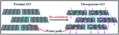
In-plane mesoporous graphene oxide (GO) sheets were prepared by a re-oxidation process and subsequently assembled into lamellar membranes. The in-plane pores significantly shortened the mass transport paths, resulting in 2–3 times higher water permeance than that of the pristine GO membrane, but continued to reject 3 nm molecules.


Membrane separation has become one of the most promising solutions for water purification technologies because of its low cost and flexible installation.1,2 To further increase their performance, lots of novel membranes have been extensively studied.3,4 Recently, graphene oxide (GO), a typical 2-dimensional (2D) atomic thick material,5 demonstrated great potential for water separation membranes.6–9 However, because of the low permeance, the separation efficiency of GO membrane is relatively low, which limits its applications.6–9 In order to further improve the separation performance of GO-related membrane, more nanochannels between the GO sheets were introduced by thermally induced wrinkles,10 intercalated nanoparticles,11 and nanostrands templates.12 Moreover, theoretical studies13–15 demonstrated that by introducing subnanometer in-plane pores in graphene sheets, the membranes could reject salt ions with over water permeance 2 orders of magnitude higher than that of the pristine GO membrane.6,7 Therefore, introducing in-plane pores is another promising way to improve the separation performance of GO-related membrane. About 0.3 nm graphene pores were realized by using ion and electron bombardment.16 A highly porous graphene sheet with pore size smaller than 5 nm was prepared by KOH activation of microwave-exfoliated GO in a large scale.17 O2 etching at high temperature was also used to introduce pores with different sizes in the pristine GO sheets.18 Palaniselvam et al. found a way to open pores on reduced GO sheets by H2O2.19 All of these reported porous GO sheets were easily aggregated, and hard to be dispersed and assembled to lamellar membrane through filtration.

Very recently we found that numerous mesopores with sizes smaller than 5 nm could be created in the GO sheets via re-oxidizing the pristine GO sheets by KMnO4.20 The high density defective domains and residual oxygen-containing functional groups on the carbon skeleton5 were easily attacked by the oxidant species, and creating pores in GO sheets.21 These porous GO sheets were dispersed readily in water after mild sonication. Based on the previous experiments7,12 and theoretical studies8,11 of the mechanism of the formation of GO membrane by filtration process, these mesoporous GO sheets could be easily assembled into GO membrane with enhanced separation performance via filtration. In this communication, similarly, mesoporous GO sheets were prepared through a re-oxidation process by KMnO4 (ref. 20) and subsequently assembled into lamellar membranes for molecule separation. As a proof of concept, the GO membranes with introduced in-plane pores demonstrated 2–3 folds enhancement of water permeance as that of the pristine GO membrane, but preserved the rejection for small molecules. This should be that the in-plane pores remarkably shortened the mass transport paths, while the inter-sheets wrinkles still determined the rejection for small molecules. In addition, it was found that these wrinkles retained their structural under low pressure while compressed under higher pressure. These compressed wrinkles were recovered after fully releasing the pressure, since the separation performance were recovered after the pressure released. Therefore, the introduced in-plane pores did not damage the mechanical property of the prepared mesoporous GO membranes.

Pristine GO sheets were obtained by the modified Hummer method.7,12,22,23 The mesoporous GO sheets with different re-oxidation degrees were prepared through re-oxidation process by KMnO4 as we reported recently20 (see detail in ESI). The pristine GO sheets, the moderately re-oxidized GO sheets and heavily re-oxidized GO sheets were named as GO-1, GO-2 and GO-3, respectively. If the re-oxidation degrees further increased, smaller GO sheets and obvious pores (Fig. S1, ESI) were observed. This indicates that the in-plane pores could be generated by the re-oxidation process. The zeta potentials (Table S1, ESI) of the GO dispersions are very close and below −40 mV at pH 7. For molecule separation, the corresponding GO membranes were typically prepared from 3.2 mL as-synthesized GO-1, GO-2, and GO-3 dispersions (0.1 mg mL−1), on porous polycarbonate membranes with a pore size of 200 nm by vacuum filtrating, respectively.

Fig. 1 and Fig. S2 and 3 show the morphologies of the prepared GO sheets and the corresponding membranes. Lots of nanoscale wrinkles are observed in Fig. 1, which built the main channels for small molecules and ions transport. AFM images shows that the GO sheets are about 1.2 nm thick (Fig. 1b), indicating that the sheets are monolayer. The tightly arranged laminar structures are clearly seen in Fig. 1d, Fig. S2d and 3d. Fig. S4, the corresponding size histograms of GO-1, GO-2 and GO-3 sheets indicate that the sizes of all these three kinds of GO sheets are almost the same of 1.5 μm Keeping the lateral sizes of the GO sheets as close as possible is very helpful to investigate the effect of the in-plane pores generated by re-oxidation on the separation performance.


image file: c4ra01495b-f1.tif
Fig. 1 (a) TEM image, (b) AFM image of GO-3 sheets on silicon substrate, (c) surface and (d) cross-section SEM images of the GO-3 membrane.

Fig. S5a (ESI), the XRD patterns of the GO-1 to 3 membranes indicate that all the three membranes are composed of GO sheets without Mn-related products. Raman (Fig. S5b, ESI) spectra show obvious D-band around 1350 cm−1. Fourier transform-infrared spectra (Fig. S5c, ESI) show oxygen-related absorption bands, including –OH, C[double bond, length as m-dash]O and C–O–C. The X-ray photoelectron spectra (XPS) (Fig. S5d and 6a, ESI) recorded from the re-oxidation products indicate all the samples exhibit the sp2 C[double bond, length as m-dash]C bonds and the oxygen-containing functional groups of C–O and C[double bond, length as m-dash]O on the GO surface, and the content of carbon–oxygen bond increases from 53.7% of pristine GO sheets to 56.2% of GO-3 sheets. There are numerous pores in the sheets of GO-2 and GO-3, which had been demonstrated in our previous work.20 Since the pores are too small and the TEM contrast of monolayer GO sheet is too low, it is very hard to distinguish the pore clearly by TEM. The pore size was further characterized by BET data. The change in pore size distribution (Fig. S5b) is obviously observed from smaller than 2 nm (GO-1) to 3–5 nm (GO-3). In addition, the pore volume increases from 0.075 cm3 g−1 of GO-1, 0.54 cm3 g−1 of GO-2 to 1.11 cm3 g−1 of GO-3.

The separation performances of the prepared GO membranes were evaluated by different molecules and ions, including [(Fe(CN6)]3− (0.9 nm × 0.9 nm), Evan's blue (EB, 1.2 nm × 3.1 nm) and Cytochrome c (Cyt. C, 2.5 nm × 2.5 nm × 3.7 nm)4,12,24 and shown in Fig. 2. As mentioned in our previous work,7 the thickness of GO membrane had significant influence on its performance. The rejection dropped along with the reduction of the thickness. In this work, the optimal thickness of the membrane was about 1 μm, because this membrane demonstrated about 90% rejection for EB molecules. Fig. 2 indicates all these three membranes demonstrate nearly 100% rejection for Cyt. C. The GO-1 membrane shows 90.5% rejection for EB with 70 L m−2 h−1 bar−1. But after introducing in-plane pores by moderate re-oxidation, GO-2 membrane presents 90% EB rejection with water permeance rose to 106 L m−2 h−1 bar−1. For the relatively heavily re-oxidized GO-3 membrane, it shows almost no degradation of the rejection for EB, while the water permeance reaches up to 191 L m−2 h−1 bar−1. Fig. 2 further shows that all of these three membranes show the rejections for [Fe(CN)6]3− are much lower than that of EB, which decreases from 40% of GO-1, to 32% of GO-2, and 26% of GO-3, because the size of [Fe(CN)6]3− is smaller and that of EB molecule. Since all of these three membranes demonstrate nearly 100% rejection for Cyt. C and close to 90% rejection for EB, it is clearly that the introduced in-plane pores significantly improve the water permeance but without obvious degradation of the rejection properties. Based on the sizes of Cyt. C and EB molecules, it could assume that the size of the nanochannels in these GO membranes is in the range of 3–5 nm, after considering the hydration layer of 0.5 nm.4 To clarify the surface charge effect, the neutral molecule, γ-cyclodextrin/adamantane (γ-Cd/Ad, 1.2 nm × 1.2 nm), was examined. Fig. 2 shows that the rejection of γ-Cd/Ad is very close to that of [Fe(CN)6]3− although it is larger than [Fe(CN)6]3−. So except the size sieving of the nanochannels, the surface charge also contributes to the rejection for charged molecules. Again, the introduced in-plane pores really enhanced the water permeance while retained the rejection performance. These are explained as illustrated in Fig. 3. The sizes of the nanochannels of three membranes are almost the same of 3–5 nm, which majorly determines the rejection and leads to the very close rejection of these membranes. However, the introduced in-plane pores on GO sheets by re-oxidation dramatically shorten the transport paths of mass, and resulted in a significant increase in water permeance, but without degradation of molecular rejection.


image file: c4ra01495b-f2.tif
Fig. 2 Separation performances of GO-1 to 3 membranes. Blue columns present the water permeance recorded during EB separation. The rejection is calculated based on the molecule concentrations in permeate and original feed.

image file: c4ra01495b-f3.tif
Fig. 3 Schematic view of mass transport through (a) pristine GO and (b) mesoporous GO membrane.

To assess the structure stability of the mesoporous GO membranes, the separation performance of GO-3 membrane was examined under different pressure. Fig. 4 shows that, at low pressure, the pure water flux increases rapidly, and then slows down when further increases the pressure. The EB rejection continuously increases from 89.5% to 96% when the pressure increased from 0.1 to 0.5 MPa. The rejection reaches the maximum of 99% at 1.0 MPa. These results indicate that the nanochannels are stable at low pressure (0.1–0.5 MPa), but start to collapse under higher pressure, leading to decline the channel size. But this is the reason that the rejection increases under high pressure. The gradual shrinkage of the nanochannels undoubtedly promoted the rejection performance but decreased the water permeance. After releasing the loading pressure back to 0.1 MPa, both the rejection and the water permeance are recovered to 89% and 250 L m−2 h−1, respectively. Therefore, the introduced in-plane pores of GO sheets by re-oxidation did not reduce the structural stability of the wrinkled nanochannels or degrade the rejection of as-prepared mesoporous GO membranes.


image file: c4ra01495b-f4.tif
Fig. 4 The separation performance of GO-3 membrane vs. pressure. Black solid squares and circles indicate the pressure loading and releasing process; the red solid triangles demonstrate the corresponding EB rejection. The black solid triangle shows the EB rejection after the loading pressure was released to 0.1 MPa.

In summary, for the first time, we successfully prepared mesoporous GO membranes by introducing in-plane mesopores of GO by re-oxidation. These membranes exhibited an excellent separation performance of 88.5% rejection for EB with a water permeance of 191 L m−2 h−1 bar−1, which was nearly 3 times higher than that of the pristine GO membrane. The in-plane pores not only increased the amount of channels but also shortened the transport paths. The pressure loading and releasing process demonstrated the excellent structural stability of the mesoporous GO membrane. Our work provides an efficient strategy to improve the separation performance of GO related membranes, which could be further extended to other 2-D material based membranes.

Acknowledgements

This work was supported by the National Natural Science Foundations of China (NSFC 21271154), Doctoral Fund of Ministry of Education of China (20110101110028), and the project-sponsored by SRF for ROCS, SEM.

Notes and references

  1. M. A. Shannon, P. W. Bohn, M. Elimelech, J. G. Georgiadis, B. J. Marinas and A. M. Mayes, Nature, 2008, 452, 301–310 CrossRef CAS PubMed.
  2. K. M. Yao, M. M. Habibian and C. R. Omelia, Environ. Sci. Technol., 1971, 5, 1105–1112 CrossRef CAS.
  3. S. Karan, S. Samitsu, X. S. Peng, K. Kurashima and I. Ichinose, Science, 2012, 335, 444–447 CrossRef CAS PubMed.
  4. X. S. Peng, J. Jin, Y. Nakamura, T. Ohno and I. Ichinose, Nat. Nanotechnol., 2009, 4, 353–357 CrossRef CAS PubMed.
  5. D. R. Dreyer, S. Park, C. W. Bielawski and R. S. Ruoff, Chem. Soc. Rev., 2010, 39, 228–240 RSC.
  6. P. Z. Sun, M. Zhu, K. L. Wang, M. L. Zhong, J. Q. Wei, D. H. Wu, Z. P. Xu and H. W. Zhu, ACS Nano, 2013, 7, 428–437 CrossRef CAS PubMed.
  7. H. B. Huang, Y. Y. Mao, Y. L. Ying, Y. Liu, L. W. Sun and X. S. Peng, Chem. Commun., 2013, 49, 5963–5965 RSC.
  8. R. R. Nair, H. A. Wu, P. N. Jayaram, I. V. Grigorieva and A. K. Geim, Science, 2012, 335, 442–444 CrossRef CAS PubMed.
  9. Y. Han, Z. Xu and C. Gao, Adv. Funct. Mater., 2013, 23, 3693–3700 CrossRef CAS.
  10. L. Qiu, X. Zhang, W. Yang, Y. Wang, G. P. Simon and D. Li, Chem. Commun., 2011, 47, 5810–5812 RSC.
  11. C. Xu, A. Cui, Y. Xu and X. Fu, Carbon, 2013, 62, 465–471 CrossRef CAS PubMed.
  12. H. Huang, Z. Song, N. Wei, L. Shi, Y. Mao, Y. Ying, L. Sun, Z. Xu and X. Peng, Nat. Commun., 2013, 4, 2979 Search PubMed.
  13. D. Cohen-Tanugi and J. C. Grossman, Nano Lett., 2012, 12, 3602–3608 CrossRef CAS PubMed.
  14. D.-E. Jiang, V. R. Cooper and S. Dai, Nano Lett., 2009, 9, 4019–4024 CrossRef CAS PubMed.
  15. H. L. Du, J. Y. Li, J. Zhang, G. Su, X. Y. Li and Y. L. Zhao, J. Phys. Chem. C, 2011, 115, 23261–23266 CAS.
  16. C. J. Russo and J. A. Golovchenko, Proc. Natl. Acad. Sci. U. S. A., 2012, 109, 5953–5957 CrossRef CAS PubMed.
  17. L. L. Zhang, X. Zhao, M. D. Stoller, Y. W. Zhu, H. X. Ji, S. Murali, Y. P. Wu, S. Perales, B. Clevenger and R. S. Ruoff, Nano Lett., 2012, 12, 1806–1812 CrossRef CAS PubMed.
  18. L. Liu, S. Ryu, M. R. Tomasik, E. Stolyarova, N. Jung, M. S. Hybertsen, M. L. Steigerwald, L. E. Brus and G. W. Flynn, Nano Lett., 2008, 8, 1965–1970 CrossRef CAS PubMed.
  19. T. Palaniselvam, M. O. Valappil, R. Illathvalappil and S. Kurungot, Energy Environ. Sci., 2014, 7, 1059–1067 CAS.
  20. Z. J. Fan, Q. K. Zhao, T. Y. Li, J. Yan, Y. M. Ren, J. Feng and T. Wei, Carbon, 2012, 50, 1699–1703 CrossRef CAS PubMed.
  21. P. Solís-Fernández, J. Paredes, S. Villar-Rodil, L. Guardia, M. Fernández-Merino, G. Dobrik, L. Biró, A. Martínez-Alonso and J. Tascón, J. Phys. Chem. C, 2011, 115, 7956–7966 Search PubMed.
  22. W. S. Hummers Jr and R. E. Offeman, J. Am. Chem. Soc., 1958, 80, 1339 CrossRef.
  23. Z. Luo, Y. Lu, L. A. Somers and A. C. Johnson, J. Am. Chem. Soc., 2009, 131, 898–899 CrossRef CAS PubMed.
  24. L. W. Sun, H. B. Huang and X. S. Peng, Chem. Commun., 2013, 49, 10718–10720 RSC.

Footnote

Electronic supplementary information (ESI) available: Experimental procedure in detail, additional SEM and TEM images, FTIR and XPS spectra. See DOI: 10.1039/c4ra01495b

This journal is © The Royal Society of Chemistry 2014
Click here to see how this site uses Cookies. View our privacy policy here.