Surface-enhanced Raman scattering (SERS) chips made from metal nanoparticle-doped polymer fibers†
Wenran
Gao
,
Gang
Chen
,
Weiqing
Xu
,
Chenggong
Yang
and
Shuping
Xu
*
State Key Laboratory of Supramolecular Structure and Materials, Jilin University, Changchun 130012, P. R. China. E-mail: xusp@jlu.edu.cn
First published on 10th April 2014
Abstract
We employed an electrospinning method to prepare metal nanoparticle (NP) doped polymer nanofiber mats, which can be easily cut to size and fixed on slides or in microfluidic channels for surface-enhanced Raman scattering (SERS) measurements. Metal NPs embedded in the composite nanofibers maintained their intrinsic shape and monodispersity, as well as their enhancement ability. In addition, they can be re-suspended in aqueous solution to recover a colloidal solution with persistent SERS activity. The SERS chips made from the metal NP-doped polymer nanofiber mats displayed high detection sensitivity and reproducibility. The microfluidic chips decorated with several metal NP-doped polymer nanofiber mats possess multi-SPR properties and can be widely used for different analytes. These composite polymer mats can be easily stored and transported, and also possess potential for use as antibacterial filters and paint for medical purposes.
1. Introduction
Metal colloidal nanoparticles (NPs) have gained great interest due to their abundant applications in the fields of biochemical sensing,1–3 optoelectronic devices,4 photocatalysis5, surface-enhanced spectroscopy,6–8 thermal medication,9,10etc. These applications are all based on the surface plasmon resonance (SPR) properties of metal colloidal NPs, which can enlarge the effective scattering cross section over their geometric shapes, causing enhanced efficiencies of light harvesting and radiation. The SPR effect behaves most obviously in terms of surface-enhanced Raman scattering (SERS), resulting in a million-fold enhancement of the Raman signal. Of course, SERS measurements strongly and strictly depend on the SPR properties of metal NPs, requiring the exact matching of the SPR band and the excitation wavelength.Besides spherical metal particles, many anisotropic metal NPs have been introduced to act as SERS substrates due to their rich SPR characteristics, such as gold nanorods,11 silver nanoprisms,12 silver nanocubes13etc. Meanwhile, a variety of techniques have strived for the controllable adjustment of the SPR properties of anisotropic metal NPs in a wide (or appropriate) spectral range, for example, changing the aspect ratio of gold rods by using different reaction conditions,14 transforming shape by adding etching reagents,15,16 restriction by the wavelength self-limiting effect in laser-induced growth17 and so on. To widen the applicability and stability of metal colloidal NPs as SERS substrates, these particles have been combined with solid-supported slides18–21 or bulky substances (e.g. vials,20 capillaries,20 optical fibers,22 porous anodic alumina,23 and others24–26) to achieve highly stable and active SERS chips. These SERS chips have distinct advantages of easy transportation and storage. Strategies for building SERS chips by using metal colloidal NPs are various. The assembly methods based on supramolecular interaction or covalent bonding are usually adopted, which require the surface modification of metal NPs or chip substrates by silicone agents18 or a polyelectrolyte.19 Owing to the different surface functional groups of various colloidal NPs, finding a universal combination strategy is still a challenge.
The electrospinning technique can satisfy the strict requirements for a universal combination strategy. It can produce composite fibrous mats with high efficiency and make metal NP combination feasible. In this study, the electrospinning technique has been employed to prepare metal NP-doped polymer nanofiber mats. Further, these composite polymer mats were cut into an appropriate size and fixed on a slide or a microfluidic chip to fabricate multi-SPR functionalized chips. These metal NP-doped polymer nanofibers can give SERS enhancement. When measuring SERS on the multi-SPR functionalized chips, the laser wavelength is chosen to match the resonance of the metal NPs, ensuring the highest SERS.
2. Experimental section
2.1 Materials
Silver nitrate (AgNO3, 99.8%) and chloroauric acid (HAuCl4·4H2O, Au content >47.8%) were purchased from Shanghai Reagent No.1 Plant. NaBH4 (96%) and polyvinyl pyrrolidone (PVP) (Mw = 30![[thin space (1/6-em)]](https://www.rsc.org/images/entities/char_2009.gif) 000) were obtained from Sinopharm Chemical Reagent Co., Ltd. Trisodium citrate (99.0%) and 4-mercaptobenzoic acid (4-MBA) were supplied by the Beijing Chemical Reagent Plant. Polyvinyl alcohol (PVA, Mw = 130000), 4-mercaptopyridine (4-MPY), Atto 610-biotin and hexadecyltrimethylammonium bromide (CTAB) were purchased from Sigma-Aldrich. Rhodamine 6G (R6G) was obtained from Exciton Chemical Co. Inc. Teflon tape was purchased from Tianjin Tiansu Science &Technology Group Co., Ltd. Polymethyl methacrylate (PMMA) was obtained from Foshan City Shunde Jundao Optical Sheet Manufacturing Co., Ltd.
000) were obtained from Sinopharm Chemical Reagent Co., Ltd. Trisodium citrate (99.0%) and 4-mercaptobenzoic acid (4-MBA) were supplied by the Beijing Chemical Reagent Plant. Polyvinyl alcohol (PVA, Mw = 130000), 4-mercaptopyridine (4-MPY), Atto 610-biotin and hexadecyltrimethylammonium bromide (CTAB) were purchased from Sigma-Aldrich. Rhodamine 6G (R6G) was obtained from Exciton Chemical Co. Inc. Teflon tape was purchased from Tianjin Tiansu Science &Technology Group Co., Ltd. Polymethyl methacrylate (PMMA) was obtained from Foshan City Shunde Jundao Optical Sheet Manufacturing Co., Ltd.
2.2 Preparation of metal NP-doped polymer fibers
Firstly, to obtain metal NP-doped polymer fibers with multiple SPR bands, we prepared several kinds of metal NP, namely Au/Ag NPs with different shapes and sizes. Colloidal Au nanospheres of different sizes were synthesized by the method of Frens.27 The size of the Au particles was controlled by the amount of trisodium citrate used. Au nanorods were prepared by the seed-mediated growth method.14 Silver nanospheres were prepared by the Lee method.28 These metal NPs were concentrated by a factor of 120 by centrifuge before further use and PVP was added as a stabilizer (0.1 g of PVP per 100 mL of metal colloid).Secondly, metal NP-doped polymer fibers were prepared by electrospinning. Electrospinning solutions containing 0.8 mL of the concentrated metal NPs and 0.08 g of PVA were prepared by stirring and heating (70 °C) on a magnetic stirring heater. In a typical electrospinning process, the electrospinning solution was kept in a plastic syringe (with a capacity of 1.0 mL) fitted with a metal needle. The applied voltage was 15 kV. A distance of 15 cm was set between the tip of the metal needle and the aluminum foil collector. The flow rate for the electrospinning solution was 0.2 mL h−1. The metal NP-doped polymer fiber mats were obtained after 4 h of electrospinning.
2.3 Fabrication of SERS chips by using metal NP-doped polymer fibers
Two types of SERS chip were fabricated for testing analytes in organic solvents (SERS chip I) and in water (SERS chip II). The type I SERS chips were constructed using the metal NP-doped polymer fiber mats (cut into 3 mm × 3 mm squares). To test the analytes in organic solvents, the metal NP-doped polymer fiber mats were immersed in sample solutions for the desired time (several hours) and then dried at room temperature before carrying out Raman measurements. SERS chip II was made by laying a piece of metal NP-doped polymer mat (3 mm × 3 mm) on a hydrophobic membrane (Teflon tape) coated glass slide. To detect the SERS signal by SERS chip II, 15 μL of the aqueous sample solution was dripped on it and then dried.In addition, several metal NP-doped polymer fiber mats were cut into narrow strips to coat the microfluidic channels to fabricate the microfluidic SERS chip with multi-SPR function. Microfluidic channels were engraved in PMMA by an engraving machine.
2.4 Characterizations
Ultraviolet-visible (UV-vis) absorption spectra of the metal colloidal solutions were measured by a Shimadzu UV-3100 and an Ocean Optics USB2000 spectrophotometer. Field-emission scanning electron microscopy (SEM) on a Hitachi SU8000 and transmission electron microscopy (TEM) on a JEOL JEM-2100F were employed to investigate the morphologies of the metal NP-doped polymer fibers. A confocal Raman system (LabRAM ARAMIS, Horiba Jobin-Yvon) with a ∼0.7 μm spatial resolution, and a 5 mW, 633 nm HeNe laser as the excitation source, was used for the collection of Raman spectra. The detection of Raman signals was carried out with a Synapse thermoelectrically cooled charge-coupled device (CCD) camera (Horiba Jobin-Yvon). Raman scattered light was collected with a 50× microscope objective lens (0.50 NA, LMPLFLN, Olympus, Japan), which was also used for focusing the light from the excitation laser. The focused laser beam on the tissue formed a spot with a diameter of 1.5 μm. The strong light from Rayleigh-scattering was then blocked by a 4-notch filter (Horiba Jobin-Yvon, Edison, NJ, USA). Extended scanning spectra with a spectral range of 300–1800 cm−1 were acquired using an integration time of 10 s and 2 accumulations. The wavenumber calibration was set by reference to the 520.7 cm−1 vibrational band of a silicon wafer.To compare the SERS signals obtained under different excitation wavelengths, a portable BWTEK Raman spectrometer with a 532 nm excitation wavelength was used. The integration time was 10 s. The laser power reaching the samples was 1.32 mW. The scan range was 300–1800 cm−1. Three different points from each sample were chosen, and the data were the average values of three repeats.
3. Results and discussion
3.1 Characterizations of metal NP-doped polymer fibers
Five types of metal NP were employed to fabricate the metal NP-doped polymer composite fibers. Three of them were Au spheres of different sizes (13, 16, and 40 nm) and the other two were Ag NPs and Au nanorods. Panel (1) of Fig. 1 shows the TEM images of the prepared colloidal metal NPs. It can be observed that the metal NPs were mostly uniform in size and shape except for the Ag NPs. When the metal NPs were combined with PVA fibers by electrospinning, they needed to be concentrated by a factor of about 125 or more to ensure that their density in the polymer fibers was high enough for the SERS enhancement requirements. However, too high a metal NP density would cause the aggregation of the metal NPs and also affect the mechanics of the PVA fibers, causing the failure of the electrospinning. Panel (2) of Fig. 1 displays the TEM images of the metal NP-doped PVA fibers. It can be observed that most of the metal NPs were well dispersed and doped in the PVA fibers and some of them were semi-embedded in the PVA fibers. Interestingly, owing to the high inducing voltage applied on these electrospinning solutions, rod or stick type metal NPs were aligned with the polymer fibers (images d-2 and e-2). Fig. 2 shows the extinction spectra of these metal colloids (solid lines) and their corresponding composite polymer nanofibers (short-dashed lines). Table 1 summarizes the details of the metal NPs' morphologies and their extinction band positions as observed in Fig. 1 and 2, respectively. It is found that the extinction band positions of the metal NP-doped polymer fiber mats are red-shifted compared to their colloid systems, with the exception of the Ag NP-doped polymer fiber mat, which shows a tiny blue-shift. These shifts are mainly because the media around the metal NPs varied when they were combined with PVA. | ||
| Fig. 1 TEM images of the five kinds of prepared metal NP colloid (left panel), the metal NP-doped PVA nanofibers obtained by electrospinning (middle panel) and the metal NPs obtained by resuspending the metal NP-doped PVA nanofibers in water (right panel). | ||
 | ||
| Fig. 2 Extinction spectra of the prepared metal NP colloids (1: solid line), metal NP-doped PVA nanofibers (2: short-dashed line) and the metal NPs obtained by resuspending the metal NP-doped PVA nanofibers in water (3: dashed line). | ||
| Metal NPs | Shape | Size/nm | Band position/nm | ||
|---|---|---|---|---|---|
| Metal colloid (1) | Metal NP-doped PVA fiber (2) | Resuspended metal colloid in water (3) | |||
| Au NPs | Sphere | 13 | 521 | 532 | 522 |
| Au NPs | Sphere | 16 | 523 | 534 | 524 |
| Au NPs | Sphere | 40 | 533 | 541 | 533 |
| Ag NPs | Irregular shape | ∼43 | 418 | 412 | 418 |
| Au rods | Rod | 31 in width, 20 in length | 535, 607 | 542, 622 | 535, 607 |
Fig. 3 shows the SEM images of the metal NP-doped polymer fibers. We can see that the average diameters of the metal NP-doped polymer fibers are 260–300 nm and the lengths extend to several millimetres or more. The metal NP-doped polymer fibers are interlaced and show a 3-dimensional network. The insets are the pictures of the metal NP-doped polymer fibers and metal NP solutions. It is observed that the metal NP-doped polymer fiber mats exhibit the same colours as their metal NP solutions, which has been proved by their extinction spectra (Fig. 2).
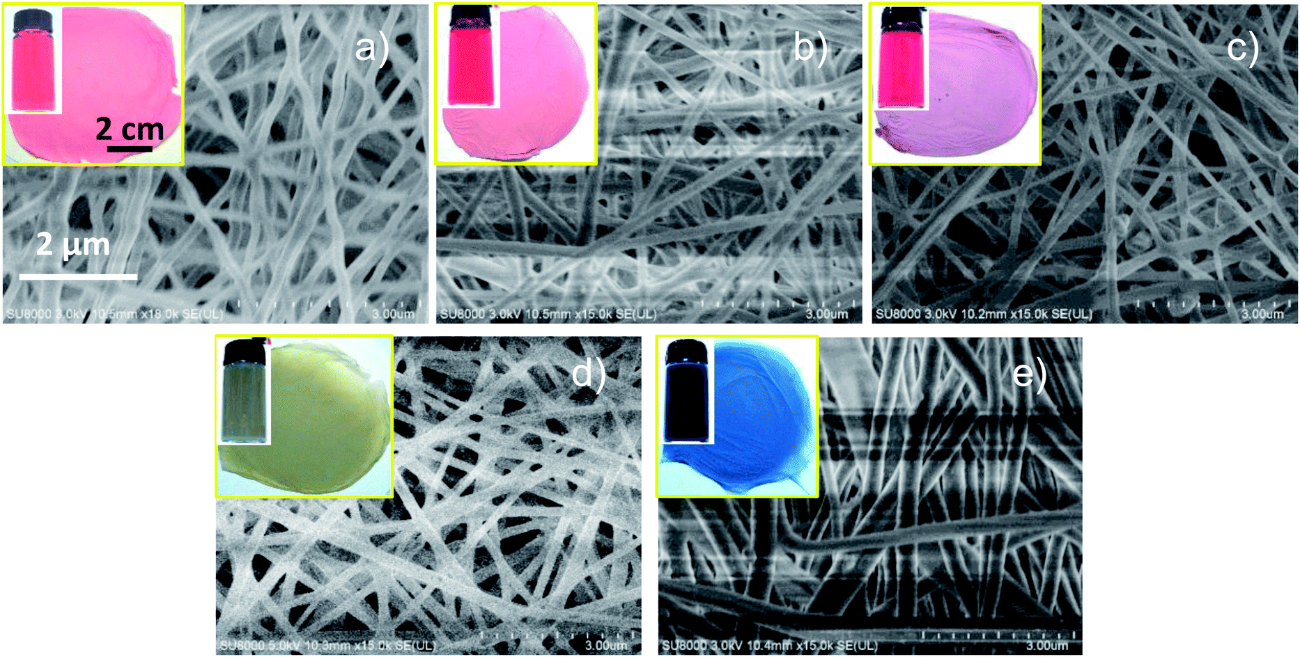 | ||
| Fig. 3 SEM images of the five kinds of metal NP-doped polymer fiber. (a)–(e) correspond to 13, 16 and 40 nm Au NPs, Ag NPs and Au nanorods, respectively. Insets show the photos of the metal colloids and metal NP-doped polymer fiber mats. | ||
The polymer we used in electrospinning was PVA, which is a water-soluble polymer. We resuspended the metal NP-doped polymer fibers into water, achieving the metal colloids again. The TEM images (panel (3) of Fig. 1) of the resuspended metal colloids prove that these metal NPs can be effectively re-dispersed in water. The extinction spectra of the resuspended metal colloids were also collected (dashed lines in Fig. 2). Compared with the freshly prepared metal colloids (solid lines), their plasmonic bands show no difference. This means that no metal aggregates are formed during the electrospinning process and their SERS activity still remains (see ESI†). These results indicate that the electrospinning process does no harm to the stability of the colloidal metal NPs. This is especially important for SERS measurements because the uncontrollable aggregation of colloidal metal NPs will lead to a poor reproducibility in SERS detections and this method avoids this.
Metal NPs combined with bulky materials show long-term stability. There are almost no changes to the metal NP-doped polymer fibers after storage for 6 months (see ESI†). The long-term stability ensures that these metal NP-doped polymer fiber mats have promise for commercial uses.
3.2 Fabrication of the SERS chips and their SERS applications
Fig. 4a–c show the SERS detections of 4-MBA using different type I SERS chips constructed with Au NPs (16 nm), Ag NPs and Au rods, at trace levels (the lowest detectable concentrations) of 1.0 × 10⁻⁵, 1.0 × 10⁻⁷ and 1.0 × 10⁻⁸ M, respectively. In the determination of 4-MPY, the trace levels of 4-MPY when using Ag NP and Au nanorod-doped polymer fibers are 1.0 × 10⁻⁸ and 1.0 × 10⁻⁹ M, respectively (Fig. 4d and e). These results show that the type I SERS chips provide acceptable SERS enhancement activities.
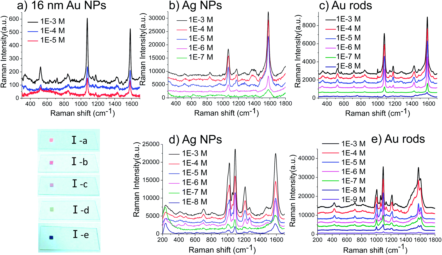 | ||
| Fig. 4 Concentration-dependant SERS detections of 4-MBA (a, b and c) and 4-MPY (d and e) using the type I SERS chips under the excitation wavelengths of 633 (a, c and e) and 532 nm (b and d). The photos of the five type I SERS chips are provided, corresponding to 13 (I-a), 16 (I-b) and 40 (I-c) nm Au NPs, Ag NPs (I-d) and Au nanorods (I-e). | ||
In addition, we investigated the reproducibility of results from a SERS chip I by comparing SERS signals from 20 different spots on the same SERS chip (Fig. 5a). Two peaks at 1078 cm−1 and 1086 cm−1 were chosen to evaluate the reproducibility of this SERS chip (Fig. 5b). The relative standard deviations of spot-to-spot Raman intensities are 10% and 8.3% in Fig. 5b, which prove that the SERS chip I exhibits greater reproducibility compared to the scientific standard of SERS substrates in which less than 20% spot-to-spot variation over 10 mm2 is required.29
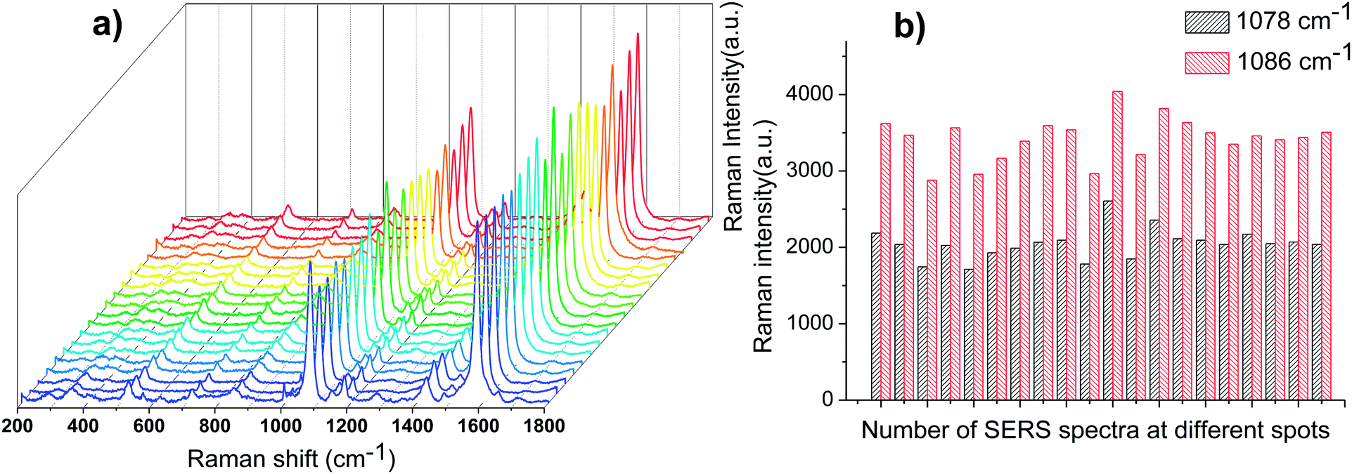 | ||
| Fig. 5 (a) SERS spectra of 4-MBA (1.0 × 10⁻⁵ M) adsorbed on the Au nanorod-doped PVA nanofibers under 633 nm excitation wavelength from 20 randomly selected spots. (b) The intensities of two different peaks at 20 spots on the same substrate. | ||
 | ||
| Fig. 6 The pictures of the SERS chip II and its SERS detections. (a) A Ag NP-doped PVA fiber mat on a hydrophobic surface. (b) 15 μL of aqueous sample solution was dropped on the Ag NP-doped PVA fiber mat. (c) The sample solution was dried and a circle of stain containing Ag NPs, PVA and analytes was left on the hydrophobic surface. (d) SERS determination of aqueous 4-MPY solutions at different concentrations (1.0 × 10⁻³ to 1.0 × 10⁻⁷ M) by the SERS chip II. (e) SERS spectra of aqueous 4-MPY solution (1.0 × 10⁻⁵ M) tested by the SERS chip II from 20 randomly selected points. The excitation wavelength of the laser was 532 nm. | ||
 | ||
| Fig. 7 (a) The UV-Vis absorption spectrum of a R6G ethanol solution (1 × 10⁻⁷ M) and the extinction spectrum of the colloidal Ag NPs. (b) SERS spectra of R6G (1 × 10⁻⁶ M, ethanol solution) obtained on the Ag NP-doped PVA nanofibers under different excitation wavelengths. (c) The UV-Vis absorption spectrum of an Atto 610-biotin ethanol solution (1 × 10⁻⁴ mg mL−1) and the extinction spectrum of the colloidal Au nanorods. (d) SERS spectra of Atto 610-biotin (1 × 10⁻⁴ mg mL−1, ethanol solution) obtained on the Au nanorod-doped PVA nanofibers under different excitation wavelengths. | ||
The metal NP-doped polymer fiber mats made by the electrospinning method can satisfy the above requirement and have the advantages of high efficiency and broad applicability for the combination of metal NPs. These composite polymer mats were cut to an appropriate size and fixed on a PMMA microfluidic chip to fabricate multi-SPR functionalized SERS chips. Fig. 8 shows the photo of the SERS microfluidic chip made using the metal NP-doped polymer fibers. We coated the microfluidic channels with different metal NP-doped polymer fibers. An example of the SERS detection of Atto 610-biotin under a 633 nm excitation wavelength with a SERS microfluidic chip made using the Au nanorod-doped polymer fibers is provided. It is noted that the solvent ethanol and the chip material PMMA both exhibit relatively weak spectral signals compared to the signal of Atto-610 (see Fig. S4 and S5 in ESI†) and show little spectral interference. This multi-SPR functionalized SERS microfluidic chip has great merits for the SERS determination of complicated and multicomponent systems.
 | ||
| Fig. 8 The pictures of a SERS microfluidic chip decorated with several kinds of metal NP-doped polymer fiber (top) and the SERS detection of Atto 610-biotin (1 × 10⁻⁴ mg mL−1, ethanol solution) in the SERS microfluidic chip (bottom). For comparison, a Raman spectrum of the microfluidic chip (PMMA) is provided in ESI.† | ||
4. Conclusions
In summary, we successfully prepared metal NP-doped polymer fibers by the electrospinning method. The electrospinning technique is a universal and flexible approach to combine metal NPs with bulky materials with high efficiency. Metal NPs were well dispersed in the polymer fibers and displayed persistent SERS activity. The metal NP-doped polymer fibers are free-standing, flexible, and extraordinarily stable. Furthermore, two types of SERS chip made using metal NP-doped polymer fiber mats (SERS chip I and SERS chip II) were fabricated for probing analytes in organic and aqueous solutions. The SERS detection results show that both types of SERS chips have good SERS enhancement activities and acceptable reproducibility. In addition, these composite polymer mats were fixed in a microfluidic chip to fabricate multi-SPR functionalized chips, which are applicable to the SERS determinations of complicated and multicomponent systems. These composite polymer mats can be easily stored and transported. They can also be applied in medical fields, e.g. as a colourful antibacterial filter and as paint. Relevant studies are underway.Acknowledgements
This work was supported by the National Natural Science Foundation of China (no. 21373096, 21073073 and 91027010), the Ministry of Education of returned overseas students to start research and fund projects and the National Instrumentation Program (NIP) of the Ministry of Science and Technology of China no. 2011YQ03012408. We thank Prof. Kun Liu, Jilin University for the supply of samples of Au nanorods and Prof. Ce Wang, Jilin University for teaching us the electrospinning technique.Notes and references
- K. A. Willets and R. P. Van Duyne, Annu. Rev. Phys. Chem., 2007, 58, 267–297 CrossRef CAS PubMed.
- C. A. Mirkin, R. L. Letsinger, R. C. Mucic and J. J. Storhoff, Nature, 1996, 382, 607–609 CrossRef CAS PubMed.
- M. Lismont and L. Dreesen, Mater. Sci. Eng., C, 2012, 32, 1437–1442 CrossRef CAS PubMed.
- J.-L. Wu, F.-C. Chen, Y.-S. Hsiao, F.-C. Chien, P. Chen, C.-H. Kuo, M. H. Huang and C.-S. Hsu, ACS Nano, 2011, 5, 959–967 CrossRef CAS PubMed.
- P. V. Kamat, J. Phys. Chem. B, 2002, 106, 7729–7744 CrossRef CAS.
- J. A. Creighton, C. G. Blatchford and M. G. Albrecht, J. Chem. Soc., Faraday Trans. 2, 1979, 75, 790–798 RSC.
- Y. C. Cao, R. C. Jin and C. A. Mirkin, Science, 2002, 297, 1536–1540 CrossRef CAS PubMed.
- M. Moskovits, Rev. Mod. Phys., 1985, 57, 783 CrossRef CAS.
- D. P. O'Neal, L. R. Hirsch, N. J. Halas, J. D. Payne and J. L. West, Cancer Lett., 2004, 209, 171–176 CrossRef PubMed.
- X. Huang, I. H. El-Sayed, W. Qian and M. A. El-Sayed, J. Am. Chem. Soc., 2006, 128, 2115–2120 CrossRef CAS PubMed.
- C.-L. Zhang, K.-P. Lv, H.-P. Cong and S.-H. Yu, Small, 2012, 8, 648–653 CrossRef CAS PubMed.
- M. Liu, Z. Wang, S. Zong, R. Zhang, D. Zhu, S. Xu, C. Wang and Y. Cui, Anal. Bioanal. Chem., 2013, 405, 6131–6136 CrossRef CAS PubMed.
- H. K. Lee, Y. H. Lee, Q. Zhang, I. Y. Phang, J. M. R. Tan, Y. Cui and X. Y. Ling, ACS Appl. Mater. Interfaces, 2013, 5, 11409–11418 CAS.
- B. Nikoobakht and M. A. El-Sayed, Chem. Mater., 2003, 15, 1957–1962 CrossRef CAS.
- J. An, B. Tang, X. L. Zheng, J. Zhou, F. X. Dong, S. P. Xu, Y. Wang, B. Zhao and W. Q. Xu, J. Phys. Chem. C, 2008, 112, 15176–15182 CAS.
- S. P. Xu, B. Tang, X. L. Zheng, J. Zhou, J. An, X. H. Ning and W. Q. Xu, Nanotechnology, 2009, 20, 415601 CrossRef PubMed.
- X. L. Zheng, W. Q. Xu, C. Corredor, S. P. Xu, J. An, B. Zhao and J. R. Lombardi, J. Phys. Chem. C, 2007, 111, 14962–14967 CAS.
- R. G. Freeman, K. C. Grabar, K. J. Allison, R. M. Bright, J. A. Davis, A. P. Guthrie, M. B. Hommer, M. A. Jackson, P. C. Smith, D. G. Water and M. J. Natan, Science, 1995, 267, 1629–1630 CrossRef CAS PubMed.
- X. L. Li, W. Q. Xu, J. H. Zhang, H. Y. Jia, B. Yang, B. Zhao, B. F. Li and Y. Ozaki, Langmuir, 2004, 20, 1298–1304 CrossRef CAS.
- http://www.rta.biz .
- http://www.pers-spec.org .
- W. Q. Xu, S. P. Xu, B. Hu, K. X. Wang, B. Zhao, Y. T. Xie and Y. G. Fan, Chem. Res. Chin. Univ., 2004, 25, 144–147 CAS.
- X. N. Wang, S. P. Xu, H. B. Li, J. L. Tao and W. Q. Xu, J. Raman Spectrosc., 2012, 43, 459–463 CrossRef CAS.
- L. Zhang, X. Gong, Y. Bao, Y. Zhao, M. Xi, C. Jiang and H. Fong, Langmuir, 2012, 28, 14433–14440 CrossRef CAS PubMed.
- M. Cao, S. Cheng, X. Zhou, Z. Tao, J. Yao and L.-J. Fan, J. Polym. Res., 2011, 19, 9810 CrossRef.
- X. Li, M. Cao, H. Zhang, L. Zhou, S. Cheng, J.-L. Yao and L.-J. Fan, J. Colloid Interface Sci., 2012, 382, 28–35 CrossRef CAS PubMed.
- J. Frens, Nature, Phys. Sci., 1973, 241, 20–22 CrossRef.
- P. C. Lee and D. Meisel, J. Phys. Chem., 1982, 86, 3391–3395 CrossRef CAS.
- M. J. Natan, Faraday Discuss., 2006, 132, 321–328 RSC.
Footnote |
| † Electronic supplementary information (ESI) available: (1) SERS spectra obtained from Au NP colloids or the dissolved Au NP-doped PVA fibers on glass substrates under a 633 nm excitation wavelength. (2) Extinction spectra of the five kinds of metal NP-doped polymer fiber before and after 6 months of storage. (3) SEM image of SERS chip (II) after an aqueous 4-MPY solution was dripped on to it and dried. (4) SERS spectrum of PMMA under a 633 nm excitation wavelength. (5) SERS spectrum of ethanol under a 633 nm excitation wavelength. See DOI: 10.1039/c4ra01432d |
| This journal is © The Royal Society of Chemistry 2014 |