A catalase-magnetic switch for cell proliferation

Sofia Municoy, Irene L. Ibañez, Hebe Durán and Martín G. Bellino*
Departamento de Micro y Nanotecnología, Comisión Nacional de Energía Atómica, Av. Gral Paz 1499 (B1650KNA), San Martín, Buenos Aires, Argentina. E-mail: mbellino@cnea.gov.ar

Received 12th February 2014 , Accepted 12th March 2014

First published on 13th March 2014


Abstract

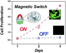
The advance of novel biotechnology requires the development of versatile control systems for cell behavior. Towards the vision of cell growth control, we report a magnetic switching in cell proliferation by a catalase–nanomagnetite complex. The system synergically exploits both the ability of catalase to scavenge cell-generated hydrogen peroxide, a messenger involved in cell cycle regulation, and magnetite manipulation to modulate cell proliferation between arrest and growth using magnetic fields. This ON/OFF switch methodology could constitute the basis of new classes of diagnostic and therapeutic strategies as well as novel integrated responsive biomaterials.


Introduction

Organisms have evolved a wide variety of mechanisms to control cell growth which are crucial to the metabolism and reproduction of living systems. Scientists are currently focused on finding ways to mimic cellular regulatory mechanisms in order to develop new methodologies with potential diagnostic and therapeutic applications.1 The ability to regulate cell proliferation in response to environmental conditions (e.g. substrate levels, exposure to growth factors and hormones) is vital to many metabolic functions.2 Controlling signaling mediators is a key feature to actuate on cell behavior. Reactive oxygen species (ROS) are generated as by-products of cell aerobic metabolism. Although hydrogen peroxide (H2O2), a non-free radical ROS, is commonly known for its cytotoxic effect, at physiological levels it plays a role as an important messenger that regulates diverse biological processes.3,4 In this sense, different regulatory proteins involved in signal transduction pathways can be oxidized by H2O2, leading to the activation or deactivation of these proteins, e.g. decreased levels of intracellular H2O2 culminate in the arrest of cell proliferation.5–8 Catalase (CAT), an enzyme highly active and specific in decomposing H2O2, has been therefore used to inhibit cell growth.5–8 Versatile methods to switch cell growth represent an evolution in biotechnological strategies, presenting a substantial scientific and engineering challenge for advanced biomaterials and biomedicine. Among addressable systems, magnetic nanoparticles (MNPs) stand out, because they can incorporate functional elements and provide magnetic control capabilities.9–13 Magnetic fields are promising for remote stimulation because they can penetrate deeply into biological tissues with negligible attenuation.14

Taking advantage of these combined features, here we report a magnetic-field-assisted switching in cell proliferation by CAT–MNP nanostructured complex to turn on and off the proliferation of a human cell line. Actuation was achieved by remote magnetic switching to spatially put together the complex and cells for growth inhibition (OFF state) or separate them for proliferation (ON state). We show that the combination of enzymatic catalysis to block cell messengers with magnetic nanoparticles lead to a versatile route to control cell behaviors.

Results and discussion

Development and characterization of CAT–MNP complex

For preparation of the CAT–MNP nanostructured complex, CAT was conjugated to iron oxide nanoparticles (nanocrystalline magnetite, Fe3O4 with an average size of 10 nm; MNP, leading to a narrow nanocluster size distribution in aqueous solution of ca. 35 nm. See ESI, Fig. S1) through glutaraldehyde crosslinking of enzymes15 in presence of MNPs.

The resulting material was a nanostructured network of fused enzyme micro-particles where MNPs clusters were physically entrapped during the cross-linking process. Further, in view of the magnetic properties of the nanoparticles, the resulting complexes can be easily separated from the reaction mixture by the application of a magnetic field, avoiding centrifugation or filtration. After magnetic separation, high catalase activity was detected in the magnetic nanocomplex. The estimated capacity for enzyme loading in the CAT–MNP complex was 7 mg enzyme per mg magnetite. Moreover, nanostructured complexes have shown to be reusable, with no performance decrease, and substantially more active than the free enzyme at extreme pHs and temperatures, indicating that the nanocomplexes had a significantly enhanced stability (see ESI Fig. S2). The CAT–MNP complexes were characterized by optical and electron microscopy to determine their morphology and size distribution as shown in Fig. 1. This analysis revealed a matrix of fused enzyme entrapping MNPs with an average size of 10 μm. The entrapped MNPs appeared as aggregates with sizes of 0.2–0.5 μm, attributable to nanocluster agglomeration during the cross-linking process.


image file: c4ra01257g-f1.tif
Fig. 1 Characterization of CAT–MNP complexes. Representative light microscopy (A) and TEM (B) images of CAT–MNP complexes. The blow-up of the TEM image shows the presence of entrapped magnetite nanoparticles.

CAT–MNP complex to control the intracellular levels of reactive oxygen species

We evaluated the ability of the CAT–MNP to modulate cell oxidation state by determining the relative level of ROS in the OFF state (in presence of CAT–MNP) compared to the ON state (CAT–MNP removed by a magnetic field). Human A375 melanoma cells were incubated with CAT–MNP complexes.

To gain evidence of the capability of CAT–MNP to remove ROS and in particular H2O2 of cell medium, intracellular ROS production was examined by dichlorofluorescein diacetate (DCFH-DA) assay and observed through fluorescence microscopy.6 In Fig. 2, we present a photographic series which illustrates the ability of the methodology to switch cell oxidation state. The presence of CAT–MNP (switch OFF) in the cell medium for 1 h decreased intracellular ROS levels, revealed by the low levels of oxidized DCF (low green fluorescence). Thus, considering that H2O2 can diffuse across membranes,4 the addition of CAT–MNP complexes to the culture medium produced a decrease in the intracellular level of H2O2 from outside to inside the cell, reaching a lower steady state concentration. On the other hand, cells exhibiting a strong green fluorescence signal were observed after the removal of the complexes by the application of a magnetic field (switch ON), associated with higher levels of ROS, comparable to control cells without treatment. Since endogenous H2O2 is involved in signal transduction pathways implicated in cell proliferation,5–8,16 this magnetic switch can be used to actuate on the cell cycle in order to regulate cell growth.


image file: c4ra01257g-f2.tif
Fig. 2 Magnetic control of intracellular ROS levels of A375 cells, determined by DCFH-DA assay. Representative images of DCF fluorescence (bottom) and light microscopy (top), photomicrographs were taken with a 100× magnification (A: non-treated control cells, B: cells incubated with CAT–MNPs complexes, C: cells after removing CAT–MNPs complexes by a magnetic field). The schemes illustrate the different conditions evaluated. (D) Quantification of DCF mean fluorescence. Data are expressed as mean ± SD. ***p < 0.001, ns: non-significant.

Magnetic cell proliferation switch by CAT–MNP complex

We then examined the ability of the method to control cell proliferation. For switching OFF proliferation, A375 cells were incubated with CAT–MNP complexes, which were observed both evenly distributed and dispersed over time. Cell proliferation was evaluated by the MTT assay.17 As shown in Fig. 3A, a significant decrease of cell proliferation rate was observed in the presence of CAT–MNP complexes as compared to non-treated cells attributable to removal of endogenous H2O2, while cell proliferation was not affected by MNPs and heat-inactivated catalase. CAT–MNP complexes reached similar levels of cell growth inhibition when compared to free catalase and CAT-complex without MNPs (see ESI, Fig. S3). These findings demonstrate that inhibition of cell proliferation is mediated by catalase activity in the magneto-enzyme complex. Results showed in Fig. 3B demonstrate the ability to magnetically activate cell proliferation (switch ON). Quantitative analysis of cell proliferation showed that cells incubated with CAT–MNPs exhibited a recovering of cell growth after exposure to a magnetic field (switch ON) whereas cells without the magnetic activation remained inhibited (switch OFF). This magnetic activation of cell proliferation did not occur when cells were treated with catalase complexes in the absence of MNPs, reinforcing the hypothesis that all the three components of the system (magnetic field, catalase and MNP) are necessary to elicit the ON/OFF switch on cell proliferation.
image file: c4ra01257g-f3.tif
Fig. 3 Magnetic switch in melanoma cells proliferation, evaluated by the MTT assay. (A) Proliferation rate of A375 cells treated with CAT–MNP complexes for 24 h, relative to non-treated control cells. (B) Comparison of proliferation of A375 cells treated with CAT–MNP complexes for 24 h or 48 h and proliferation of A375 cells treated with CAT–MNP complexes for 24 h followed by removal of these complexes by magnetic field exposure for 30 min or by replacing the medium with fresh one and leaving these cells grown for another 24 h. The schemes on top illustrate the different conditions evaluated. Data are expressed as mean ± SD. ***p < 0.001, ns: non-significant. (C) A375 cell proliferation vs. time under treatment with CAT–MNPs complexes (□) or following their removal by a magnetic field after 24 hours of treatment (○).

The efficacy of reversing the inhibition of proliferation was higher for the magnetic switch than for the removal of the complexes by replacing the medium with fresh one (Fig. 3B). This could be explained by the maintenance of the conditioned culture medium (containing growth factors released by the cells)18 when CAT–MNP was removed by using the magnetically activated system. This comparison illustrates an advantage of being magnetically switched ON to activate cell proliferation in a remotely and non-invasively way. As shown in Fig. 3C the remote magnetic activation was able to actuate the on/off cell growth, with the ON state producing on average 35-fold higher cell proliferation than the OFF state after 48 h. Furthermore, after 48 h of magnetic activation, non-significant differences in cell proliferation rates were found as compared to non-treated cells.

Ageing effects of the CAT–MNP complexes should be carefully studied, in order to extract an activity lifetime window necessary for practical applications. So far, preliminary results showed that CAT–MNPs could maintain a significant arrest of cell proliferation up to 10 days incubation, thus suggesting that long-term inhibition of cell growth can be reached.

Conclusions

In summary, we have designed and demonstrated a novel and versatile methodology to turn cell proliferation on and off by means of magnetic control in a temporarily controlled manner. This switching cell growth system is a proof of concept of the potential use of enzyme–magnetite complexes to scavenge mediators of signal pathways to switch cell responses by the application of a magnetic field. Our approach could activate cells uniformly across a large volume, making it feasible for in vivo applications.

A platform based on catalase–magnetite complexes has been developed. Cell proliferation was turned OFF by catalytically removing cell-produced H2O2, a messenger implicated in cell proliferation. The magnetic switch turned into its ON mode when a magnetic field was applied to separate nanomagneto-catalase complexes from cells, promoting proliferation. Since control cell proliferation is one of the main bases in regenerative medicine,19 this development would be of great importance for improving tissue engineering and integration of prosthesis and implants.

This system provides additional opportunities for controlling other cell behaviors, which have H2O2 mediators as key features, so dissimilar that span from immune regulation and vascular remodeling to stomatal closure.20 Furthermore, the compatibility of this approach with the vast library of enzymes that are potentially available to head off cell-generated mediators may create new approaches to integrated systems for controlling cellular activities, such as differentiation, metabolism and apoptosis. This simple yet robust methodology has therefore the potential to open a new avenue for exploring novel diagnostic and therapeutic scenarios. Finally, considering that products of interest are synthesized by cells depending on different growth states,21 a straightforward combination of this control system with cell-integrated materials to allow the design of novel classes of devices with magnetic-responsive behavior can be envisioned for a broad range of applications.

Acknowledgements

This work was supported by CONICET (PIP 00186) and ANPCyT (PICT 2969). M.G.B., I.L.I. and H.D. are members of CONICET. S.M. acknowledges CONICET for a doctoral scholarship. We thank Dr P. Catalano for proof-reading the manuscript. Both S.M. and I.L.I. contributed equally to this work. We thank Dr. A. Wolosiuk for assistance with DLS measurements.

Notes and references

  1. M. Forlin, R. Lentini and S. S. Mansy, Curr. Opin. Chem. Biol., 2012, 16, 586 CrossRef CAS PubMed.
  2. J. K. Heath, Index, in Principles of Cell Proliferation, Blackwell Publishing Ltd, Oxford, UK, 2008 Search PubMed.
  3. J. Hong, D. A. Heller, M. Kalbacova, J.-H. Kim, J. Zhang, A. A. Boghossian, N. Maheshri and M. S. Strano, Nat. Nanotechnol., 2010, 5, 302 CrossRef PubMed.
  4. J. R. Stone and S. Yang, Antioxid. Redox Signaling, 2006, 8, 243 CrossRef CAS PubMed.
  5. L. Policastro, B. Molinari, F. Larcher, P. Blanco, O. L. Podhajcer, C. S. Costa, P. Rojas and H. Durán, Mol. Carcinog., 2004, 39, 103 CrossRef CAS PubMed.
  6. I. L. Ibañez, L. L. Policastro, I. Tropper, C. Bracalente, M. A. Palmieri, P. A. Rojas, B. L. Molinari and H. Durán, Cancer Lett., 2011, 305, 58 CrossRef PubMed.
  7. R. S. Arnold, J. Shi, E. Murad, A. M. Whalen and C. Q. Sun, et al., Proc. Natl. Acad. Sci. U. S. A., 2001, 98, 5550 CrossRef CAS PubMed.
  8. I. L. Ibañez, C. Bracalente, C. Notcovich, I. Tropper, B. L. Molinari, L. L. Policastro and H. Durán, PLoS One, 2012, 7, e44502 Search PubMed.
  9. M. Arruebo, R. Fernandez-Pacheca, M. R. Ibarra and S. Santamaria, Nano Today, 2007, 2, 22 CrossRef.
  10. H. Huang, S. Delikanli, H. Zeng, D. M. Ferkey and A. Pralle, Nat. Nanotechnol., 2010, 5, 602 CrossRef CAS PubMed.
  11. C. Hoffmann, E. Mazari, S. Lallet, R. Le Borgne, V. Marchi, C. Gosse and Z. Gueroui, Nat. Nanotechnol., 2013, 8, 199 CrossRef CAS PubMed.
  12. M. H. Cho, E. J. Lee, M. Son, J.-H. Lee, D. Yoo, J.-w. Kim, S. W. Park, J.-S. Shin and J. Cheon, Nat. Mater., 2012, 11, 978 CrossRef PubMed.
  13. M. Nair, R. Guduru, P. Liang, J. Hong, V. Sagar and S. Khizroev, Nat. Commun., 2013, 4, 1707 CrossRef PubMed.
  14. Y. Namiki, et al., Nat. Nanotechnol., 2009, 4, 598 CrossRef CAS PubMed.
  15. I. Migneault, C. Dartiguenave, M. J. Bertrand and K. C. Waldron, BioTechniques, 2004, 37, 790–802 CAS.
  16. P. M. Burch and N. H. Heintz, Antioxid. Redox Signaling, 2005, 7, 741 CrossRef CAS PubMed.
  17. Y. Liu, D. A. Peterson, H. Kimura and D. Schubert, J. Neurochem., 1997, 69, 581 CrossRef CAS PubMed.
  18. P. Dowling and M. Clynes, Proteomics, 2011, 11, 794 CrossRef CAS PubMed.
  19. A. Abo and H. Clevers, Nat. Biotechnol., 2012, 30, 835 CrossRef CAS PubMed.
  20. E. A. Veal, A. M. Day and B. A. Morgan, Mol. Cell, 2007, 26, 1 CrossRef CAS PubMed.
  21. M. Perullini, M. Jobbagy, G. J. A. A. Soler-Illia and S. A. Bilmes, Chem. Mater., 2005, 17, 3806 CrossRef CAS.

Footnote

Electronic supplementary information (ESI) available: Detailed nanocomplex preparation and characterization. Experimental procedures for cell culture-treatments and analysis. See DOI: 10.1039/c4ra01257g

This journal is © The Royal Society of Chemistry 2014
Click here to see how this site uses Cookies. View our privacy policy here.