Facile fabrication of graphene composite microwires via drying-induced size reduction of hydrogel filaments

Hyung-Jun Koo , Sung-Kon Kim and Paul V. Braun*
Materials Science and Engineering Department, University of Illinois at Urbana-Champaign, Urbana, IL 61801, USA. E-mail: pbraun@illinois.edu

Received 10th February 2014 , Accepted 28th April 2014

First published on 28th April 2014


Abstract

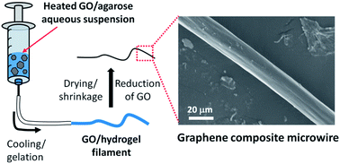
Graphene based electrical conductors are under consideration for numerous applications in energy storage, energy conversion devices, electronics and sensors. Here, we report a facile and versatile method for fabrication of graphene based composite microwires ranging from ∼20 to 250 μm in diameter via size reduction during dehydration of extruded larger diameter graphene oxide-loaded agarose hydrogel fibers. The graphene oxide is effectively reduced to graphene by chemical and thermal treatment. After dehydration and reduction, the resulting graphene composite microwires exhibit high electrical conductivities of up to 1.8 S m−1.


Graphene, a one-atom-thick 2D single layer of sp2-bonded carbon, is attracting great interest due to its outstanding properties, including its good electrical conductivity, exceptionally high surface area (theoretically 2765 m2 g−1), high charge carrier mobility (>20 m2 V−1 s−1 at room temperature), strong mechanical strength (∼1 TPa), and high thermal conductivity (∼5000 W m−1 K−1).1,2 Recently, studies on fabricating graphene-based materials with various shapes, including spheres,3 fibers,4 and films5 have been attempted for a range of applications such as energy storage/conversion materials, electronic devices, polymer composites, and bio/chemical sensors and actuators.6–8 Incorporation of 2D graphene nanosheets into micron-sized wires with smaller feature size has been suggested to have advantages in terms of electrical conductivity and surface area.9–11 Compared with conventional planar structures, electrical microwires are also attractive as component for space-limited applications such as portable consumer electronics, roll-up displays, wearable electronic textiles and implantable systems,12 as they efficiently use space and could be integrated into various shape-conformable structures. However, there has been little research on the fabrication of the graphene based composite microwires, and it is currently a challenge to control the feature size of the composite materials without sacrificing the intrinsic properties of graphene. We of course are not the first to create conductive microwires. This work was inspired, for example, by Lewis' demonstration of the direct-ink writing of silver microwires starting from both nanoparticle13 and reactive silver inks.14 Here, we report a facile and efficient method to fabricate graphene embedded microwires starting from larger diameter agarose hydrogel–graphene oxide composite which are subsequently induced to shrink.

Hydrogels, highly hydrated polymer networks, are an effective matrix component for fabrication of composites which include water soluble or dispersible materials.15 Agarose gel, a representative hydrogel, is naturally derived, abundant, environmentally friendly and biocompatible.16 Since the sol–gel transition of agarose gel is thermally reversible, various shapes or patterns can be easily realized in the gel with corresponding templates.15,17 Volume reduction of the hydrogel during drying can be utilized for miniaturization of various shapes or patterns.18

To make a graphene based microwire, we start with a hot liquefied suspension of agarose and water-soluble graphene oxide (GO). This mixture solidifies by gelation and forms a wire while being extruded through a 0.19–1 mm diameter orifice. The diameter of the extruded hydrogel wires are primarily defined by the tube inner diameter. High volume fractions of water in the agarose gel (∼98 wt%) enables the diameter of the wires to drastically decrease by 76–90% after drying. The composite microwires are subsequently treated with hydrazine to reduce the GO to graphene. The surface morphology of the composite microwires and the degree of defects in the graphene lattice after the chemical reduction step are investigated by scanning electron microscope (SEM) and Raman spectroscopy, respectively. The conductivity of the composite microwires with the reduced graphene oxide (rGO) is one order of magnitude higher than that of the graphene oxide composite film. To further improve the electrical conductivity of the composite microwires, thermal treatments at various temperatures were applied. Fig. 1(a) outlines the agarose–reduced graphene oxide (A–rGO) microwire fabrication procedure. First, agarose gel with embedded GO was prepared similarly to the TiO2 particle loaded heat-set agarose gel described previously.15 Briefly, aqueous suspensions of GO (Angstron Materials) with various contents, ranging from 0.01 to 0.5 wt%, were heated close to ∼100 °C, then agarose (Acros Organics) was added. This heated agarose–graphene oxide (A–GO) mixture was transferred to a syringe and extruded through a tygon tube (inner diameter: 0.5 mm, unless mentioned otherwise). The A–GO mixture cooled and gelled while being extruded through the tube. Within the range of tube inner diameters of our experiment (0.19–1 mm), the extruded A–GO hydrogel maintains the filamentary shape with a diameter corresponding to the tube inner diameter. After hydrogel dehydration, an A–GO microwire with a significantly reduced diameter was obtained. The microwire was placed in a closed chamber with hydrazine (35 wt% in water, Aldrich) at 90 °C for 4 h to reduce the embedded GO to rGO.


image file: c4ra01154f-f1.tif
Fig. 1 (a) A–rGO composite microwire fabrication process. Images of the extrusion process are shown in Fig. S1. (b) Phase diagram of the hydrated A–GO composite as a function of weight fraction of agarose and GO. The symbols “O” and “X” indicate where the resulting phase is gel and sol, respectively. The insets show the gelled and fluid nature of the A–GO composite at the indicated compositions. Scale bar = 1 cm. The dotted line is approximately where we estimate the sol–gel phase transition to occur.

Successful fabrication of the A–GO composite wires requires the heated A–GO aqueous suspension to cool and gel before leaving the tube. The reported minimum concentration of agarose for hydrogel formation is ∼0.15 wt%,16 however, we observe a higher agarose concentration is necessary when GO is embedded. Fig. 1(b) shows the phase diagram of the A–GO composite as a function of the agarose, GO, and water content (water is the remaining content). The agarose content up to 3 wt% is tested for ease of handling of the composite when liquefied. At 2–3 wt% of agarose concentration, the maximum content of the embedded GO for hydrogel formation is ∼0.1 wt%, and it decreases as the agarose concentration decreases. For microwire fabrication, we prepared A–GO aqueous mixtures with 2 wt% of agarose and 0.1 or 0.01 wt% of GO.

Optical micrographs of A–GO composite wires extruded from 0.1 and 0.01 wt% GO before and after drying are shown in Fig. 2(a)–(d). The diameters of both wires after extrusion are ∼0.5 mm, the same as the inner diameter of the tube. After the A–GO hydrogel wires are dried, the diameter decreases to ∼80 μm due to the volume reduction. This value is close to the theoretical diameter of 74 μm assuming no contraction along the wire axis.19 The ratio of the wet and dry diameter for the GO containing wires is almost the same as that for a GO-free agarose hydrogel filament (Fig. S3). The diameters of the A–GO microwires are easily controlled by varying the tube inner diameter. Fig. S4 shows the dependence of the diameters of the dried A–GO microwires on the tube inner diameters. For the hydrated microwires with the large diameters of 0.76 and 1 mm, the dried diameters are much larger than the calculated values under the assumption of only radial contraction. This is presumably because the contraction along the wire axis direction becomes more significant as the diameter of the hydrated microwire increases, possibly larger than 0.5 mm. Microwires with diameters as small as 18 μm were obtained by extruding through 0.19 mm inner diameter tubes. It should be noted that this diameter is not the minimum limit of the method and may be reduced by using a tube with smaller inner diameter. After chemical reduction with hydrazine, the colour of the wires becomes darker and no significant change in the diameter was observed (Fig. 2(e) and (f)). It is known that the brownish yellow GO turns dark black after reduction. There is no change in colour when a GO-free agarose microwire is treated with hydrazine (Fig. S3).


image file: c4ra01154f-f2.tif
Fig. 2 Optical micrographs of the extruded A–GO composite wires with (a) 0.1 wt% and (b) 0.01 wt% of GO. The agarose content is 2 wt% and water content is ∼98 wt% for both wires. Low magnification images of (a) and (b) are in Fig. S2. (c and d) The wires of (a) and (b) after drying, respectively. (e and f) The wires in (c) and (d) after chemical reduction, respectively.

Fig. 3(a) and (b) presents the surface morphology of the A–rGO composite microwire. The 1D ridging on the surface is probably wrinkling due to the dominant volume reduction perpendicular to the wire axis. No significant aggregation of the rGO on the surface of the dried microwire is observed. To confirm the hydrazine reduction of GO to rGO, Raman scattering was collected after each step (Fig. 3(c)). In the Raman, two characteristic peaks appear at 1350 cm−1 and 1590 cm−1, corresponding to the D band and G band, respectively. The D band, associated with the A1g breathing vibration of a graphene, is proportional to the defect density of the graphene, whereas the G band, resulting from the E2g in-plane stretching vibration, is not affected by the presence of defects.20 As such, the Raman peak intensity of the D band relative to the G band is associated with the relative defect density on the GO. The Raman intensity ratio, ID/IG, decreases from 1.11 to 0.99 after the hydrazine reduction, indicating a decrease of the number of the defects.


image file: c4ra01154f-f3.tif
Fig. 3 (a and b) SEM images of A–rGO composite microwires prepared from an A–GO hydrogel containing 0.01 wt% GO. Similar morphology is observed for microwires with 0.1 wt% GO in Fig. S5. (c) Raman spectra of the dried A–GO composites before and after chemical reduction and thermal treatment (400 °C for 30 min). GO content is 0.1 wt% before drying.

Due to the superior electrical conductivity of a graphene, the A–rGO microwire may have potential to be used as a conductor in microelectronics. We measured the electrical conductivity of the A–rGO microwire (1D), and compared it to the A–rGO film (2D) to investigate the benefit of the one dimensional shape. Fig. 4(a) presents the current–voltage (IV) characteristics of the A–rGO microwire and film. The values on y axes in the IV curves are a current times distance between two probe electrodes divided by a cross-sectional area of the wire or the film of the A–rGO composites. The electrical conductivity (unit: mS m−1) of each sample can be directly derived from the slopes of the plots. All samples exhibit the linear IV curves over the −0.1 to +0.1 V voltage range, indicating ohmic behaviour. The conductivity of the A–rGO microwire is 1 mS m−1, one order of magnitude higher than that of the A–rGO film (0.08 mS m−1). We speculate that the higher conductivity of the A–rGO wire is because the graphene sheets are aligned preferentially in the wires by the extrusion process. The A–GO microwire or the hydrazine-treated agarose microwire without rGO did not show a detectable electrical conductivity within our measurement limit (∼10−15 Amps). However, the conductivity value of the A–rGO composite wire is still low compared to the values of other graphene-based composite materials,6,21,22 primarily because of the high volumetric ratio of the insulating agarose.


image file: c4ra01154f-f4.tif
Fig. 4 (a) IV curves of A–rGO microwire and film after chemical reduction. (b) Effect of annealing temperature on the conductivity of A–rGO composite microwires. (c) IV curves of A–rGO microwire and film after annealing at 400 °C for 30 min. IV plots of A–rGO microwires after annealing at 200 and 300 °C are shown in Fig. S8. All samples were prepared from the A–GO hydrogel with 0.1 wt% GO content.

The electrical conductivity of the microwire could be improved by decreasing the amount of the insulating agarose polymer. Since the reduced form of GO is relatively thermally stable (mass loss is ∼10% at 400 °C in air),23,24 the polysaccharide agarose polymer can be selectively removed in the A–rGO composite by thermal treatment. The decomposition temperature of the neat agarose polymer and the A–rGO composite under air was determined by thermogravimetric analysis (TGA). Fig. S6 shows that the major mass loss of agarose occurs at about 300 °C, where it loses ∼80% of its initial weight. The electrical conductivity of the A–rGO microwires was measured after thermal treatments at temperatures ranging from 200–600 °C, for 30 min (Fig. 4(b)). The electrical conductivity of the A–rGO microwire increases with increasing thermal treatment temperature up to 400 °C, and the wire shape is preserved. However, at 500 °C or higher, the microwire collapsed, perhaps because of the severe decomposition of the agarose matrix. The A–rGO microwire after the thermal treatment at 400 °C for 30 min (Fig. 4(c)) exhibits an electrical conductivity of 1.8 S m−1, three orders of magnitude greater than the value before the thermal treatment. This value is comparable to or one order of magnitude higher than previously reported conductive graphene based composites.6,21,22 The highly increased electrical conductivity results from the further reduction of the graphene oxide and the densification of the A–rGO wire during the agarose decomposition. The Raman spectra are compared before and after the thermal treatment at 400 °C in Fig. 3(c). The Raman intensity ratio, ID/IG, of the A–rGO decreases from 0.99 to 0.93 after the thermal treatment at 400 °C, indicating the decrease of the defect sites in the embedded graphene. The SEM images of the A–rGO microwire after the thermal treatment at 400 °C (Fig. S7) show that the microwire retains its initial shape and the diameter decreases from 80 to 24 μm. No significant deformation or morphology change is observed. Overall, the thermal treatment significantly improved the electrical conductivity of the A–rGO microwire, and decreases the wire diameter.

In conclusion, we presented a simple and general method to fabricate a conducting microwire, starting from aqueous agarose–GO mixtures with the appropriate compositions for gel formation. The heated (fluid) mixture can be extruded from an orifice, at which point it gels, forming a hydrogel filament. The diameter of this filament is then reduced by up to 90% by drying. After drying, the microwire has a fibril surface morphology due to the preferential volume shrinkage perpendicular to the wire axis. When the A–GO is treated with the hydrazine the GO is reduced to graphene form, and the microwire becomes conductive. Thermal treatment of the microwire in air further reduces the embedded graphene and densifies the A–rGO microwire, resulting in an electrical conductivity comparable to or even higher than the values of other recently reported graphene based materials. An important attribute of this approach is the ease by which materials with small feature sizes can be made. For example, the 0.5 mm diameter A–GO composite wire decreases to only 24 μm after the drying and the thermal treatment steps. The approach reported here should be generally applicable for fabrication of composite microwires containing other hydrophilic fillers. Along with simple one dimensional shapes, more complicated shapes can be formed via the controlled evaporation of water from complex starting shapes, something which is now underway.

Acknowledgements

The work was supported by the Defense Threat Reduction Agency under award number HDTRA 1-12-1-0035 (microwire fabrication) and the U.S. Department of Energy, Office of Basic Energy Sciences, Division of Materials Sciences and Engineering under Award # DE-FG02-07ER46471 (electrical property studies). The authors thank Tae Wook Lim and Dr Chanwoo Park for TGA measurements.

Notes and references

  1. A. K. Geim and K. S. Novoselov, Nat. Mater., 2007, 6, 183–191 CrossRef CAS PubMed; S. Park and R. S. Ruoff, Nat. Nanotechnol., 2009, 4, 217–224 CrossRef PubMed.
  2. E. V. Castro, H. Ochoa, M. I. Katsnelson, R. V. Gorbachev, D. C. Elias, K. S. Novoselov, A. K. Geim and F. Guinea, Phys. Rev. Lett., 2010, 105, 266601 CrossRef CAS; A. A. Balandin, S. Ghosh, W. Bao, I. Calizo, D. Teweldebrhan, F. Miao and C. N. Lau, Nano Lett., 2008, 8, 902–907 CrossRef PubMed.
  3. S.-M. Yoon, W. M. Choi, H. Baik, H.-J. Shin, I. Song, M.-S. Kwon, J. J. Bae, H. Kim, Y. H. Lee and J.-Y. Choi, ACS Nano, 2012, 6, 6803–6811 CrossRef CAS PubMed.
  4. Q. Bao, H. Zhang, J.-x. Yang, S. Wang, D. Y. Tang, R. Jose, S. Ramakrishna, C. T. Lim and K. P. Loh, Adv. Funct. Mater., 2010, 20, 782–791 CrossRef CAS.
  5. G. Eda and M. Chhowalla, Nano Lett., 2009, 9, 814–818 CrossRef CAS PubMed.
  6. Y. Xu, Z. Lin, X. Huang, Y. Wang, Y. Huang and X. Duan, Adv. Mater., 2013, 25, 5779–5784 CrossRef CAS PubMed.
  7. S. H. Che Man, S. C. Thickett, M. R. Whittaker and P. B. Zetterlund, J. Polym. Sci., Part A: Polym. Chem., 2013, 51, 47–58 CrossRef CAS; T. K. Das and S. Prusty, Polym.-Plast. Technol. Eng., 2013, 52, 319–331 CrossRef; J. R. Potts, D. R. Dreyer, C. W. Bielawski and R. S. Ruoff, Polymer, 2011, 52, 5–25 CrossRef PubMed.
  8. O. Parlak, A. P. F. Turner and A. Tiwari, Adv. Mater., 2014, 26, 482–486 CrossRef CAS PubMed; J. Tyagi and R. Kakkar, Adv. Mater. Lett., 2013, 4, 721–736 Search PubMed; M. N. Muralidharan and S. Ansari, Adv. Mater. Lett., 2013, 4, 927–932 Search PubMed; O. Parlak, A. Tiwari, A. P. F. Turner and A. Tiwari, Biosens. Bioelectron., 2013, 49, 53–62 CrossRef PubMed; G. Alarcon-Angeles, G. A. Álvarez-Romero and A. Merkoçi, in Advanced Carbon Materials and Technology, ed. A. Tiwari and S. K. Shukla, Wiley-Scrivener, USA, 2014, pp. 87–128 Search PubMed.
  9. Z. Yang, H. Sun, T. Chen, L. Qiu, Y. Luo and H. Peng, Angew. Chem., Int. Ed., 2013, 52, 7545–7548 CrossRef CAS PubMed.
  10. H.-P. Cong, X.-C. Ren, P. Wang and S.-H. Yu, Sci. Rep., 2012, 2, 613 Search PubMed.
  11. Z. Xu and C. Gao, Nat. Commun., 2011, 2, 571 CrossRef PubMed.
  12. J. Bae, M. K. Song, Y. J. Park, J. M. Kim, M. Liu and Z. L. Wang, Angew. Chem., Int. Ed., 2011, 50, 1683–1687 CrossRef CAS PubMed; Y. H. Kwon, S.-W. Woo, H.-R. Jung, H. K. Yu, K. Kim, B. H. Oh, S. Ahn, S.-Y. Lee, S.-W. Song, J. Cho, H.-C. Shin and J. Y. Kim, Adv. Mater., 2012, 24, 5192–5197 CrossRef PubMed; K. Jost, C. R. Perez, J. K. McDonough, V. Presser, M. Heon, G. Diona and Y. Gogotsi, Energy Environ. Sci., 2011, 4, 5060–5067 Search PubMed.
  13. J. J. Adams, E. B. Duoss, T. F. Malkowski, M. J. Motala, B. Y. Ahn, R. G. Nuzzo, J. T. Bernhard and J. A. Lewis, Adv. Mater., 2011, 23, 1335–1340 CrossRef CAS PubMed.
  14. S. B. Walker and J. A. Lewis, J. Am. Chem. Soc., 2012, 134, 1419 CrossRef CAS PubMed.
  15. H.-J. Koo and O. D. Velev, J. Mater. Chem. A, 2013, 1, 11106–11110 CAS.
  16. P. Serwer, Electrophoresis, 1983, 4, 375–382 CrossRef CAS.
  17. H.-J. Koo and O. D. Velev, Sci. Rep., 2013, 3, 2357 Search PubMed.
  18. A. Lal[thin space (1/6-em)]Das, R. Mukherjee, V. Katiyer, M. Kulkarni, A. Ghatak and A. Sharma, Adv. Mater., 2007, 19, 1943–1946 CrossRef.
  19. For the A–GO wire with 2 wt% of agarose and 0.1 wt% of GO, V′ = π(d′/2)2 × l′ = V × 0.021 = π(d/2)2 × l where V, d and l are the volume, diameter and length of the A–GO wire, respectively. V′, d′ and l′ are the values of the dried A–GO wire. Under the assumption of no contraction along the wire axis, l′ = l, therefore d′ = (0.021)1/2 × d. Since the d is same as the tube inner diameter (0.02 inch = 508 μm), d′ = (0.021)1/2 × 508 μm = ∼74 μm.
  20. D. C. Kim, D.-Y. Jeon, H.-J. Chung, Y. Woo, J. K. Shin and S. Seo, Nanotechnology, 2009, 20, 375703 CrossRef PubMed.
  21. S. Stankovich, D. A. Dikin, G. H. B. Dommett, K. M. Kohlhaas, E. J. Zimney, E. A. Stach, R. D. Piner, S. T. Nguyen and R. S. Ruoff, Nature, 2006, 442, 282–286 CrossRef CAS PubMed.
  22. M. Yoonessi and J. R. Gaier, ACS Nano, 2010, 4, 7211–7220 CrossRef CAS PubMed.
  23. S. Stankovich, D. A. Dikin, R. D. Piner, K. A. Kohlhaas, A. Kleinhammes, Y. Jia, Y. Wu, S. T. Nguyen and R. S. Ruoff, Carbon, 2007, 45, 1558–1565 CrossRef CAS PubMed.
  24. E.-Y. Choi, T. H. Han, J. Hong, J. E. Kim, S. H. Lee, H. W. Kim and S. O. Kim, J. Mater. Chem., 2010, 20, 1907–1912 RSC.

Footnotes

Electronic supplementary information (ESI) available: More optical and SEM images of the composite microwires, plot of the resulting diameters of the composite microwires as a function of the tube inner diameters, TGA data for agarose and A–rGO composite and IV curves of A–rGO microwires after thermal treatment at different temperatures. See DOI: 10.1039/c4ra01154f
Authors equally contributed.

This journal is © The Royal Society of Chemistry 2014
Click here to see how this site uses Cookies. View our privacy policy here.