Design, synthesis and anticancer activity of benzofuran derivatives targeting VEGFR-2 tyrosine kinase

Omaima M. Abdelhafez*a, Kamelia M. Aminb, Hamed I. Alic, Mohamed M. Abdallad and Eman Y. Ahmeda
aChemistry of Natural Products Dept., National Research Center, Dokki, Egypt. E-mail: dromaima45@gmail.com; Fax: +20 202 33370931; Tel: +20 202 37608284
bPharmaceutical Chemistry Dept., Faculty of Pharmacy, Cairo University, Egypt
cPharmaceutical Chemistry Dept., Faculty of Pharmacy, Helwan University, Egypt
dResearch Unit, Mapco Pharmaceutical Industries, Balteim, Egypt

Received 2nd February 2014 , Accepted 4th February 2014

First published on 19th February 2014


Abstract

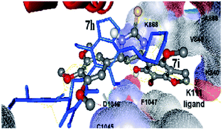
Two series of chalcone and thiopyrimidine benzofuran derivatives were designed, synthesized and evaluated in vitro for their vascular endothelial growth factor receptor (VEGFR-2) inhibitory activity, their cytotoxicity on seventeen human cancer cell lines and their in vivo antiprostate cancer activity. The highest anti-VEGFR-2 activity was demonstrated by 1-(6-hydroxy-4-methoxybenzofuran-5-yl)-3-(4-nitrophenyl)prop-2-en-1-one (6d) exhibiting an IC50 value (1.00 × 10⁻³ μM) higher than the reference drug Sorafenib (IC50 = 2.00 × 10⁻³ μM). On the other hand, most of the synthesized compounds showed potent cytotoxicity against most of the tested cell lines and were more potent than the reference drugs, in particular, bromovisnagin (4) exhibited the best activity on the majority of the cell lines with IC50 values ranging from 3.67 × 10⁻¹³ to 7.65 × 10⁻⁷ μM. Moreover, the synthesized compounds showed significant in vivo antiprostate cancer activity. The docking experiments were performed using the GOLD program on (VEGFR-2) kinase which introduced new information about the enzyme–inhibitor interaction and the potential therapeutic application of the benzofuran scaffold.


Introduction

Cancer is a very complex disease which affects different organs and systems of the body. Its burden is increasing across the world dramatically, and it is considered as the first leading cause of deaths in economically developed countries and the second leading cause of deaths in developing countries.1,2

Most signal transduction pathways are mediated by protein kinases, and aberrant kinase signaling leads to proliferation of cancer cells as well as angiogenesis and growth of solid tumors such as prostate, colon, breast, and gastric cancers.3–5 The vascular endothelial growth factor (VEGF) family of tyrosine kinase receptors consists of three protein receptors (VEGFR-1, VEGFR-2, and VEGFR-3).6–8 The (VEGFR-2), (also known as Flk-1 for fetal liver kinase-1 or KDR for kinase insert domain-containing receptor)9–11 is a receptor for (VEGF) and a key angiogenic factor and is secreted by malignant tumors, it induces the proliferation and the migration of vascular endothelial cells.12–15 There is much evidence that direct inhibition of the kinase activity of (VEGFR-2) will result in a reduction in angiogenesis, the suppression of this signaling pathway has become an inhibiting method of tumor growth. Therefore, inhibition of (VEGFR-2) is an attractive strategy in the treatment of cancers.16

Indeed, some small molecule inhibitors of (VEGFR-2) such as sunitinib17 and sorafenib,18 have been approved by the Food and Drug Administration for treating tumors, also a number of compounds inhibiting the biological activity of (VEGFR-2) has been identified as promising anticancer agents, e.g., chromone bioisosteres,19 benzofuran derivatives,20 mannich base derivatives,21 chalcones22 and pyrimidines.23

The aforementioned studies encouraged us to design a large array of benzofuran derivatives with chalcones and thiopyrimidines as bioactive moieties using visnagin (chromone derivative) as a starting material, all known for their anti-VEGFR-2 (ref. 19–23) and anticancer activity,24–27 in order to evaluate their biological effect in vitro and in vivo. Finally the docking experiments were carried out on (VEGFR-2) kinase crystallographic structures to analyze the structural requirements for anti-VEGFR-2 activity and selectivity.

Chemistry

The chalcones (6a–i) and thiopyrimidine derivatives (7a–i) were efficiently synthesized according to the protocols outlined in the Scheme 1. All the starting materials were prepared according to the reported methods, while the general reaction conditions, procedures and compounds characterization of the newly synthesized derivatives were described in the Experimental section.
image file: c4ra00943f-s1.tif
Scheme 1

Refluxing visnagin (1) in aqueous potassium hydroxide afforded the visnaginone derivative (2),28 which then reacted with piperidine in ethanol in the presence of formaline under reflux to give the mannich base (3),29 Moreover, bromination of visnagin (1) in glacial acetic acid at room temperature gave the corresponding bromovisnagin derivative (4),30 which upon cleavage with aqueous potassium hydroxide yielded the bromovisnaginone derivative (5).30

The chalcones (6a–i), were prepared by the reaction of visnaginone (2), visnaginone methyl piperidine (3) and bromovisnaginone (5) in aqueous sodium hydroxide and ethanol at room temperature with different aromatic aldehydes namely, 4-bromobenzaldehyde, 4-nitrobenzaldehyde and 3,4,5-trimethoxybenzaldehyde.

Furthermore, refluxing the obtained chalcones (6a–i) with thiourea in ethanol and in the presence of aqueous potassium hydroxide afforded the thiopyrimidine derivatives (7a–i).31

Results and discussion

1 Biology

In vitro anti-VEGFR-2 screening. Studying the anti-VEGFR-2 activity of the tested compounds (Table 1) showed that 1-(6-hydroxy-4-methoxybenzofuran-5-yl)-3-(4-nitrophenyl)prop-2-en-1-one (6d) was the most active compound (IC50 = 1.00 × 10⁻³ μM) and even showed a higher activity than the reference drug (IC50 = 2.00 × 10⁻³ μM).
Table 1 Anti-VEGFR-2 activity of the synthesized compounds
Compound Enzymatic inhibition (IC50/μM)
VEGFR-2
3 1.20 × 10⁻²
4 3.40 × 10⁻²
5 5.60 × 10⁻²
6a 2.30 × 10⁻²
6b 3.50 × 10⁻²
6c 3.40 × 10⁻²
6d 1.00 × 10⁻³
6e 7.30 × 10⁻²
6f 8.70 × 10⁻²
6g 5.40 × 10⁻²
6h 3.30 × 10⁻²
6i 3.60 × 10⁻²
7a 7.60 × 10⁻²
7b 5.40 × 10⁻²
7c 4.30 × 10⁻²
7d 3.20 × 10⁻²
7e 1.20 × 10⁻²
7f 3.20 × 10⁻²
7g 2.10 × 10⁻²
7h 2.20 × 10⁻²
7i 3.40 × 10⁻²
Sorafenib 2.00 × 10⁻³


In vitro cytotoxicity screening. The cytotoxicity of the tested compounds (3, 4, 5, 6a–i, 7a–i) was determined on seventeen different human cancer cell lines (Table 2a–d).
Table 2 (a–d) Cytotoxicity of the synthesized compounds
a
Compound IC50
KB SKOV-3 SF-268 NCI H 460 RKOP27
3 4.30 × 10⁻⁴ 5.50 × 10⁻⁵ 5.50 × 10⁻³ 3.30 × 10⁻³ 4.80 × 10⁻³
4 3.45 × 10⁻⁷ 2.34 × 10⁻⁷ 6.52 × 10⁻¹⁰ 8.88 × 10⁻¹³ 7.61 × 10⁻¹³
5 5.30 × 10⁻⁴ 7.00 × 10⁻⁵ 9.90 × 10⁻⁵ 8.60 × 10⁻³ 6.20 × 10⁻³
6a 5.21 × 10⁻⁵ 2.41 × 10⁻³ 4.90 × 10⁻³ 8.00 × 10⁻³ 8.00 × 10⁻⁴
6b 1.70 × 10⁻⁵ 3.30 × 10⁻⁵ 4.21 × 10⁻⁴ 5.68 × 10⁻³ 2.15 × 10⁻⁴
6c 8.60 × 10⁻⁵ 6.14 × 10⁻⁵ 7.83 × 10⁻⁵ 5.90 × 10⁻³ 7.50 × 10⁻³
6d 9.90 × 10⁻⁴ 5.67 × 10⁻⁴ 5.43 × 10⁻⁵ 9.87 × 10⁻³ 7.65 × 10⁻⁵
6e 7.53 × 10⁻⁵ 6.79 × 10⁻³ 8.97 × 10⁻⁴ 8.65 × 10⁻³ 9.86 × 10⁻³
6f 6.50 × 10⁻⁵ 8.00 × 10⁻³ 5.40 × 10⁻³ 7.60 × 10⁻³ 9.80 × 10⁻³
6g 5.40 × 10⁻⁴ 7.77 × 10⁻⁵ 6.60 × 10⁻³ 5.50 × 10⁻³ 3.50 × 10⁻³
6h 5.20 × 10⁻⁵ 8.63 × 10⁻³ 8.30 × 10⁻³ 8.93 × 10⁻³ 1.40 × 10⁻³
6i 4.30 × 10⁻⁵ 7.50 × 10⁻³ 8.71 × 10⁻⁴ 8.90 × 10⁻⁵ 6.30 × 10⁻³
7a 6.88 × 10⁻³ 8.76 × 10⁻⁵ 5.43 × 10⁻³ 6.78 × 10⁻⁵ 7.89 × 10⁻³
7b 8.56 × 10⁻⁵ 7.25 × 10⁻⁴ 7.50 × 10⁻⁴ 2.31 × 10⁻⁵ 4.40 × 10⁻³
7c 7.25 × 10⁻⁴ 9.00 × 10⁻⁵ 2.37 × 10⁻³ 2.51 × 10⁻⁴ 5.10 × 10⁻⁵
7d 7.80 × 10⁻⁵ 6.60 × 10⁻³ 8.90 × 10⁻³ 7.70 × 10⁻³ 7.80 × 10⁻³
7e 4.73 × 10⁻⁵ 5.50 × 10⁻⁵ 9.90 × 10⁻³ 8.70 × 10⁻⁴ 9.40 × 10⁻⁵
7f 5.27 × 10⁻⁵ 9.87 × 10⁻³ 7.90 × 10⁻³ 6.54 × 10⁻⁴ 9.87 × 10⁻⁴
7g 6.14 × 10⁻⁴ 4.00 × 10⁻⁵ 6.60 × 10⁻⁴ 7.70 × 10⁻⁴ 6.60 × 10⁻⁶
7h 8.00 × 10⁻⁴ 5.60 × 10⁻³ 6.60 × 10⁻³ 6.60 × 10⁻³ 3.80 × 10⁻⁵
7i 5.72 × 10⁻⁴ 4.00 × 10⁻⁴ 4.00 × 10⁻⁵ 5.50 × 10⁻⁵ 6.60 × 10⁻⁵
Fluorouracil 4.46 × 10⁻³        
Doxorubicin   4.16 × 10⁻³      
Cytarabine     7.68 × 10⁻³    
Gemcitabine       2.13 × 10⁻³  
Capecitabine         4.33 × 10⁻³

b
Compound IC50 μM
Leukemia Melanoma
HL60 U937 K561 G361 SK-MEL-28
3 2.48 × 10⁻⁴ 9.60 × 10⁻⁵ 8.63 × 10⁻³ 7.50 × 10⁻³ 3.00 × 10⁻³
4 5.32 × 10⁻⁷ 4.56 × 10⁻⁸ 7.65 × 10⁻⁷
5 5.30 × 10⁻³ 6.30 × 10⁻³ 7.70 × 10⁻³ 8.80 × 10⁻³ 9.90 × 10⁻⁵
6a 5.30 × 10⁻³ 6.00 × 10⁻³ 4.00 × 10⁻³ 6.43 × 10⁻³ 3.00 × 10⁻⁵
6b 4.50 × 10⁻⁵ 3.56 × 10⁻³ 1.38 × 10⁻⁴ 4.80 × 10⁻⁵ 8.06 × 10⁻³
6c 4.86 × 10⁻³ 5.90 × 10⁻⁴ 8.00 × 10⁻⁴ 6.64 × 10⁻³ 2.60 × 10⁻⁵
6d 3.52 × 10⁻⁴ 8.80 × 10⁻⁵ 3.30 × 10⁻³ 9.90 × 10⁻³ 1.00 × 10⁻⁴
6e 6.30 × 10⁻³ 5.80 × 10⁻³ 3.88 × 10⁻³ 7.70 × 10⁻³ 9.63 × 10⁻⁵
6f 4.50 × 10⁻⁵ 8.46 × 10⁻³ 3.50 × 10⁻³ 6.04 × 10⁻³ 8.00 × 10⁻⁴
6g 7.00 × 10⁻⁵ 7.00 × 10⁻³ 7.00 × 10⁻⁴ 7.00 × 10⁻⁵ 9.70 × 10⁻³
6h 6.40 × 10⁻³ 9.00 × 10⁻³ 5.00 × 10⁻³ 8.50 × 10⁻³ 9.89 × 10⁻⁵
6i 7.60 × 10⁻³ 5.50 × 10⁻³ 4.30 × 10⁻⁴ 5.50 × 10⁻⁴ 6.90 × 10⁻⁴
7a 6.60 × 10⁻⁴ 5.50 × 10⁻⁵ 3.30 × 10⁻⁵ 8.00 × 10⁻³ 8.40 × 10⁻³
7b 8.46 × 10⁻³ 9.10 × 10⁻³ 8.60 × 10⁻³ 6.00 × 10⁻³ 9.70 × 10⁻⁵
7c 4.50 × 10⁻⁵ 5.00 × 10⁻³ 6.60 × 10⁻² 6.60 × 10⁻³ 6.60 × 10⁻³
7d 7.50 × 10⁻⁵ 7.70 × 10⁻³ 4.30 × 10⁻³ 6.60 × 10⁻³ 3.25 × 10⁻³
7e 3.64 × 10⁻⁴ 6.00 × 10⁻⁵ 4.00 × 10⁻³ 7.70 × 10⁻³ 7.50 × 10⁻³
7f 7.00 × 10⁻⁵ 4.40 × 10⁻³ 7.70 × 10⁻⁴ 3.40 × 10⁻⁵ 4.20 × 10⁻³
7g 6.59 × 10⁻⁴ 8.30 × 10⁻⁵ 3.80 × 10⁻⁵ 3.80 × 10⁻³ 8.30 × 10⁻³
7h 5.50 × 10⁻⁴ 6.00 × 10⁻⁵ 7.70 × 10⁻³ 8.80 × 10⁻³ 9.90 × 10⁻³
7i 4.50 × 10⁻⁵ 6.60 × 10⁻³ 7.00 × 10⁻³ 8.80 × 10⁻³ 9.90 × 10⁻³
Doxorubicin 1.13 × 10⁻³ 4.45 × 10⁻³ 6.66 × 10⁻³    
Aldesleukin       6.66 × 10⁻³ 3.45 × 10⁻³

c
Compound IC50 μM
Neuroblastoma
GOTO NB-1
3 7.60 × 10⁻⁴ 5.00 × 10⁻⁵
4 5.69 × 10⁻⁹
5 9.90 × 10⁻⁴ 6.59 × 10⁻⁵
6a 4.90 × 10⁻⁵ 8.86 × 10⁻⁶
6b 7.30 × 10⁻⁵ 5.77 × 10⁻⁵
6c 2.85 × 10⁻⁵ 9.70 × 10⁻⁴
6d 7.52 × 10⁻⁴ 7.50 × 10⁻⁴
6e 4.70 × 10⁻⁵ 8.63 × 10⁻⁵
6f 3.40 × 10⁻⁵ 2.80 × 10⁻³
6g 9.74 × 10⁻⁴ 7.56 × 10⁻⁵
6h 2.80 × 10⁻⁵ 5.77 × 10⁻³
6i 8.03 × 10⁻⁵ 5.77 × 10⁻³
7a 5.80 × 10⁻⁴ 7.25 × 10⁻³
7b 3.00 × 10⁻⁵ 6.98 × 10⁻³
7c 8.00 × 10⁻⁴ 9.78 × 10⁻⁵
7d 8.60 × 10⁻⁵ 6.40 × 10⁻⁶
7e 6.58 × 10⁻⁵ 6.20 × 10⁻⁵
7f 7.44 × 10⁻⁵ 8.56 × 10⁻⁵
7g 9.00 × 10⁻⁴ 8.67 × 10⁻⁵
7h 4.00 × 10⁻⁴ 7.46 × 10⁻³
7i 8.60 × 10⁻⁴ 3.56 × 10⁻⁴
Doxorubicin 4.73 × 10⁻³ 5.15 × 10⁻³

d
Compound IC50 μM
HeLa (cervical) MCF-7 (breast) HT1080 (fibrosarcoma) HepG2 (liver) PC-3
3 9.74 × 10⁻⁴ 8.00 × 10⁻⁶ 8.00 × 10⁻³ 1.46 × 10⁻³ 7.25 × 10⁻⁵
4 4.45 × 10⁻⁹ 3.67 × 10⁻⁷ 6.45 × 10⁻⁷ 4.46 × 10⁻⁷ 6.34 × 10⁻⁴
5 5.87 × 10⁻⁴ 8.38 × 10⁻⁵ 6.40 × 10⁻⁵ 9.86 × 10⁻³ 6.45 × 10⁻⁴
6a 6.34 × 10⁻⁵ 6.30 × 10⁻⁶ 6.60 × 10⁻³ 7.95 × 10⁻³ 9.00 × 10⁻⁵
6b 8.50 × 10⁻⁵ 8.00 × 10⁻⁵ 9.73 × 10⁻⁴ 9.70 × 10⁻⁴ 7.60 × 10⁻⁵
6c 5.86 × 10⁻⁵ 9.80 × 10⁻⁵ 6.40 × 10⁻⁵ 9.00 × 10⁻³ 7.00 × 10⁻⁵
6d 9.00 × 10⁻⁴ 7.00 × 10⁻⁴ 9.79 × 10⁻⁵ 9.70 × 10⁻³ 5.36 × 10⁻⁴
6e 3.26 × 10⁻⁵ 7.90 × 10⁻⁵ 9.79 × 10⁻⁴ 7.90 × 10⁻⁵ 5.63 × 10⁻⁵
6f 8.70 × 10⁻⁶ 7.40 × 10⁻³ 9.80 × 10⁻³ 2.35 × 10⁻³ 8.00 × 10⁻⁵
6g 7.00 × 10⁻⁴ 5.70 × 10⁻⁵ 7.08 × 10⁻³ 7.90 × 10⁻⁴ 7.00 × 10⁻⁴
6h 8.70 × 10⁻⁶ 8.56 × 10⁻³ 6.30 × 10⁻³ 8.70 × 10⁻⁴ 7.00 × 10⁻⁵
6i 7.90 × 10⁻⁵ 5.88 × 10⁻³ 5.00 × 10⁻⁴ 4.50 × 10⁻⁵ 4.23 × 10⁻⁵
7a 8.60 × 10⁻⁴ 5.70 × 10⁻³ 9.70 × 10⁻⁴ 7.50 × 10⁻⁴ 7.50 × 10⁻⁴
7b 6.80 × 10⁻⁵ 8.00 × 10⁻⁴ 8.36 × 10⁻⁴ 2.41 × 10⁻⁵ 3.50 × 10⁻⁵
7c 7.09 × 10⁻⁴ 8.00 × 10⁻⁵ 3.00 × 10⁻³ 7.99 × 10⁻³ 8.00 × 10⁻⁵
7d 8.08 × 10⁻⁵ 8.60 × 10⁻⁶ 7.90 × 10⁻³ 5.70 × 10⁻³ 2.50 × 10⁻⁵
7e 8.00 × 10⁻⁵ 9.70 × 10⁻⁵ 4.70 × 10⁻³ 6.30 × 10⁻³ 4.20 × 10⁻⁵
7f 6.56 × 10⁻⁵ 7.60 × 10⁻⁵ 9.94 × 10⁻⁵ 8.56 × 10⁻³ 7.60 × 10⁻⁵
7g 4.70 × 10⁻⁴ 5.00 × 10⁻⁵ 7.43 × 10⁻⁴ 4.20 × 10⁻⁴ 5.70 × 10⁻⁴
7h 7.00 × 10⁻⁵ 4.70 × 10⁻³ 8.65 × 10⁻³ 6.00 × 10⁻³ 5.32 × 10⁻⁴
7i 9.85 × 10⁻⁴ 5.20 × 10⁻⁴ 3.35 × 10⁻⁵ 7.50 × 10⁻⁵ 6.79 × 10⁻⁴
Paclitaxel 1.18 × 10⁻⁸        
Epirubicin   2.22 × 10⁻⁹      
Imatinib     13.24 × 10⁻⁵    
Gemcitabine       3.44 × 10⁻³  
Bicalutamide         8.22 × 10⁻⁴


Screening the cytotoxicity of the tested compounds on cervical carcinoma (KB) and On ovarian carcinoma (SKOV-3) cell lines (Table 2a), showed that compound (6b) has a very remarkable activity (IC50 = 1.70 × 10⁻⁵ μM) and (IC50 = 3.30 × 10⁻⁵ μM) respectively which decreased upon cyclization to the corresponding thiopyrimidine derivative (7b) (IC50 = 8.56 × 10⁻⁵ μM) and (IC50 = 7.25 × 10⁻⁴ μM) respectively.

On the CNS cancer (SF-268) cell line, compound (7i) was significantly potent (IC50 = 4.00 × 10⁻⁵ μM) and was more potent than its chalcone derivative (6i) (IC50 = 8.71 × 10⁻⁴ μM) (Table 2a).

Regarding the non-small lung cancer (NCI H460) cell line (Table 2a), it was found that compound (7b) (IC50 = 2.31 × 10⁻⁵ μM) demonstrated a potent cytotoxicity among the other synthesized benzofuran derivatives and was more potent than its chalcone derivative (6b) which displayed a lower IC50 value (5.68 × 10⁻³ μM) even lower than the reference drug.

Estimation of the cytotoxicity on colonoadenocarcinoma (RKOP27) cell line revealed that compound (7g) showed a very pronounced potency (IC50 = 6.60 × 10⁻⁶ μM) compared to the reference drug and the other benzofuran derivatives (Table 2a).

The study of the cytotoxicity on leukemia (HL60) cell line indicated that, compounds (6b, 6f, 7c and 7i) displayed significant and the same IC50 value (IC50 = 4.50 × 10⁻⁵ μM) (Table 2b).

On leukemia (U937) cell line, compound (7a) (IC50 = 5.50 × 10⁻⁵ μM) was the most potent, whereas, compound (7b) showed the least bioactivity (IC50 = 9.00 × 10⁻³ μM) (Table 2b).

As for the leukemia (K562) cell line, compounds (7a) and (7g) (IC50 = 3.30 × 10⁻⁵ μM) and (IC50 = 3.80 × 10⁻⁵ μM) respectively were the most potent among the other synthesized benzofuran derivatives (Table 2b).

Evaluation of the cytotoxicity on melanoma (G361) cell line (Table 2b) showed that compound (7f) exhibited significantly higher IC50 (3.40 × 10⁻⁵ μM) than its chalcone derivative (6f) (IC50 = 6.04 × 10⁻³ μM).

On melanoma (SK-MEL-28) cell line, compound (6c) demonstrated a remarkable cytotoxicity (IC50 = 2.60 × 10⁻⁵ μM) which decreased dramatically upon cyclization to compound (7c) (IC50 = 6.60 × 10⁻³ μM) (Table 2b).

Screening the cytotoxicity results on neuroblastoma (GOTO) cell line, compound (6c) was the most potent compound with (IC50 = 2.85 × 10⁻⁵ μM), on the other hand, on neuroblastoma (NB-1) cell line, compound (7d) showed a very promising activity (IC50 = 6.40 × 10⁻⁶ μM) (Table 2c).

All the tested compounds on cervical carcinoma (HeLa) cell line (Table 2d) showed a very pronounced activity with IC50 ranging from (6.78 10−6 to 9.74 × 10⁻⁵ μM).

Among the tested compounds on breast carcinoma (MCF-7) cell line (Table 2d), compound (6a) showed a remarkably significant activity (IC50 = 6.30 × 10⁻⁶ μM) which decreased upon cyclization to the corresponding thiopyrimidine derivative (7a) (IC50 = 5.70 × 10⁻³ μM).

Compound (7i) displayed a significantly high IC50 value (3.35 × 10⁻⁵ μM) on fibrosarcoma (HT1080) cell line which was higher than its chalcone derivative (6i) (IC50 = 5.00 × 10⁻⁴ μM) (Table 2d).

Comparing the results of the liver carcinoma (HepG2) cell line, compound (7b) (IC50 = 2.41 × 10⁻⁵ μM) was significantly active compared to the other benzofuran derivatives (Table 2d).

All the synthesized compounds when tested against the prostate cancer cell line (PC-3) (Table 2d) showed pronounced and higher activity (IC50 = 2.50 × 10⁻⁵ to 7.50 × 10⁻⁴ μM) compared to the reference drug Bicalutamide (IC50 = 8.22 × 10⁻⁴ μM).

The previous screening of the bioactivity of the tested compounds on the presented panel of cell lines (Table 2a–d) indicated that, the majority of the synthesized compounds were more significantly potent than the comparative reference drugs used. Bromovisnagin (4) was the most potent compound against (KB, SKOV-3, SF-268, NCI H460, RKOP27, HL60, G361, SK-MEL-28, NB-1, HeLa, MCF-7, HT1080 and HePG2) cell lines compared to the reference drugs and to the synthesized benzofuran derivatives.

In vivo antiprostate cancer activity. The results of the in vivo antiprostate cancer activity (Table 3) proved that the most significantly high ED50 value was observed for compound (7b) (ED50 = 2.64 μM) which was higher than its chalcone derivative (6b) (ED50 = 8.80 μM).
Table 3 In vivo antiprostate cancer activity of the synthesized compoundsa
Compound ED50 μM
a Each ED50 value is the mean (three significant digits) ± SEM from five experiments. Level of statistical significance: P < 0.01 with respect to ED50 value of Flutamide as determined by ANOVA/Dunnett's.
3 3.40 ± 0.006
4 21.49 ± 0.005
5 4.51 ± 0.004
6a 11.71 ± 0.004
6b 8.80 ± 0.003
6c 9.68 ± 0.006
6d 8.28 ± 0.005
6e 9.11 ± 0.006
6f 3.09 ± 0.005
6g 5.46 ± 0.006
6h 4.97 ± 0.004
6i 4.10 ± 0.005
7a 3.97 ± 0.004
7b 2.64 ± 0.003
7c 3.51 ± 0.003
7d 12.13 ± 0.003
7e 13.34 ± 0.004
7f 10.02 ± 0.005
7g 6.01 ± 0.005
7h 7.27 ± 0.006
7i 6.61 ± 0.004
Flutamide 11.60 ± 0.09


2 Docking studies

The GOLD docking results are reported in terms of the values of fitness which means the higher the fitness the better the docked interaction of the complexes. The top 10 poses for each docked compound were saved based on the GOLD fitness score. The fitness score is taken as the negative of the sum of the component energy terms, such as protein–ligand hydrogen bond energy (external H-bond), protein–ligand van der Waals (vdw) energy (external vdw), ligand internal van der Waals energy (internal vdw), and ligand torsional strains energy (internal torsion) so that larger fitness scores are better. GOLD v 5.1 molecular docking was performed to explore the putative binding mode and to define the perfect orientation of the inhibitors.

Initially, evaluation of docking performance and accuracy into (VEGFR-2) kinase was performed. Where the results indicated by flexible docking involving GOLD 5.1 seems to be accurate as the docking result of the natively embedded ligand of (VEGFR-2) being of small RMSD (root mean square deviation) values exhibited high resemblance to the biological co-crystallization.32 As cited in literature33 if the RMSD of the best docked conformation of the native ligand is ≤2.0 Å from the experimental one, the used scoring function is successful.

Docking of our novel compounds into the (VEGFR-2) kinase, indicated that many compounds revealed high fitting scores (GoldScore). The best Gold fitness score (GoldScore) values were obtained for compounds (6d, 6e, 6f, 7c, 7d, 7e, 7f, 7i), showing RMSD of 2.22–8.67 Å (Table 4).

Table 4 The flexible docking results (GOLD 5.1), regarding the GoldScore Fitness and external vdw of compounds docked into VEGFR-2 kinase (3EWH)
Comp. GoldScore External vdw Hydrogen bonds between atoms of compounds and amino acids of VEGFR-2 RMSDa (Å)
Atoms of comp. Amino acids (Å) Distance (Å) Angle (°)
a Root mean square deviation.b N-[4-({3-[2-(Methylamino) pyrimidin-4-yl]pyridin-2-yl}oxy)naphthalen-1-yl]-6-(trifluorometh-yl)-1H-benzimidazol-2-amine.
3 57.89 39.98 Furan O HN of K868 2.37 171.5 3.86
6-O HN of D1046 1.85 171.3
4 48.52 30.79 Furan O HN of D1046 1.76 170.4 3.12
4-CH3O HN of K868 2.07 169.7
5 46.21 24.09 5-C[double bond, length as m-dash]O HN of K868 2.21 174.6 2.25
6a 53.85 38.52 Furan O 1HN of K868 2.28 121.6 3.10
5-C[double bond, length as m-dash]O 2HN of K868 2.00 141.9
6b 66.94 49.47 Furan O HN of D1046 2.33 162.2 4.52
6-O HN of K868 1.44 140.3
6c 53.98 35.60 5-C[double bond, length as m-dash]O 1HN of K868 2.37 144.1 2.35
6-HO 2HN of K868 2.17 157.7
6d 52.92 35.71 6-HO HN of K868 1.94 165.4 2.60
6e 65.96 47.58 6-OH O[double bond, length as m-dash]C of D1046 1.75 133.7 5.06
4′-COO HN of K868 2.27 106.8
6f 53.93 37.27 5-C[double bond, length as m-dash]O HN of K868 2.46 105.9 2.90
6g 61.06 39.09 5-C[double bond, length as m-dash]O HN of K868 1.99 128.8 3.88
6h 69.39 49.14 6-O HN of D1046 2.23 159.9 4.64
3′-CH3O HN of R1051 2.48 139.2
6i 59.14 37.75 5-C[double bond, length as m-dash]O HN of D1046 1.70 158.8 5.72
4′-CH3O HN of R1051 2.32 177.4
7a 59.91 39.27 6-HO HN of K868 2.07 170.3 2.13
7b 64.11 46.20 Furan O HN of K868 1.40 118.1 4.94
6-O HN of D1046 2.31 173.3
7c 57.18 41.82 7-Br HO of T916 2.40 142.9 1.99
7d 60.02 39.24 6-HO HN of K868 2.01 165.1 1.93
7e 79.68 58.29 3′-C[double bond, length as m-dash]S HN of N1033 1.69 131.5 8.67
4′-NH O[double bond, length as m-dash]C of D1046 2.38 129.1
7f 55.26 43.98 6-OH O[double bond, length as m-dash]C of D1046 2.25 107.1 2.86
7g 63.73 42.78 3′-C[double bond, length as m-dash]S HN of D1046 1.79 148.9 7.78
4′-NH O[double bond, length as m-dash]C of E885 2.16 150.2
7h 68.42 50.58 Furan O HN of K868 2.18 151.9 5.67
6-OH O[double bond, length as m-dash]C of D1046 1.88 134.9
6-O HN of D1046 1.43 161.1
7i 66.55 44.66 6-HO HN of K868 2.31 174.0 2.22
6-OH O[double bond, length as m-dash]C of F1047 2.22 120.0
K111b 80.74 60.92 Pyrimidine-N1 1HN of K868 2.46 139.4 1.83
Naphthyloxy-O 2HN of K868 2.34 114.7
Naphthyl-NH OH of T916 1.90 154.9


All the docked compounds show the common hydrogen bond interactions (1–3) with Asp1046 (NH, C[double bond, length as m-dash]O), K868 (NH), E885 (C[double bond, length as m-dash]O), and R1051 (NH), where K868 (NH) is one of the potential hydrogen bond donor, exactly similar to the native co-crystallized ligand. This reveals that the interactions between the K868 (NH) and the small molecules will be crucial to inhibit the (VEGFR-2) kinase activity.

As illustrated in (Fig. 1), compound (7i) possess a high potential fitness (GoldScore: 66.55, RMSD of 2.22 Å) into the binding site of the 3D macromolecule. Its high affinity is presumably attributed to its two hydrogen bonds formed between its 6-OH group and K868 (NH) and F1047 (C[double bond, length as m-dash]O) amino acids. Compound (7h) has a better potential fitness (GoldScore: 68.42) forming three hydrogen bonds between the furan O and 6-OH and K868 (NH) and D1046 (NH and C[double bond, length as m-dash]O) amino acids. In addition, both compounds revealed (external vdw) of 44.66 and 50.58, respectively.


image file: c4ra00943f-f1.tif
Fig. 1 The binding mode of compound 7i (balls and sticks) and 7f (blue sticks) involving flexible GOLD into VEGFR-2 kinase. They exhibited two hydrogen bonds with K868 (NH) and F1047 (C[double bond, length as m-dash]O) and three hydrogen bonds with K868 (NH), D1046 (NH, C[double bond, length as m-dash]O), respectively; shown as green dashed lines. They are shown superimposed onto K111 ligand (yellow line) within RMSD of 2.22 and 5.67 Å, respectively.

In the analysis of GOLD docking results, a high correlation (R2 = 0.806) between IC50 of (VEGFR-2) enzymatic inhibition and the GoldScore fitness for compounds (5, 6c, 6d, 6g, 7b, 7c, 7d, 7e, 7f and 7i) into (VEGFR-2) kinase is shown in (Fig. 2).


image file: c4ra00943f-f2.tif
Fig. 2 The correlation between IC50 of VEGFR-2 enzymatic inhibition and the GoldScore fitness for compounds 5, 6c, 6d, 6g, 7b, 7c, 7d, 7e, 7f and 7i into VEGFR-2 kinase.

Moreover, a fair overall correlation exists between the biological results (IC50 (μM) against lung carcinoma cell line (NCI H460) and the corresponding GoldScore fitness, many compounds, namely (3, 5, 6b, 6c, 6g, 6h, 7c, 7e, 7f, 7h and 7i) revealed a reasonable correlation coefficient (R2) of 0.653 as represented in (Fig. 3A). Whereas, compounds (3, 6a, 6c, 6d, 6e, 6g, 6h, 7a, 7c and 7h, 7i) revealed better correlation coefficient (R2) between (IC50 (μM)) against glioblastoma cell line (SF268) and GoldScore fitness into (VEGFR-2) kinase (R2), being of 0.723 as represented in (Fig. 3B). Also well correlated results are shown between (IC50 (μM) against prostate cancer cell line (PC-3) and GoldScore fitness of compounds (3, 4, 5, 6b, 6d, 6e, 6h, 6i, 7b, 7c and 7d) (Fig. 4).


image file: c4ra00943f-f3.tif
Fig. 3 (A) The correlation between IC50 against lung carcinoma cell line (NCI H460) and the GoldScore fitness for compounds 3, 5, 6b, 6c, 6g, 6h, 7c, 7e, 7f, 7h and 7i. (B) The correlation between IC50 against glioblastoma cell line (SF268) and the GoldScore fitness for compounds 3, 6a, 6c, 6d, 6e, 6g, 6h, 7a, 7c and 7h, 7i into VEGFR-2 kinase.

image file: c4ra00943f-f4.tif
Fig. 4 The correlation between IC50 against prostate cancer cell line (PC-3) and the GoldScore fitness for compounds 3, 4, 5, 6b, 6d, 6e, 6h, 6i, 7b, 7c and 7d.

On other hand, the correlations between the biological results (IC50 (μM)) against different tumor cell lines namely: (G361, HL60, K561, KB, RKOP27, SKOV-3, and U937) were variable and revealing correlation coefficients (R2) of 0.54, 0.55, 0.69, 0.83, 0.60, 0.60, and 0.64, respectively.

Conclusion

Among the synthesized chalcones (6a–i) and thiopyrimidine benzofuran derivatives (7a–i), compound (6d) exhibited a significantly high anti-VEGFR-2 activity (Table 1), where most of the compounds showed very high potency against most of the tested cell lines compared to the reference drugs, particularly, bromovisnagin (4) demonstrated the best IC50 values on the majority of the cell lines (Table 2a–d). In addition, the target compounds displayed significant in vivo antiprostate cancer activity (Table 3).

Moreover, in the analysis of GOLD docking results, a high overall correlation exists between IC50 of (VEGFR-2) enzymatic inhibition and the GoldScore fitness for most of the compounds (Fig. 2), also a fair correlation was shown between the biological results (IC50 (μM)) and the corresponding GoldScore fitness predicted by GOLD into (VEGFR-2) kinase against lung carcinoma cell line (NCI H460), glioblastoma cell line (SF268) (Fig. 3A and B) and prostate cancer cell lines (PC-3) (Fig. 4).

From the aforementioned correlation between the biological activity and molecular docking results, we can conclude that our compounds in this study are proposed to act via inhibition of the (VEGFR-2) kinase, which is considered to be the causative protein for many cancers.

Experimental

1 Chemistry

Melting points were determined on Electrothermal IA 9000 apparatus and were uncorrected. The infrared (IR) spectra were recorded using Nexus 670 FT-IR FT-Raman spectrometer as potassium bromide discs, at National Research Centre. The proton nuclear resonance (1H NMR) spectra were determined on Varian mercury 500 MHz spectrometer, using tetramethylsilane (TMS) as the internal standard, at National Research Centre. The mass spectra were performed on JEOL JMS-AX500 mass spectrometer at National Research Centre. The reactions were followed by thin layer chromatography (TLC) (Silica gel, aluminum sheets 60 F254, Merck) using benzene[thin space (1/6-em)]:[thin space (1/6-em)]ethyl acetate (8[thin space (1/6-em)]:[thin space (1/6-em)]2 v/v) as eluent and sprayed with iodine–potassium iodide reagent. The purity of the newly synthesized compounds was assessed by elemental analysis and was found to be higher than 95%.
Preparation of (6-hydroxy-4-methoxybenzofuran-5-yl)ethanone (2), 1-(6-hydroxy-4-methoxy-7-(piperidin-1-ylmethyl)benzofuran-5-yl)-3-(5-methylfuran-2-yl)prop-2-en-1-one (3), bromo-4-methoxy-7-methyl-5H-furo[3,2-g]chromen-5-one (4), 1-(7-bromo-6-hydroxy-4-methoxy benzofuran-5-yl)ethanone (5). Visnaginone (2),28 visnaginone methyl piperidine (3),29 bromovisnagin (4),30 bromovisnaginone (5) (ref. 30) were prepared according to the reported methods.
General procedure for the preparation of different chalcones (6a–i). To a solution of visnaginone (2), visnaginone methyl piperidine (3) and/or bromovisnaginone (5) (0.0018 mole) in 10 ml ethanol, one gram of sodium hydroxide in 3 ml water was added followed by the addition of the appropriate aromatic aldehyde namely (4-bromobenzaldehyde, 4-nitrobenzaldehyde, 3,4,5-trimethoxybenzaldehyde), the reaction mixture was stirred for one hour at room temperature and left overnight. After acidification with diluted hydrochloric acid, the formed solid was filtered off, washed with water, air dried then crystallized from ethanol.
3-(4-Bromophenyl)-1-(6-hydroxy-4-methoxybenzofuran-5-yl)prop-2-en-1-one (6a). Yield: 94%; mp: 132–133 °C; mol. wt: 373. IR (cm−1, KBr): 3464 (OH aromatic), 1724 (C[double bond, length as m-dash]O), 1593 (C[double bond, length as m-dash]C), 627 (C–Br), 1H NMR (DMSO-d6, δ, ppm): 3.94 (3H, s, OCH3), 6.71 (1H, s, CH), 7.12 (1H, d, H-3 furan), 7.21–7.50 (6H, m, Ar-H and CH[double bond, length as m-dash]CH), 7.56 (1H, d, H-2 furan), 10.37 (1H, s, OH, D2O exchangeable). EIMS; m/z (R.A.%): 373/375 (M+/M+ + 2) (16%, 15%), 217 (13%), 190 (100%), 146 (35%).
3-(4-Bromophenyl)-1-(6-hydroxy-4-methoxy-7-(piperidine-1-ylmethyl)benzofuran-5-yl)prop-2-en-1-one (6b). Yield: 71%; mp: 247–249 °C; mol. wt: 470. IR (cm−1, KBr): 3440 (OH aromatic), 2915 (CH aliphatic stretching), 1682 (C[double bond, length as m-dash]O), 1583 (C[double bond, length as m-dash]C), 755 (C–Br), 1H NMR (DMSO-d6, δ, ppm): 1.45–2.3 (10H, m, CH2–piperidine), 3.61 (2H, s, CH2–N), 3.71 (3H, s, OCH3), 6.67 (1H, d, H-3 furan), 7.17–7.81 (6H, m, Ar-H and CH[double bond, length as m-dash]CH), 7.52 (1H, d, H-2 furan), 9.93 (1H, s, OH, D2O exchangeable). EIMS; m/z (R.A.%): 470/472 (M+/M+ + 2) (39%), 402 (40%), 155 (37%), 75 (100%).
1-(7-Bromo-6-hydroxy-4-methoxybenzofuran-5-yl)-3-(4-bromophenyl)prop-2-en-1-one (6c). Yield: 85%; mp: 170–171 °C; mol. wt: 452. 1H NMR (DMSO-d6, δ, ppm): 3.72 (3H, s, OCH3), 6.65 (1H, d, H-3 furan), 7.21–7.81 (6H, m, Ar-H and CH[double bond, length as m-dash]CH), 7.52 (1H, d, H-2 furan), 9.84 (1H, s, OH, D2O exchangeable).
1-(6-Hydroxy-4-methoxybenzofuran-5-yl)-3-(4-nitrophenyl)prop-2-en-1-one (6d). Yield: 90%; mp: 160–162 °C; mol. wt: 339. IR (cm−1, KBr): 3464 (OH aromatic), 1737 (C[double bond, length as m-dash]O), 1526 (C[double bond, length as m-dash]C), 1H NMR (DMSO-d6, δ, ppm): 3.74 (3H, s, OCH3), 6.61 (1H, s, CH), 6.66 (1H, d, H-3 furan), 7.56 (1H, d, H-2 furan), 7.57–8.13 (6H, m, Ar-H and CH[double bond, length as m-dash]CH), 10.33 (1H, s, OH, D2O exchangeable). EIMS; m/z (R.A.%): 339 (M+) (20%), 217 (15%) 190 (100%), 146 (35%).
1-(6-Hydroxy-4-methoxy-7-(piperidin-1-ylmethyl)benzofuran-5-yl)-3-(4-nitrophenyl)prop-2-en-1-one (6e). Yield: 67%; mp: 118–121 °C; mol. wt: 436.1H NMR (DMSO-d6, δ, ppm): 1.50–2.25 (10H, m, CH2–piperidine), 3.65 (2H, s, CH2–N), 3.72 (3H, s, OCH3), 6.66 (1H, d, H-3 furan), 7.52 (1H, d, H-2 furan), 7.60–8.20 (6H, m, Ar-H and CH[double bond, length as m-dash]CH), 9.83 (1H, s, OH, D2O exchangeable).
1-(7-Bromo-6-hydroxy-4-methoxybenzofuran-5-yl)-3-(4-nitrophenyl)prop-2-en-1-one (6f). Yield: 80%; mp: 191–193 °C; mol. wt: 418. IR (cm−1, KBr): 3446 (OH aromatic), 1704 (C[double bond, length as m-dash]O), 1605 (C[double bond, length as m-dash]C), 759 (C–Br), 1H NMR (DMSO-d6, δ, ppm): 3.74 (3H, s, OCH3), 6.71 (1H, d, H-3 furan), 7.17–7.81 (6H, m, Ar-H and CH[double bond, length as m-dash]CH), 7.52 (1H, d, H-2 furan), 9.85 (1H, s, OH, D2O exchangeable). EIMS; m/z (R.A.%): 418/420 (M+/M+ + 2) (100%, 99%), 386 (47%), 337 (45%), 261 (20%), 163 (15%).
1-(6-Hydroxy-4-methoxybenzofuran-5-yl)-3-(3,4,5-trimethoxyphenyl)prop-2-en-1-one (6g). Yield: 95%; mp: 150–153 °C; mol. wt: 384. IR (cm−1, KBr): 3465 (OH aromatic), 1724 (C[double bond, length as m-dash]O), 1583 (C[double bond, length as m-dash]C), 1H NMR (DMSO-d6, δ, ppm): 3.73–3.83 (12H, m, OCH3), 6.60 (1H, s, H-7 benzofuran), 6.66 (1H, d, H-3 furan), 6.9 (2H, s, Ar-H), 7.54 (1H, d, H-2 furan), 7.56–7.9 (2H, d, CH[double bond, length as m-dash]CH), 10.38 (1H, s, OH, D2O exchangeable). EIMS; m/z (R.A.%): 384 (M+) (64%), 217 (10%), 190 (100%), 194 (90%), 181 (95%), 146 (30%).
1-(6-Hydroxy-4-methoxy-7-(piperidin-1-ylmethyl)benzofuran-5-yl)-3-(3,4,5-trimethoxyphenyl)prop-2-en-1-one (6h). Yield: 75%; mp: 113–114 °C; mol. wt: 481. IR (cm−1, KBr): 3412 (OH aromatic), 2931 (CH aliphatic stretching) 1672 (C[double bond, length as m-dash]O), 1607 (C[double bond, length as m-dash]C). 1H NMR (DMSO-d6, δ, ppm): 1.45–2.3 (10H, m, CH2–piperidine), 3.62 (2H, s, CH2–N), 3.71–3.85 (12H, m, OCH3), 6.66 (1H, d, H-3 furan), 6.78 (2H, s, Ar-H), 7.52 (1H, d, H-2 furan), 7.58–7.99 (2H, d, CH[double bond, length as m-dash]CH), 9.93 (1H, s, OH, D2O exchangeable). EIMS; m/z (R.A.%): 481 (M+) (5%), 216 (10%), 189 (20%), 75 (100%).
1-(7-Bromo-6-hydroxy-4-methoxybenzofuran-5-yl)-3-(3,4,5-trimethoxyphenyl)prop-2-en-1-one (6i). Yield: 85%; mp: 145–147 °C; mol. wt: 463. IR (cm−1, KBr): 3400 (OH aromatic), 1651 (C[double bond, length as m-dash]O), 1600 (C[double bond, length as m-dash]C), 760 (C–Br), 1H NMR (DMSO-d6, δ, ppm): 3.71–3.81 (12H, m, OCH3), 6.77 (2H, s, CH), 6.66 (1H, d, H-3 furan), 7.54 (1H, d, H-2 furan), 7.56–7.9 (2H, d, CH[double bond, length as m-dash]CH), 9.83 (1H, s, OH, D2O exchangeable). EIMS; m/z (R.A.%): 463/465 (M+/M+ + 2) (30%, 28%), 337 (5%), 255 (100%), 261 (37%).
General procedure for the preparation of different thiopyrimidines (7a–i)31. A mixture of chalcone (6a–i) (0.001 mole) and thiourea (0.004 mole) in 25 ml ethanol, 0.2 g of potassium hydroxide in 2 ml water was refluxed for six hours, the alcohol was evaporated under vacuum till dryness and the residue was acidified with diluted hydrochloric acid. The solid formed was filtered off, washed with water, air dried and crystallized from methanol to give the title compounds.
4-(4-Bromophenyl)-6-(6-hydroxy-4-methoxybenzofuran-5-yl)-4,5-dihydropyrimidine-2(1H)-thione (7a). Yield: 88%; mp: 145–147 °C; mol. wt: 431. IR (cm−1, KBr): 3500 (OH aromatic), 3407 (NH), 1591 (C[double bond, length as m-dash]N), 1200 (C[double bond, length as m-dash]S), 618 (C–Br), 1H NMR (DMSO-d6, δ, ppm): 1.89 (1H, s, H-5 thiopyrimidine), 2.00 (1H, s, NH, D2O exchangeable), 3.75 (3H, s, OCH3), 4.00 (1H, s, H-4 thiopyrimidine), 6.65 (1H, d, H-3 furan), 6.7 (1H, s, CH), 7.00–7.31 (4H, m, Ar-H), 7.52 (1H, d, H-2 furan), 10.10 (1H, s, OH, D2O exchangeable). EIMS; m/z (R.A.%): 431/433 (M+/M+ + 2) (17%, 18%), 386 (13%), 216 (7%), 168 (20%), 98 (100%).
4-(4-Bromophenyl)-6-(6-hydroxy-4-methoxy-7-(piperidin-1-ylmethyl) benzofuran-5-yl)-4,5-dihydropyrimidine-2(1H)-thione (7b). Yield: 68%; mp: above 300 °C; mol. wt: 528. IR (cm−1, KBr): 3490 (OH aromatic), 3423 (NH), 3087 (CH aromatic stretching), 1681 (C[double bond, length as m-dash]N), 1235 (C[double bond, length as m-dash]S), 1H NMR (DMSO-d6, δ, ppm): 1.5–2.25 (10H, m, CH2–piperidine) 1.89 (1H, s, H-5 thiopyrimidine), 2.00 (1H, s, NH, D2O exchangeable), 3.61 (2H, s, CH2–N) 3.75 (3H, s, OCH3), 4.00 (1H, s, H-4 thiopyrimidine), 6.68 (1H, d, H-3 furan), 7.00–7.31 (4H, m, Ar-H), 7.52 (1H, d, H-2 furan), 10.00 (1H, s, OH, D2O exchangeable).
6-(7-Bromo-6-hydroxy-4-methoxybenzofuran-5-yl)-4-(4-bromophenyl)-4,5-dihydropyrimidine-2(1H)-thione (7c). Yield: 71%; mp: 160–163 °C; mol. wt: 510. IR (cm−1, KBr): 3352 (OH aromatic), 3126 (NH), 2937 (CH aliphatic stretching), 1591 (C[double bond, length as m-dash]N), 1242 (C[double bond, length as m-dash]S), 1H NMR (DMSO-d6, δ, ppm): 1.89 (1H, s, H-5 thiopyrimidine), 2.00 (1H, s, NH, D2O exchangeable), 3.75 (3H, s, OCH3), 4.00 (1H, s, H-4 thiopyrimidine), 6.68 (1H, d, H-3 furan), 7.00–7.31 (4H, m, Ar-H), 7.52 (1H, d, H-2 furan), 10.00 (1H, s, OH, D2O exchangeable). EIMS; m/z (R.A.%): 510/512 (M+/M+ + 2) (20%, 21%), 368 (5%), 216 (7%), 168 (14%), 98 (100%).
6-(6-Hydroxy-4-methoxybenzofuran-5-yl)-4-(4-nitrophenyl)-4,5-dihydropyrimidine-2(1H)-thione (7d). Yield: 85%; mp: above 300 °C; mol. wt: 397. 1H NMR (DMSO-d6, δ, ppm): 1.99 (1H, s, H-5 thiopyrimidine), 2.00 (1H, s, NH, D2O exchangeable), 3.74 (3H, s, OCH3), 3.90 (1H, s, H-4 thiopyrimidine), 6.66 (1H, d, H-3 furan), 6.68 (1H, s, CH), 7.51 (1H, d, H-2 furan), 7.60–8.31 (4H, m, Ar-H), 9.81 (1H, s, OH, D2O exchangeable).
6-(6-Hydroxy-4-methoxy-7-(piperidin-1-ylmethyl)benzofuran-5-yl)-4-(4-nitrophenyl)-4,5-dihydropyrimidine-2(1H)-thione (7e). Yield: 65%; mp: above 300 °C; mol. wt: 494.1H NMR (DMSO-d6, δ, ppm): 1.51–2.24 (10H, m, CH2–piperidine) 1.81 (1H, s, H-5 thiopyrimidine), 2.00 (1H, s, NH, D2O exchangeable), 3.62 (2H, s, CH2–N) 3.75 (3H, s, OCH3), 3.90 (1H, s, H-4 thiopyrimidine), 6.67 (1H, d, H-3 furan),7.52 (1H, d, H-2 furan), 7.60–8.31 (4H, m, Ar-H), 10.00 (1H, s, OH, D2O exchangeable).
6-(7-Bromo-6-hydroxy-4-methoxybenzofuran-5-yl)-4-(4-nitrophenyl)-4,5-dihydropyrimidine-2(1H)-thione (7f). Yield: 70%; mp: 180–181 °C; mol. wt: 476.1H NMR (DMSO-d6, δ, ppm): 1.85 (1H, s, H-5 thiopyrimidine), 2.00 (1H, s, NH, D2O exchangeable), 3.73 (3H, s, OCH3), 3.90 (1H, s, H-4 thiopyrimidine), 6.68 (1H, d, H-3 furan), 7.50–8.20 (4H, m, Ar-H), 7.52 (1H, d, H-2 furan), 9.70 (1H, s, OH, D2O exchangeable).
6-(6-Hydroxy-4-methoxybenzofuran-5-yl)-4-(3,4,5-trimethoxyphenyl)-4,5-dihydropyrimidine-2(1H)-thione (7g). Yield: 90%; mp: 175–177 °C; mol. wt: 442. IR (cm−1, KBr): 3500 (OH aromatic), 3426 (NH), 2922 (CH aliphatic stretching), 1589 (C[double bond, length as m-dash]N), 1234 (C[double bond, length as m-dash]S), 1H NMR (DMSO-d6, δ, ppm): 1.89 (1H, s, H-5 thiopyrimidine), 2.00 (1H, s, NH, D2O exchangeable), 3.71–3.73 (12H, m, OCH3), 3.75 (3H, s, OCH3), 4.00 (1H, s, H-4 thiopyrimidine), 6.68 (1H, d, H-3 furan), 7.00–7.31 (2H, s, Ar-H), 7.52 (1H, d, H-2 furan), 10.00 (1H, s, OH, D2O exchangeable). EIMS; m/z (R.A.%): 442 (M+) (17), 255 (7%), 191 (5%), 64 (100%).
6-(6-Hydroxy-4-methoxy-7-(piperidin-1-ylmethyl) benzofuran-5-yl)-4-(3,4,5-trimethoxyphenyl)-4,5-dihydropyrimidine-2(1H)-thione (7h). Yield: 68%; mp: above 300 °C; mol. wt: 539.1H NMR (DMSO-d6, δ, ppm): 1.50–2.24 (10H, m, CH2–piperidine) 1.81 (1H, s, H-5 thiopyrimidine), 2.00 (1H, s, NH, D2O exchangeable), 3.61 (2H, s, CH2–N), 3.71–3.74 (12H, m, OCH3), 3.90 (1H, s, H-4 thiopyrimidine), 6.10 (2H, m, Ar-H), 6.66 (1H, d, H-3 furan),7.50 (1H, d, H-2 furan), 9.99 (1H, s, OH, D2O exchangeable).
6-(7-Bromo-6-hydroxy-4-methoxybenzofuran-5-yl)-4-(3,4,5-trimethoxyphenyl)-4,5-dihydropyrimidine-2(1H)-thione (7i). Yield: 75%; mp: 190–192 °C; mol. wt: 521.1H NMR (DMSO-d6, δ, ppm): 1.85 (1H, s, H-5 thiopyrimidine), 2.00 (1H, s, NH, D2O exchangeable), 3.71–3.73 (12H, m, OCH3), 3.90 (1H, s, H-4 thiopyrimidine), 6.10 (2H, s, Ar-H), 6.68 (1H, d, H-3 furan), 7.52 (1H, d, H-2 furan), 9.70 (1H, s, OH, D2O exchangeable).

2 Biology

VEGFR-2 kinase activity assays by ELISA34. The assay was performed in 96-well plates pre-coated with 20 μg ml−1 poly (Glu, Tyr) 4[thin space (1/6-em)]:[thin space (1/6-em)]1 as a substrate. In each well, 85 μl of an 8 μM ATP solution and 10 μl of the compound were added at varying concentrations. Sorafenib was used as a positive control, and 0.1% (v/v) DMSO was the negative control. Experiments at each concentration were performed in triplicate. The reaction was initiated by adding 5 μl of VEGFR-2 kinase. After incubation for 1 h at 37 °C, the plate was washed three times with PBS containing 0.1% Tween 20 (T-PBS). Next, 100 μl of anti-phosphotyrosine (PY99; 1[thin space (1/6-em)]:[thin space (1/6-em)]500 dilution) antibody was added. After 1 h of incubation at room temperature, the plate was washed three times. Goat anti-mouse IgG horseradish peroxidase (100 μl; 1[thin space (1/6-em)]:[thin space (1/6-em)]2000 dilution) diluted in T-PBS containing 5 mg ml−1 BSA was added. The plate was reincubated at room temperature for 1 h, and washed as before. Finally, 100 μl of developing solution (0.03% H2O2, 2 mg ml−1 o-phenylenediamine in citrate buffer 0.1 M, pH 5.5) was added and incubated at room temperature until color emerged. The reaction was terminated by the addition of 100 μl of 2 M H2SO4, and A492 was measured using a multiwell spectrophotometer (VERSAmax™).

The inhibition rate (%) was calculated using the equation:

Inhibition rate (%) = [1 − ( A492/A492 control)] × 100%.

In vitro cytotoxicity screening35. Compounds were subjected to in vitro disease-oriented primary antitumor screening. Different tumor cell lines were utilized. The human tumor cell lines of the cancer screening panel were grown in RPMI 1640 medium containing 5% fetal bovine serum and 2 mM L-glutamine. For a typical screening experiment, cells were inoculated into 96-well micro-titer plates in 100 μl at plating densities ranging from 5000 to 40[thin space (1/6-em)]000 cells per well depending on the doubling time of individual cell lines. After cell inoculation, the micro-titer plates were incubated at 37 °C, 5% CO2, 95% air, and 100% relative humidity for 24 h prior to addition of experimental drugs. After 24 h, two plates of each cell line were fixed in situ with tricarboxylic acid (TCA), to represent a measurement of the cell population for each cell line at the time of drug addition. Experimental drugs were solubilized in dimethyl sulfoxide at 400-fold the desired final maximum test concentration and stored frozen prior to use. At the time of drug addition, an aliquot of frozen concentrate was thawed and diluted to twice the desired final maximum test concentration with complete medium containing 50 μg ml−1 gentamicin. Additional four 10-fold or 1/2 log serial dilutions were made to provide a total of five drug concentrations plus control. Aliquots of 100 μl of these different drug dilutions were added to the appropriate microtiter wells already containing 100 μl of medium, resulting in the required final drug concentrations. Following drug addition, the plates were incubated for an additional 48 h at 37 °C, 5% CO2, 95% air, and 100% relative humidity. For adherent cells, the assay was terminated by the addition of cold TCA. Cells were fixed in situ by the gentle addition of 50 μl of cold 50% (w/v) TCA (final concentration, 10% TCA) and incubated for 60 min at 4 °C. The supernatant was discarded, and the plates were washed five times with tap water and air dried. Sulforhodamine B (SRB) solution (100 μl) at 0.4% (w/v) in 1% acetic acid was added to each well, and plates were incubated for 10 min at room temperature. After staining, unbound dye was removed by washing five times with 1% acetic acid and the plates were air dried. Bound stain was subsequently solubilized with 10 mM Trizma base, and the absorbance was read on an automated plate reader at a wavelength of 515 nm. For suspension cells, the methodology was the same except that the assay was terminated by fixing settled cells at the bottom of the wells by gently adding 50 μl of 80% TCA (final concentration, 16% TCA). The parameter used here is IC50 and was calculated for each cell line.
In vivo evaluation of antiprostate cancer activitiy36. Male Wistar rats treated with androgen were obtained from the Animal House Colony, Research Institute of Ophthalmology, Giza, Egypt. Prepubertal male rats aged 3 weeks were castrated by the scrotal route under ether anesthesia. Three days after the castration, testosterone propionate (TP, 0.5 mg kg−1, s.c.) was administered once daily for 5 days alone or in combination with the tested compounds (10–30 mg kg−1, p.o.). TP was dissolved in cotton seed oil containing 5% ethanol. The tested compounds were suspended with 0.5% methylcellulose. The rats were sacrificed by excessive chloroform anesthesia 6 h after final dosing, and both ventral prostates and seminal vesicles-coagulate glands were removed and weighed. The prostate cancer suppressing activities activity was expressed as a percentage of inhibition of the TP effect (TP-treated rats were arbitrarily assigned a value of 0% and vehicle-treated rats a value of 100%).

3 Molecular docking

GOLD (Genetic Optimization for Ligand Docking) software package, version 5.1 (Cambridge Crystallographic Data Centre, Cambridge, U.K.),37 for docking and virtual screening, was used. The Hermes visualizer in the GOLD Suite was used to further prepare the receptors for docking. The region of interest used for GOLD docking was defined as all the protein residues within the 10 Å of the reference ligands.

Default values of speed settings and all other parameters were used for both pose selection and enrichment studies. The structurally conserved water molecule was set ‘on’ with spin orientation enabled, and the set atom types function was ‘on’ for ligand and ‘off’ for the protein. The fitness function was set to the GoldScore fitness function with default input and annealing parameters. The GoldScore was opted to select the best docked conformations of the PTK inhibitors in the active site. The annealing parameters of van der Waals and H-bond interactions were considered within 4.0 and 2.5 Å, respectively.38,39

Hydrophobic fitting points were calculated to facilitate the correct starting orientation of the compound for docking by placing the hydrophobic atoms appropriately in the corresponding areas of the active site. The best docking poses are selected based on the Gold fitness score and the critical interactions reported in the literatures. GoldScore “Allow early termination” and soft potentials were turned off, and 200% search efficiency was employed to allow maximal exploration of ligand conformation. When the top three solutions attained root-mean-square deviation (RMSD) values within 1.5 Å, docking was terminated. With respect to ligand flexibility, special care was taken by including options such as flipping of all planar RNR1R2, ring NH-R ring, flip protonated carboxylic acids –(O[double bond, length as m-dash]C)–OH. As well as torsion angle distribution and post process rotatable bonds as default.

We used 10 genetic algorithm (GA) docking runs with internal energy offset. For pose reproduction analysis, the radius of the binding pocket was set as the maximal atomic distance from the geometrical center of the ligand plus 3 Å. The top ranked docking pose was retained for the 3D cumulative success rate analysis. Rescoring was conducted with the GOLD rescore option, in which poses would be optimized by the program. The genetic algorithm default settings were accepted as population size 100, selection pressure 1.1, number of operations 100[thin space (1/6-em)]000, number of islands 5, niche size 2, migrate 10, mutate 95, and crossover 95. All other parameters accepted the default settings.

The target crystal structure of the (VEGFR-2) kinase domain in complex with N-[4-({3-[2-(methylamino) pyrimidin-4-yl] pyridin-2-yl}oxy)naphtha en-1-yl]-6-(trifluoromethy-l)-1H-benzimidazol-2-amine (K111) bound ligand (pdb code: 3EWH). This was retrieved from the Protein Data Bank (PDB). For the docking target, crucial amino acids of the active site and flexible residues were identified using data in PDBsum, The binding site was defined by including all residues within the flood fill radius 10 Å of the origin: 13.237, −2.016 and 11.23 of the co-crystallized ligand coordinates. The selected flexible residues were: E885, T916, and D1046, all of free Rotamer Library Operation was set at 0(180) 0 (180). Gold flexible ligand docking generated 10 poses of each ligand, which were ranked using the GoldScore scoring function. Default values were used for all other docking parameters. The ligands were energetically minimized by using MOPAC with 100 iterations and minimum RMS gradient of 0.10. The top ranked pose with highest GoldScore fitness was analyzed using Accelrys Discovery studio to reveal the hydrogen bond interaction and binding mode within the binding domain.

Abbreviations

IC50Half maximal inhibitory concentration
ED50Median effective dose
TLCThin layer chromatography
MOPACMolecular orbital package

References

  1. S. P. Gupta, Chem. Rev., 1994, 94, 1507–1551 CrossRef CAS.
  2. A. Jemal, F. Bray, M. M. Center, J. Ferlay, E. Ward and D. Forman, Ca-Cancer J. Clin., 2011, 61, 69–90 CrossRef PubMed.
  3. P. Cohen, Eur. J. Biochem., 2001, 268, 5001–5010 CrossRef CAS.
  4. E. Zwick, J. Bange and A. Ullrich, Trends Mol. Med., 2002, 8, 17–23 CrossRef CAS.
  5. P. Traxler, G. Bold, E. Buchdunger, G. Caravatti, P. Furet, P. Manley, T. O'Reilly, J. Wood and J. Zimmermann, Med. Res. Rev., 2001, 21, 499–512 CrossRef CAS PubMed.
  6. N. Rahimi, Exp. Eye Res., 2006, 83, 1005–1016 CrossRef CAS PubMed.
  7. M. Lohela, M. Bry, T. Tammela and K. Alitalo, Curr. Opin. Cell Biol., 2009, 21, 154–165 CrossRef CAS PubMed.
  8. N. M. Pandaya, N. S. Dhalla and D. D. Santani, Vasc. Pharmacol., 2006, 44, 265–274 CrossRef PubMed.
  9. M. Kowanetz and N. Ferrara, Clin. Cancer Res., 2006, 12, 5018–5022 CrossRef CAS PubMed.
  10. K. A. Houck, N. Ferrara, J. Winer, G. Cachianes, B. Li and D. W. Leung, Mol. Endocrinol., 1991, 5, 1806–1814 CrossRef CAS PubMed.
  11. P. N. Bernatchez, S. Soker and M. G. Sirois, J. Biol. Chem., 1999, 274, 3147–3154 CrossRef PubMed.
  12. K. J. Kim, J. Winer, M. Armanini, N. Gillett, H. S. Phillips and N. Ferrara, Nature, 1993, 362, 841–844 CrossRef CAS PubMed.
  13. W. Kolch, G. Martiny-Baron, A. Kieser and D. Marme, Breast Cancer Res. Treat., 1995, 36, 139–155 CrossRef CAS.
  14. W. P. J. Leenders, Int. J. Exp. Pathol., 1998, 79, 339–346 CrossRef CAS.
  15. L. M. Strawn, G. McMahon, H. App, R. Schreck, W. R. Kuchler, M. P. Longhi, T. H. Hui, C. Tang, A. Levitzki, A. Gazit, I. Chen, G. Keri, L. Orfi, W. Risau, I. Flamme, A. Ullrich, K. P. Hirth and L. K. Shawver, Cancer Res., 1996, 56, 3540–3545 CAS.
  16. N. Ferrara, K. J. Hillan, H. P. Gerber and N. Novotny, Nat. Rev. Drug Discovery, 2004, 3, 391–400 CrossRef CAS PubMed.
  17. E. Cabebe and H. Wakelee, Drugs Today, 2006, 42, 387–398 CrossRef CAS PubMed.
  18. R. C. Kane, A. T. Farrell, H. Saber, S. Tang, G. Williams, J. M. Jee, C. Liang, B. Booth, N. Chidambaram, D. Morse, R. Sridhara, P. Garvey, R. Justice and R. Pazdur, Clin. Cancer Res., 2006, 12, 7271–7278 CrossRef CAS PubMed.
  19. J. Lee, S. Choi, Y. Lee, H. Lee, K. Kim, K. Ahn, H. Bae, H. Lee, E. Lee, K. Ahn, S. Ryu, J. Lü and S. Kim, Mol. Cancer Ther., 2010, 9, 389–399 CrossRef CAS PubMed.
  20. Y. Song, F. Dai, D. Zhai, Y. Dong, J. Zhang, B. Lu, J. Luo, M. Liu and Z. Yi, Angiogenesis, 2012, 15, 421–432 CrossRef CAS PubMed.
  21. Z. Y. Xiang, M. Sc. thesis, Guangdong College of Pharmacy, 2009.
  22. S. U. F. Rizvi, H. L. Siddiqui, M. Nisar, N. Khan and I. Khan, Bioorg. Med. Chem. Lett., 2012, 22, 942–944 CrossRef CAS PubMed.
  23. Y. Oguro, N. Miyamoto, K. Okada, T. Takagi, H. Iwata and Y. Awazu, Bioorg. Med. Chem., 2010, 18, 7260–7273 CrossRef CAS PubMed.
  24. S. M. Sh. Atta, D. S. Farrag, A. M. K. Sweed and A. H. Abdel-Rahman, Eur. J. Med. Chem., 2010, 45, 4920–4927 CrossRef CAS PubMed.
  25. S. Syam, S. I. Abdelwahab, M. A. Al mamary and S. Mohan, Molecules, 2012, 17, 6179–6195 CrossRef CAS PubMed.
  26. A. T. Taher and A. A. Helwa, Chem. Pharm. Bull., 2012, 60(4), 521–530 CrossRef CAS.
  27. J. M. Pezzuto, Biochem. Pharmacol., 1997, 53, 121–133 CrossRef CAS.
  28. E. Spath and W. Gruber, Ber. Dtsch. Chem. Ges., 1941, 74, 1492–1500 CrossRef.
  29. F. A. Ragab, S. L. El Ansary and A. B. Hassan, Egypt. J. Pharm. Sci., 1992, 33, 931–942 CAS.
  30. N. A. Starkowsky, Egypt. J. Chem., 1959, 2, 111–117 Search PubMed.
  31. K. M. Amin, M. M. Kamel, M. M. Anwar, M. Khedr and Y. M. Syam, Eur. J. Med. Chem., 2010, 45, 2117–2131 CrossRef CAS PubMed.
  32. V. J. Cee, A. C. Cheng, K. Romero, S. Bellon, C. Mohr, D. A. Whittington, A. Bak, J. Bready, S. Caenepeel, A. Coxon, H. L. Deak, J. Fretland, Y. Gu, B. L. Hodous, X. Huang, J. L. Kim, J. Lin, A. M. Long, H. Nguyen, P. R. Olivieri, V. F. Patel, L. Wang, Y. Zhou, P. Hughes, P. Geuns and S. Meyer, Bioorg. Med. Chem. Lett., 2009, 19, 424–427 CrossRef CAS PubMed.
  33. R. Wang, Y. Wang and S. Lu, J. Med. Chem., 2003, 46, 2287–2303 CrossRef CAS PubMed.
  34. L. Xi, J. Zhang, Z. Liu, J. Zhang, J. Yan, Y. Jin and J. Lin, Org. Biomol. Chem., 2013, 11, 4367–4378 CAS.
  35. M. R. Grever, S. A. Schepartz and B. A. Chabner, Semin. Oncol., 1992, 19, 622–638 CAS.
  36. I. Kinoyama, N. Taniguchi, E. Kawaminami, E. Nozawa, H. Koutoku, T. Furutani, M. Kudoh and M. Okada, Chem. Pharm. Bull., 2005, 53, 402–409 CrossRef CAS.
  37. J. C. Cole, J. W. M. Nissink and R. Taylor, in Virtual Screening in Drug Discovery, ed. B. Shoichet and J. Alvarez, Taylor and Francis CRC Press, Boca Raton, FL, USA, 2005, pp. 379–416 Search PubMed.
  38. G. Jones and P. Willett, Curr. Opin. Biotechnol., 1995, 6, 652–656 CrossRef CAS.
  39. G. Jones, P. Willett, R. C. Glen, A. R. Leach and R. Taylor, J. Mol. Biol., 1997, 267, 727–748 CrossRef CAS PubMed.

This journal is © The Royal Society of Chemistry 2014