Rapid production of silver nanowires based on high concentration of AgNO3 precursor and use of FeCl3 as reaction promoter

Jingjing Ma and Maosheng Zhan*
School of Materials Science and Engineering, Beihang University, Beijing 100191, P.R. China. E-mail: zhanms@buaa.edu.cn; Tel: +86 010 82338557

Received 24th January 2014 , Accepted 14th April 2014

First published on 14th April 2014


Abstract

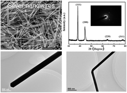
Massive silver nanowires with high yields have been synthesized within 10 minutes by a facile and effective two-step dropwise addition polyol method due to high reaction rates by virtue of high AgNO3 concentrations and anti-oxidation properties of the reaction promoter FeCl3. In particular, pure nanowires could be obtained by varying the AgNO3 concentrations from 0.1 M to 0.6 M. It was found that the concentrations of Fe3+, Cl, and PVP, bubbling atmosphere, reaction temperature, and time were important factors for controlling the morphology of the products. The results showed that a FeCl3 concentration of 0.1–0.15 mM, a PVP concentration of 0.3–0.45 M, air bubbling atmosphere, a temperature of 150–160 °C, and a reaction time of 10–120 min were appropriate conditions for the synthesis of nanowires, where the diameters could be tuned from 80 nm to 323 nm, and the length could be tuned from 3.7 μm to 14.3 μm. Moreover, Fe3+ could accelerate and facilitate the formation of silver nanowires by preventing their oxidative etching, while excessive Fe3+ also etched silver nanostructures. Furthermore, the rapid growth of nanowires due to a sufficient silver source provided by the second rapid dropwise addition and etching of other particles by Cl/O2 occurred simultaneously under air, thereby producing high-quality silver nanowires. Additionally, the diameter of the silver nanowires decreased and their surfaces became rougher, which is ascribed to etching effects with the time. More specifically, two silver nanowires could fuse together to form long nanowires arising from the existence of high nitrate anion concentrations. The size-dependent UV-VIS absorption spectra displayed that the surface plasmon resonance (SPR) peak broadened and red shifted from 384 nm to 458 nm with an increase in diameter from 80 nm to 323 nm. Meanwhile, the number of SPR peaks decreased from two to one. In consideration of morphology controllability, less time and energy cost, and synthesis of massive nanowires, the method is promising for realizing the industrial production of silver nanowires.


Introduction

In past decades, one-dimensional (1-D) silver nanowires have drawn a significant amount of interest due to their unique size-dependent optical,1–3 electrical,4 and thermal properties,5,6 in addition to their various applications in nanoscale electronic devices such as surface-enhanced Raman scattering (SERS),7 biological sensors,8,9 and transparent conducting electrodes.10,11 Recently, many researchers are exploring and utilizing these unique properties, which necessitates rapid and large-scale production of silver nanowires.12–14 Particularly, it is easy to form networks15,16 of silver nanowires because of their high aspect ratio and therefore, a low concentration of load can enable good performance matrices.17,18 From an industrial point of view, silver powders are widely used in the electronics industry, particularly for producing conductive adhesives where high concentrations (about 80 wt%) of load are used to form a conductive pattern in the matrix to obtain low resistivity at an order of 10−4 ohm cm.19 Wu et al. demonstrated that a combination of silver particles and nanowires would drastically lower the threshold of filling concentration to reach high conductivity.20 Afterward, Yu et al. demonstrated that the mass ratio of silver nanowires to silver nanoparticles was 1[thin space (1/6-em)]:[thin space (1/6-em)]8 for the PVA matrix when the volume electrical resistivity reached around 10−2 ohm cm,21 which means that the lower filling of silver nanowires in polymer composites is used to form an ideal conductive path. Hence, it would be highly desirable to produce large-scale, long silver nanowires within a short time via a facile and rapid industrial method.

A variety of methods have been introduced to produce silver nanowires. Many hard or soft templates are widely used to obtain silver nanowires, in which the hard templates usually exhibit one-dimensional structures, such as carbon nanorods,22 and mesoporous silica,23 and the soft templates are generally soft molecules, such as DNA chains24 and polymer-capping agents.25 For instance, Wehrspohn et al. synthesized monodisperse silver nanowires with diameters of 180–400 nm and a length of 30 μm using porous alumina as templates.26 Meanwhile, Zhao et al. produced silver nanowires with diameters of 10–70 nm using tetrabutyl titanate (TBT) and acetylacetone (AcAc) as templates based on 0.005 M AgNO3 concentration.27 Although template techniques are effective in synthesizing silver nanowires with controllable sizes and shapes, the removal of the templates is difficult, and nanowires are damaged during the removal. Moreover, the templates are usually fragile, small in size and difficult to produce. Hence, such a method is unsuitable for the mass production of silver nanowires. Chemical reduction methods are also widely used to produce silver nanowires by template-free synthesis. For instance, Giersig et al. synthesized silver nanowires with a diameter of 100 nm and a length of 1.2 μm in 0.03 M AgNO3 solution in N,N-dimethylformamide (DMF).28 Afterward, Mdluli et al. reported an improved solution chemistry route for the synthesis of long silver nanowires with a diameter of 40 nm using DMF as a reducing agent in the presence of polyvinylpyrolidone (PVP) for 30 days based on 0.05 M AgNO3 concentration.29 More recently, Chen et al. synthesized silver nanowires with diameters of 50–500 nm and length up to tens of micrometers using cuprous oxide nanospheres as reductants and directional agents, in which the AgNO3 concentration was in the range of 0.01–0.1 M.30 However, low production yield due to low AgNO3 concentration, long reaction time and uncontrollable structure restricted the development of these strategies. Recently, Du et al. reported a simple solvent-thermal method to yield silver microwires with a diameter of 2 μm and a length of 10 μm using ethanol as a reductant in the presence of ammonia at 250 °C for 10 h in a 0.05 M AgNO3 solution.31 Nevertheless, due to the obvious disadvantages, such as low manufacturing efficiency, long reaction time and harsh operating conditions, this method was not suitable for the mass production of silver nanowires. In contrast, the polyol process is an effective and facile approach for the preparation of silver nanowires in high yields.32 Xia et al. first produced silver nanowires with diameters of 30–40 nm and a length of 50 μm by this process using ethylene glycol (EG) as a reductant in the presence of seeds based on 0.02 M AgNO3 concentration.33 Afterward, numerous efforts have been undertaken to improve the polyol synthesis. For instance, Wang et al. prepared silver nanowires with diameters of 40–460 nm in 90% yield via a seed-catalysis-polyol process based on 0.03 M AgNO3 concentration.34–36 Afterward, microwave irradiation has been proposed to accelerate the preparation of silver nanowires.37–39 Tsuji et al. produced silver nanowires with diameters of 40–107 nm and lengths of 0.98–1.45 μm in 62% yield using a microwave-polyol method, in which the AgNO3 concentration was 0.005–0.011 M.40 Meanwhile, Gou et al. synthesized silver nanowires with a diameter of 45 nm and lengths of 4–12 μm in 80% yield under microwave irradiation in the presence of NaCl and PVP based on 0.026 M AgNO3 concentration.41 Recently, Kou et al. fabricated silver nanowires with diameters of 100–700 nm using glycerol as a reductant under microwave irradiation with aid of sodium dodecyl sulfate (SDS) based on 0.0375 M AgNO3 concentration.42 More recently, Cheng et al. used 2-ethoxy ethanol instead of EG to produce silver nanowires with a diameter of 80 nm and lengths of 2–10 μm in 75% yield based on 0.03 M AgNO3 concentration.43 Particularly, Chang et al. produced silver nanowires with diameter of 40 nm and lengths of 3–12 μm via an aging polyol process in the presence of HCl based on 0.04 × 10⁻³ M AgNO3 concentration.44 However, the strategies previously referred to still have limitations in the mass production of silver nanostructures due to the relatively low AgNO3 concentration, in which only milligrams of products will be obtained at a time, whereas silver nanostructures are in great demand with the rapid development of nanotechnology.45,46 As a result, it becomes a great challenge to produce silver nanowires based on high AgNO3 concentration precursor by a facile and efficient method. Recently, Hu et al. have proposed a new two-step, injection solution-based polyol method using NaCl as a control agent to prepare uniform silver nanowires with a diameter of 80 nm based on 0.15 M AgNO3 concentration within 20 min.47 As we know, metal ions in various valences48,49 such as Fe3+/Fe2+, Cu2+/Cu+ and Mn7+/Mn2+ can markedly influence the morphology and yield of silver nanowires by removing the oxygen from the surface of silver atoms; therefore, the introduction of Fe3+ would accelerate the rate of reduction and improve the selectivity of silver nanowires in high precursor concentrations. A detailed and systematic investigation of the effects of experimental parameters on the final morphology of the products based on the two-step dropwise addition polyol process with high precursor concentrations and reaction promoter FeCl3, which provides great theoretical aid for realizing the industrial production of silver nanowires, is of great significance for optimizing silver nanowire growth to realize morphology control.

In this paper, we report a facile and effective two-step dropwise addition method for the mass production of silver nanowires based on high AgNO3 concentration and FeCl3 as a reaction promoter. In this method, the first slow dropwise addition facilitated the formation of pentagonal twinned seeds, and the second rapid dropwise addition provided a sufficient silver source, which led to the rapid growth of silver nanowires. Particularly, high-quality silver nanowires could be synthesized within 10 min and several grams could be produced at a time, which would save time, energy, and manpower. Detailed and systematic parametric studies on the dependence of the morphology of the resulting products on the AgNO3, FeCl3, Cl, Fe3+, and PVP concentrations, gas bubbling atmosphere, temperature, and reaction times were elucidated. The as-synthesized silver nanowires were analyzed by scanning electron microscopy (SEM), transmission electron microscopy (TEM), X-ray diffraction (XRD), and UV-visible absorption spectra. Furthermore, the optical properties of silver nanowires with various diameters were investigated. Our detailed research on rapid production of silver nanowires based on high AgNO3 concentration precursors and FeCl3 as a reaction promoter will play an important theoretical guiding role in realizing the industrial production of silver nanowires.

Experimental

Materials

AgNO3 (≧99.0% purity), NaCl (≧99.5% purity), FeCl3·6H2O (≧99.0% purity), and ethanol were all supplied by Beijing Finechem, China. EG (≧99.5% purity) and PVP (Mw ≈ 40[thin space (1/6-em)]000, ≧99.0% purity) were purchased from Xilong Chemical Industry Incorporated Co., Ltd., China. All chemicals were of analytical grade and used without further purification.

Preparation of silver nanowires

Generally, EG was used as both a solvent and a reducing agent, PVP served as the capping agent, chloride salts served as the control agent, and AgNO3 was used as the precursor in traditional polyol process.50 In this typical synthesis procedure, 40 mL 0.56 M PVP solution in EG was added to a 250 mL volume flask at approximately 25 °C to which a stir bar was added. The mixture was then heated to 160 °C by oil bath heating until the temperature was steady. Afterward, 100 μL of a 0.05 M FeCl3 solution in EG was injected into the heated flask. After 5 minutes, 100 μL of a 0.15 M NaCl solution in EG was also injected into the aforementioned mixture. The solution was then heated for an additional 5 min. Then 10 mL 1.5 M AgNO3 solution in EG was added into the dropping funnel and then added dropwise into the flask at the rate of 15 μL s−1 at first. Once the reaction solution turned grey, which indicated the presence of silver nanoparticles and pentagonal twinned decahedron particles, all of the remaining AgNO3 solution was added into the flask at once. For those syntheses that produced high-concentration silver nanowires under air, the following color changes were observed: initially transparent and clear to light yellow (within 2 min), to yellow (within 1 min), to red-orange, to cloudy, to grey, and finally, to silver-white and glistening (within 10 min). After 2 h, the reaction was quenched and the reaction solution was then allowed to cool to room temperature. Then, in order to separate excess PVP from the silver nanowires, the suspension was diluted with ethanol (at a ratio of 1[thin space (1/6-em)]:[thin space (1/6-em)]10) and centrifuged three times at 8000 rpm for 20 min. The final products were dispersed in ethanol and stored at room temperature for further characterization.

To investigate the effects of the concentrations of raw materials, including AgNO3, FeCl3, Cl, Fe3+, and PVP, and technological conditions, including a bubbling atmosphere (air or N2), temperature and reaction time, on the final morphologies of silver nanowires, we performed a series of experiments in which the concentrations of AgNO3, FeCl3, Cl, Fe3+, and PVP, temperature, and reaction time were varied from 0.1 M to 0.3 M, from 0.05 mM to 0.25 mM, from 0.15 mM to 0.6 mM, from 0.05 mM to 0.2 mM, from 0.3 M to 0.9 M, from 130 °C to 170 °C, and from 10 min to 150 min, respectively. The detailed conditions of these experiments are summarized in Table 1.

Table 1 Detailed conditions of all experiments in this study
Samples Raw materials Technology conditions
AgNO3 (M) PVP (M) FeCl3 (mM) NaCl (mM) Atmosphere Temperature (°C) Reaction time (min)
a1 0.1 0.45 0.1 0.3 Air 160 120
b1 0.15 0.45 0.1 0.3 Air 160 120
c1(c4, b5, e6, d7, e8) 0.3 0.45 0.1 0.3 Air 160 120
d1 0.6 0.45 0.1 0.3 Air 160 120
a2 0.3 0.45 0.05 0 Air 160 120
b2(c6) 0.3 0.45 0.1 0 Air 160 120
c2 0.3 0.45 0.15 0 Air 160 120
d2(e4) 0.3 0.45 0.20 0 Air 160 120
e2 0.3 0.45 0.25 0 Air 160 120
a3 0.3 0.45 0 0.15 Air 160 120
b3(a6) 0.3 0.45 0 0.3 Air 160 120
c3 0.3 0.45 0 0.45 Air 160 120
d3(a4) 0.3 0.45 0 0.6 Air 160 120
b4 0.3 0.45 0.05 0.45 Air 160 120
d4 0.3 0.45 0.3 0.3 Air 160 120
a5 0.3 0.3 0.1 0.3 Air 160 120
c5 0.3 0.6 0.1 0.3 Air 160 120
d5 0.3 0.9 0.1 0.3 Air 160 120
b6 0.3 0.45 0 0.3 N2 160 120
d6 0.3 0.45 0.1 0 N2 160 120
f6 0.3 0.45 0.1 0.3 N2 160 120
a7 0.3 0.45 0.1 0.3 Air 130 120
b7 0.3 0.45 0.1 0.3 Air 140 120
c7 0.3 0.45 0.1 0.3 Air 150 120
e7 0.3 0.45 0.1 0.3 Air 170 120
a8 0.3 0.45 0.1 0.3 Air 160 10
b8 0.3 0.45 0.1 0.3 Air 160 30
c8 0.3 0.45 0.1 0.3 Air 160 60
d8 0.3 0.45 0.1 0.3 Air 160 90
f8 0.3 0.45 0.1 0.3 Air 160 150


Characterization

Scanning electron microscopy (SEM) images were obtained with a FEI field emission microscope (Apollo 300) operated at an accelerating voltage of 10 kV, and the samples were prepared by drop-casting a drop of the aqueous suspension of silver nanowires over a smooth aluminum wafer. Transmission electron microscopy (TEM) images were obtained from a JEM-2100 operated at 200 kV, and the samples were prepared by placing small droplets of the diluted suspension of samples on copper grids coated with amorphous carbon film. X-ray powder diffraction (XRD) measurements were taken by a D/max 2200 PC X-ray diffractometer operated at a voltage of 40 kV and a current of 40 mA with Cu Kα radiation (λ = 0.15406 nm) at a scanning rate of 0.02° s−1 in 2θ ranging from 20° to 80°, and the samples were prepared by dropping a suspension of silver nanowires on a slide. All the samples were dried under vacuum at 50 °C for 3 h. The diameters and lengths of the silver nanowires were determined by individually measuring the diameter of 100 nanostructures by the aid of SISAC-IAS image analyzer from the SEM and TEM images. The UV-visible absorption spectrum was recorded at room temperature with a UV-3600SHIMADZU spectrometer using a 1 cm optical path quartz cuvette by diluting solutions with ethanol, in which the wavelength was in the range of 200–800 nm.

Results and discussion

Effects of raw materials

Effects of AgNO3 concentration. In this paper, the AgNO3 concentration is approximately more than 10 times higher than the concentration generally reported in the literature. To investigate the effects of AgNO3 concentration on the morphology of products, a series of experiments with varying AgNO3 concentration were carried out. Representative samples were imaged by SEM with an enlargement of 10[thin space (1/6-em)]000 times to characterize the morphologies. The SEM images of the as-synthesized silver nanowires with AgNO3 concentrations of 0.1 M, 0.15 M, 0.3 M, and 0.6 M while maintaining the other parameters in the typical polyol process constant are displayed in Fig. 1(a1)–(d1), respectively. Fig. 1(e1) shows the changes in nanowire diameter and length as a function of the AgNO3 concentration. High-yield silver nanowires can be clearly observed from all images. It is well documented that the slow reduction at the initial stage facilitated the formation of pentagonal twinned decahedron particles, which could grow into silver nanowires quickly at a sufficient silver source supply.32,51 In this experiment, the first slow dropwise addition and high mole ratio of PVP to silver ions favored the formation of twinned seeds and the fast dropwise addition provided a sufficient silver source for fast anisotropic growth, and the high concentration of in situ generated nitric acid could etch the other irregular silver particles,47 resulting in high-concentration silver nanowires in high yields. Another important insight from Fig. 1(e1) is that the diameter of silver nanowires increased from 142 nm to 290 nm, while the length gradually decreased from 14.3 μm to 7.8 μm, as the AgNO3 concentration was varied from 0.1 M to 0.6 M. In this case, the diameter and amount of pentagonal twinned seeds increased with AgNO3 concentration, leading to the larger diameter. Meanwhile, the number of silver atoms for the growth of wider nanowires decreased accordingly, resulting in the decreasing length. Our data indicate that the size still can be controlled in high AgNO3 concentrations. Particularly, compared with the previous report, which showed that the diameter of silver nanowires increased from 98 nm to 121 nm as the AgNO3 concentration was varied from 0.024 M to 0.026 M,52 our results indicated that the variation in the AgNO3 concentration was effective for controlling the sizes of silver nanowires over a wider window of sizes.
image file: c4ra00711e-f1.tif
Fig. 1 SEM images of silver nanowires synthesized with different concentrations of AgNO3: (a1) 0.1 M, (b1) 0.15 M, (c1) 0.3 M, (d1) 0.6 M. (e1) Changes in nanowire diameter and length as a function of the AgNO3 concentration.
Effects of FeCl3 concentration. It has been found that the control agents play an important role in the production of silver nanowires.48,49 In this section, we tested FeCl3 as a control agent. In particular, the concentration of FeCl3 was varied from 0.05 mM to 0.25 mM, while all the other parameters were constant. Fig. 2(a2)–(e2) depict the SEM images of products with FeCl3 in various amounts. The changes in nanowire diameter and length as a function of the FeCl3 concentration are presented in Fig. 2(f2). When small amounts of FeCl3 (0.05 mM) were applied, some nanorods with a diameter of 257 nm and lengths of 2–3 μm could be noticed in addition to spherical nanoparticles (Fig. 2(a)), and the yield of nanorods was about 65%. When the concentration of FeCl3 was increased to 0.1 mM and 0.15 mM (Fig. 2(b2) and (c2)), pure silver nanowires were observed. In this case, the diameter decreased from 154 nm to 139 nm, while the length increased from 4.2 μm to 8.0 μm. Namely, the products had a higher aspect ratio at CFeCl3 = 0.15 mM than at CFeCl3 = 0.1 mM. Further increasing the concentration of FeCl3 to 0.2 mM, besides obtaining wide nanorods with a diameter of 270 nm, a small amount of silver nanoparticles such as spheres and triangles appeared. With excess amounts of FeCl3, 0.25 mM, spherical nanoparticles, cubes, and bipyramids were obtained as main products, and almost no nanowires could be viewed, as depicted in Fig. 2(e2). The significant differences among the five images proved that FeCl3 was an important morphology-controlling factor. Compared with previously reported results of low AgNO3 concentrations, which showed that the diameter of nanowires increased from 80 nm to 100 nm as FeCl3 concentration was varied from 18 μM to 24 μM,53 the variation in diameter and aspect ratio with FeCl3 concentrations shown in Fig. 2(f2) was highly irregular, which proved that Fe3+ and Cl played different roles in controlling the final morphology of the as-synthesized products. Our data indicated that 0.05–0.15 mM FeCl3 concentration was an appropriate condition in high AgNO3 concentrations.
image file: c4ra00711e-f2.tif
Fig. 2 SEM images of silver nanowires synthesized with different amounts of FeCl3: (a2) 0.05 mM, (b2) 0.10 mM, (c2) 0.15 mM, (d2) 0.20 mM, (e2) 0.25 mM. (f2) Changes in nanowire diameter and length as a function of the FeCl3 concentration.

To improve the understanding of the role of FeCl3 in this process, a number of control experiments were conducted to separate the effects of the concentrations of Cl and Fe3+. It has been demonstrated that chloride ions played a crucial role in the formation of silver nanowires.50,53,54 Fe(II) and Fe(III)-containing salts could not induce the formation of silver nanowires in the absence of chloride ions.53 Fig. 3(a3)–(d3) show the SEM images of the nanowires synthesized with different NaCl amounts of 0.15 mM, 0.3 mM, 0.45 mM, and 0.6 mM, respectively. Fig. 3(e3) shows the changes in nanowire diameter and length as a function of Cl concentration. It can be observed from Fig. 3(a3)–(c3) that silver nanowires are dominant. Herein, as the concentration of NaCl increases, average diameter of nanowires increases from 80 nm to 139 nm, while their length decreases from 9.4 μm to 5.5 μm, as indicated in Fig. 3(e3). Further increasing the NaCl concentration to 0.6 mM leads to the formation of irregular micrometer nanoparticles besides a small amount of nanorods and nanowires (Fig. 3(d3)). According to the surface-energy minimization, it is favorable to form twinned nuclei when the particle size reaches a critical value, thus five twin boundaries generate the pentagonal twinned seeds.32,55,56 It is well documented that Cl reacts with Ag cation to form AgCl colloids that could serve as a seed for twinned nuclei, which also slows the rate of reduction.57,58 Twinned seeds then grow into silver nanowires rapidly by the selective etching of Cl/O2, in which the etched irregular particles serve as the silver sources.44,47,52 Scheme 1 shows the schematic illustration of the role of Cl. In our experiment, the reaction rate decreased due to oxidative etching and the size of twinned nuclei increased with Cl concentration, which led to wider silver nanowires. The increase in diameter of nanowires and the amount of AgCl solids resulted in insufficient silver atoms for further growth, leading to shorter nanowires. While excessive Cl resulted in a large amount of large AgCl precipitate particles and excessive Cl/O2 could strongly etch the formed silver nanowires and twinned seeds, leading to the formation of a large amount of nanoparticles as shown in Scheme 1. These observations are consistent with previously reported results in low AgNO3 concentrations.57 Compared with the report52 that the diameter increased from 65 nm to 305 nm as Cl concentration was varied from 0.014 mM to 0.1 mM, the variation in sizes of the silver nanowires in our study was within a relatively narrow range. Our data indicated that 0.15–0.45 mM Cl concentration was appropriate for the growth of nanowires, while excessive Cl resulted in the formation of nanoparticles.


image file: c4ra00711e-f3.tif
Fig. 3 SEM images of silver nanowires synthesized with different amounts of NaCl: (a3) 0.15 mM, (b3) 0.30 mM, (c3) 0.45 mM, (d3) 0.60 mM. (e3) Changes in nanowire diameter and length with the concentration of NaCl.

image file: c4ra00711e-s1.tif
Scheme 1 Schematic illustration of the role of Cl in the formation of silver nanowires.

To investigate the effect of Fe3+ on the two-step dropwise addition polyol reduction in high AgNO3 concentration precursors, a series of experiments were conducted, in which the Fe3+ amount was varied from 0 mM, 0.05 mM, 0.1 mM, 0.15 mM to 0.2 mM while the total amount of Cl was kept unchanged at 0.6 mM. Fig. 4(a4)–(e4) show SEM images of the products. The changes in nanowire diameter and length as a function of the Fe3+ concentration are depicted in Fig. 4(f4). Besides silver nanowires, almost no particles could be obtained as shown in Fig. 4(b4). The difference between Fig. 4(a4) and (b4) indicated that a small amount of Fe3+ could promote the formation of silver nanowires. Moreover, the rate of color changes in the synthesis process accelerated in the presence of Fe3+. When the Fe3+ concentration was increased from 0.05 mM to 0.15 mM, the average diameter increased from 135 nm to 296 nm, and the length increased from 7.1 μm to 8.5 μm. Further increases in Fe3+ to 0.2 mM again started the formation of a small amount of nanoparticles. The observations validated the theory48 that iron could remove oxygen from the surfaces of twinned seeds and prevent their oxidative etching, accelerating and facilitating the formation of pure silver nanowires. In this case, EG reduced Fe3+ to Fe2+, then Fe2+ rapidly scavenged the absorbed oxygen, leading to the oxidation of Fe2+ to Fe3+.48 It has been demonstrated that excess Fe3+ also etched the silver nanostructures due to its oxidation, leading to the formation of nanoparticles.53 Scheme 2 shows the schematic illustration of the role of Fe3+. Our data indicated that Fe3+ played a promoting role in the morphology control of silver nanowires, in which the appropriate concentration was in the range of 0.05–0.15 mM when 0.6 mM Cl was added. In this case, the more Fe3+ was added, the more obvious the inhibition of oxidative etching for the surface of silver nanowires and twinned particles occurred; the twinned seeds were larger, leading to the wider nanowires. Moreover, the reduction acceleration promoted long nanowires.


image file: c4ra00711e-f4.tif
Fig. 4 SEM images of silver nanowires synthesized with different proportions of FeCl3 and NaCl amounts: (a4) 0 mM and 0.6 mM, (b4) 0.05 mM and 0.45 mM, (c4) 0.1 mM and 0.3 mM, (d4) 0.15 mM and 0.15 mM, (e4) 0.2 mM and 0 mM. (f4) Changes in nanowire diameter and length as a function of the Fe3+ concentration.

image file: c4ra00711e-s2.tif
Scheme 2 Schematic illustration of the role of Fe3+ in the formation of silver nanowires.
Effects of PVP concentration. Fig. 5(a5)–(d5) show the SEM images of as-synthesized products with different PVP amounts of 0.3 M, 0.45 M, 0.6 M, and 0.9 M, respectively. The changes in nanowire diameter and length as a function of the PVP concentration are shown in Fig. 5(e5). When the PVP concentration was increased from 0.3 M to 0.6 M, nanowire diameter decreased from 287 nm to 212 nm, and the length decreased from 8.5 μm to 3.6 μm as indicated in Fig. 5(e5). Moreover, besides short and wide nanorods, some spherical nanoparticles were also observed in Fig. 5(c5). When the concentration of PVP was increased further to 0.9 M, the yields of other nanoparticles were drastically improved (Fig. 5(d5)). It has been demonstrated that PVP molecules could be absorbed on the surface of silver nanoparticles through Ag–O coordination.59,60 Because the surface energy of the (100) facets is higher than that of the (111) facets for twinned seeds, the PVP molecules are tightly absorbed on the (100) planes, according to the surface-energy minimization, leading the rapid anisotropic growth along the (110) direction.32,55,61 Scheme 3 shows the schematic illustration of the role of PVP. In our experiment, a low concentration of PVP caused larger seeds and insufficient passivation of the (100) facets, thus the growth along both (100) facets and (110) facets appeared, leading to nanorods with larger diameters. On the other hand, high concentration of PVP covered all surfaces of seeds, which blocked the anisotropic growth of silver nanowires. Compared with the previous results that the diameter decreased from 300 nm to 80 nm as PVP concentration was varied from 0.12 M to 0.3 M at 0.04 M AgNO3 concentration,57 our data were within a narrow range. Additionally, by comparing the results of the effects of AgNO3 concentration with that of the PVP concentration, we suggested that the molar ratio of PVP to AgNO3 was no longer the important factor for morphology control of silver nanowires in high AgNO3 concentration precursors, which was not consistent with the report.33,53,57 Our data indicate that the 0.45 M PVP concentration was an optimum condition for the synthesis of nanowires (Scheme 3).
image file: c4ra00711e-f5.tif
Fig. 5 SEM images of silver nanowires synthesized with different PVP concentrations: (a5) 0.3 M, (b5) 0.45 M, (c5) 0.6 M, (d5) 0.9 M. (e5) Changes in nanowire diameter and length as a function of the PVP concentration.

image file: c4ra00711e-s3.tif
Scheme 3 Schematic illustration of the role of PVP in the formation of silver nanowires.

Effects of technological conditions

Effects of gas-bubbling atmosphere. The gas-bubbling atmosphere is an essential factor for the synthesis of silver nanowires.51,62 The yields and sizes of products under different shielding atmospheres are presented in Table 2. Fig. 6(a6), (c6) and (e6) depict typical SEM images of the as-synthesized products obtained with different control agents under air. Silver nanostructures obtained under N2 are shown in Fig. 6(b6), (d6) and (f6). It was noteworthy that silver nanowires were produced in high yields (>90%) under air. Obviously, a significant decrease in the sizes of silver nanowires occurred when the bubbling gas was changed from air to N2. Particularly, the rate of color changes became faster during the synthesis process under N2. Additionally, small amounts of nanocubes, bipyramids, and nanotriangles appeared in addition to silver nanowires with a diameter of 94 nm under N2 when 0.3 mM NaCl was used as a control agent, whereas the yields of the nanowires were still more than 90% in the presence of Fe3+ as shown in Fig. 6(d6) and (f6). Scheme 4 shows the schematic illustration of the formation mechanism of silver nanostructures synthesized in air and N2. It was known that the reduction rate slowed down due to the etching and dissolution by Cl/O2 at the initial stage, leading to fewer and larger twinned particles.44,47,52 On the other hand, rapid crystal growth and oxidative etching occurred competitively after fast injection at a later stage. In this condition, a sufficient amount of silver source was supplied, making the growth rates higher than the etching rates by Cl/O2 for twinned particles, thus rapid and selective growth of silver nanowires occurred.51 Meanwhile, the etching rates of cubes, triangles and bipyramids were higher than the growth rates in the presence of O2, thus the initially formed cubes were dissolved and served as the silver source for the anisotropic growth of nanowires.32,51 On the contrary, the accelerated rates of seed formation by the protection of N2 and the absence of the etching ability of Cl/O2 led to the formation of other nanoparticles, such as the cubes shown in Fig. 6(b6). In particular, Fe3+ could also etch cubes and bipyramids in the absence of O2,48 supporting the phenomenon that silver nanowires in high yields were observed in Fig. 6(d6) and (f6). This observation further validated the conclusion that the addition of Fe3+ could accelerate and facilitate the growth of silver nanowires.
Table 2 Yields and average sizes of products under different shielding atmospheres
Control agents Atmosphere products Yields of wires (%) Diameter of wires (nm) Length of wires (μm)
0.3 mM NaCl Air Wires 93 118 6.8
0.3 mM NaCl N2 Wires, spheres, cubes, triangular bipyramids 72 94 6.2
0.1 mM FeCl3 Air Wires 92 154 4.2
0.1 mM FeCl3 N2 Wires 90 110 4.4
0.3 mM NaCl, 0.1 mM FeCl3 Air Wires 93 236 7.8
0.3 mM NaCl, 0.1 mM FeCl3 N2 Wires 90 125 7.2


image file: c4ra00711e-f6.tif
Fig. 6 SEM images of silver nanowires synthesized under different shielding atmospheres. Air: (a6), (c6), (e6); N2: (b6), (d6), (f6). In this case, silver nanowires synthesized with different control agents: (a6, b6) 0.3 mM NaCl, (c6, d6) 0.1 mM FeCl3, (e6, f6) 0.3 mM NaCl, and 0.1 mM FeCl3.

image file: c4ra00711e-s4.tif
Scheme 4 Schematic illustration of the formation mechanism of silver nanostructures synthesized in air and N2.
Effects of reaction temperature. It is known that temperature is an important factor for the morphology control of silver nanowires in low-concentration precursors.33,57,62 Fig. 7(a7)–(e7) depict the SEM images of the products obtained at 130 °C, 140 °C, 150 °C, 160 °C and 170 °C, respectively, while maintaining the other parameters constant. The changes in nanowire diameter and length as a function of temperature are shown in Fig. 7(f7). When the temperature was 130 °C, a large amount of other nanoparticles besides nanowires were obtained (Fig. 7(a7)). When the temperature increased from 140 °C to 160 °C, the yield of silver nanowires increased and almost no nanoparticles were observed at 160 °C. Moreover, the diameter decreased from 276 nm to 235 nm, while the length increased from 5.8 μm to 7.8 μm with the temperature increase. Upon the increase in the reaction temperature to 170 °C, some spherical particles were again observed besides the wide rods. It has been demonstrated that the conversion of EG to glycolaldehyde occurred above 140 °C with the aid of O2, as shown in eqn (1).63
 
2HOCH2CH2OH + O2 → 2HOCH2CHO + 2H2O (1)

image file: c4ra00711e-f7.tif
Fig. 7 SEM images of silver nanowires synthesized at different temperatures: (a7) 130 °C, (b7) 140 °C, (c7) 150 °C, (d7) 160 °C, (e7) 170 °C. (f7) Changes in nanowire diameter and length as a function of the temperature.

In this case, the higher the temperature, the more the reducing agents generated, leading to faster reduction and more twinned seeds in smaller sizes; thus, thinner and longer nanowires appeared. On the other hand, lower or higher temperatures would block the formation of the twinned particles due to the unfavorable thermal energy.32,53,57 We suggested that a temperature of 150–160 °C was an appropriate condition for the synthesis of nanowires. A previously studied report by Sun et al. claimed that silver nanowires with a diameter of 39 nm and length of 1.9 μm could be produced at 185 °C.33 In another study, Coskun et al. produced silver nanowires with a diameter of 170 nm and length of 10 μm at 190 °C.57 In contrast, the temperature in our study is lower for the synthesis of silver nanowires in high yields with high precursor concentrations.

Effects of reaction time. The SEM images of the silver nanowires synthesized with reaction times of 10, 30, 60, 90, 120, and 150 min while keeping all the other parameters constant are shown in Fig. 8(a8)–(f8), respectively. The changes in nanowire diameter and length with respect to the reaction time are depicted in Fig. 8(g8). Silver nanorods with a diameter of 323 nm were the dominant products when the reaction time was only 10 min (Fig. 8(b8)). When the reaction continued to 30 minutes, the aspect ratio of nanowires increased, and some curved nanowires appeared (Fig. 8(b8)). Moreover, the proportion of curved nanowires with rougher surfaces increased with prolonged reaction time (Fig. 8(c8) and (d8)), which provided direct evidence that the presence of high concentrations of nitrate anions could dramatically promote the self-assembly growth for the formation of long, silver nanowires.64 Straight nanowires with a diameter of 235 nm and length of 7.8 μm would be the major products when the reaction time was 120 min, as shown in Fig. 8(e8), but a longer reaction time, such as 150 min, did not improve the aspect ratio remarkably (Fig. 8(f8)). As a whole, the diameter decreased from 323 nm to 229 nm, and the length increased from 4.4 μm to 8.2 μm, as the time was varied from 10 min to 150 min. Scheme 5 shows the schematic illustration of the formation mechanism of silver nanowires in high precursor concentrations. It is believed that the strong corrosion of Cl/NO3, Cl/O2 and Cl/Fe3+ for the surface of silver nanowires led decreasing diameters and rougher surfaces with the reaction time. Additionally, in high concentrations, nitrate anions could interact with the AgCl colloids on the surfaces of silver nanorods, as well as the pyrrolidone ring of PVP through cooperative electrostatic interactions to form PVP–NO3–AgCl, which could serve as a template for the attachment between the ends of the silver nanowires and nanoparticles.64 The ends of nanowires could then be sensitized, thus attaching other silver nanowires to lengthen themselves. Afterwards, the curved nanowires would become straight due to the thermodynamic stability of the nanowires.47
image file: c4ra00711e-f8.tif
Fig. 8 SEM images of silver nanowires synthesized with different reaction times: (a8) 10 min, (b8) 30 min, (c8) 60 min, (d8) 90 min, (e8) 120 min, (f8) 150 min. (g8) Changes in nanowire diameter and length with reaction time.

image file: c4ra00711e-s5.tif
Scheme 5 Schematic illustration of the formation mechanism of silver nanowires in high precursor concentrations.

A two-step dropwise addition polyol process based on high AgNO3 concentration precursors and Fe3+ as reaction promoter, as reported in this work, has achieved structural control of silver nanowires through varying the experimental parameters. To further demonstrate the structure of the as-synthesized silver nanowires, TEM and XRD were used to characterize the representative sample c2. Fig. 9a shows the TEM image of a single silver nanowire, in which the nanowire exhibits a diameter of 200 nm and a pentagonal cross section. The inset displays the model of the pentagonal cross section of the silver nanowires. The XRD pattern is presented in Fig. 9(b), indicating that the silver nanowires in this study exhibit FCC crystal structure (JCPDS File 04-0783).58 The lattice constant calculated from this XRD pattern, according to the lattice spacing of the (111) planes is 4.102 Å, which is highly similar to the reported data value of 4.0862 Å. Obviously, the ratio of intensity between the (111) and (200) peaks shows a relatively high value of 2.88 (the theoretical ratio is 2.5),58 suggesting the enrichment of the (111) crystalline planes in the silver nanowires. Furthermore, the inset shows the selected area electron diffraction (SAED) of a single silver nanowire, which is consistent with the XRD results.


image file: c4ra00711e-f9.tif
Fig. 9 Typical structural characterization of the products. (a) TEM image of single silver nanowire. (b) XRD pattern of the silver nanowires. Inset (a) shows the morphology of cross section of silver nanowires. Inset (b) shows the selected area electron diffraction (SAED) of a single silver nanowire.

Optical properties of silver nanowires with different diameters

Silver nanostructures show unique optical properties dependent upon their morphologies due to their surface plasmon response (SPR).55 The UV-visible absorption spectra of silver nanowires with various diameters are shown in Fig. 10. In particular, (A) shows the variation of absorption with respect to wavelength in the range of 345–460 nm and (B) shows the red-shift of SPR peak. As depicted in this graph, nanowires with diameters of 80 nm, 139 nm, and 178 nm have two peaks appearing at 352/384 nm, 356/402 nm, and 359/406 nm, respectively. The two SPR peaks can be ascribed to the quadrupole resonance excitation (the weaker peak) and the transverse plasmon resonance (the stronger peak) of the silver nanowires.35 Typically, when the diameter increases to 235 nm, 296 nm, and 323 nm, there is only one SPR peak appearing at 422 nm, 442 nm, and 458 nm, respectively. It is noteworthy that the SPR peak of these six samples red shifts and broadens with the diameter (shown in Fig. 10(B)). The decrease in number of SPR peaks can be attributed to the fact that the symmetry of the nanowires increases with the diameter, thus the quadrupole resonance excitation gradually disappears.34 Additionally, the red shift and width broadening of the SPR peaks are consistent with the greater charge separation during plasmon resonance and the inhomogeneous polarization of nanowires with the diameter.34,35 The observations are in agreement with the previous results, which showed that the SPR peak red shifted by a maximum amount from 378 nm to 467 nm as the diameter was varied from 77 nm to 584 nm.52 Our data further demonstrated that the properties of the silver nanowires were strongly dependent upon their sizes.
image file: c4ra00711e-f10.tif
Fig. 10 UV-vis absorption spectra of silver nanowires with different diameters: (a3) 80 nm, (c2) 139 nm, (b1) 178 nm, (c1) 235 nm, (d4) 296 nm, (a8) 323 nm. (A) shows the variation of the absorption with respect to the wavelength in the range of 345–460 nm, and (B) shows the red shift of SPR peak.

Conclusions

Detailed and systematic parametric studies on the synthesis of massive silver nanowires in high yields via a facile, two-step, dropwise addition polyol method using high AgNO3 concentrations and FeCl3 as the reaction promoter were presented. Based on the experimental results, the following conclusions can be drawn:

(1) When the other parameters were kept constant, the diameter of silver nanowires increased from 142 nm to 290 nm and the aspect ratio gradually decreased as AgNO3 concentration was increased from 0.1 M to 0.6 M.

(2) A Cl concentration of 0.15–0.45 mM was an appropriate condition in the absence of Fe3+ for the growth of nanowires in which the diameter could be tuned from 80 nm to 139 nm. When the Cl concentration was 0.6 mM, Fe3+ in the range 0.05–0.15 mM could accelerate and promote the growth of nanowires, and excessive Fe3+ resulted in the formation of nanoparticles due to its etching ability.

(3) The diameter of silver nanowires decreased from 287 nm to 212 nm as the PVP concentration was varied from 0.3 M to 0.6 M, and excessive PVP resulted in the formation of nanoparticles.

(4) The rapid growth of nanowires and the etching of cubes and bipyramids by Cl/O2 occurred simultaneously at later stages under air, thereby producing high-quality silver nanowires.

(5) A temperature of 150–160 °C was an appropriate condition for the growth of nanowires, in which the diameter decreased from 276 nm to 235 nm. Temperatures lower or higher than this range were unfavorable for the formation of twinned seeds, leading to the formation of nanoparticles.

(6) The diameter decreased from 323 nm to 230 nm with time due to the etching effects. The existence of high-nitrate anion concentrations could promote self-assembly growth by two silver nanowires fusing together to form curved nanowires.

(7) When the diameter increased, the number of surface plasmon response (SPR) peaks decreased, and the SPR peak red shifted and broadened, which is attributed to the greater charge separation during plasmon resonance and the inhomogeneous polarization of the nanowires.

References

  1. M. Chen, I. Y. Phang, M. R. Lee, J. K. W. Yang and X. Y. Ling, Langmuir, 2013, 29, 7061–7069 CrossRef CAS PubMed.
  2. X. Ma, X. Zhu, F. You, J. Feng, M. C. Wang and X. Zhao, J. Alloys Compd., 2014, 592, 57–62 CrossRef CAS PubMed.
  3. S. Mehra, M. G. Christoforo and P. Peumans, et al., Nanoscale, 2013, 5, 4400–4403 RSC.
  4. Y. Li, P. Cui, L. Y. Wang, H. Lee, K. Lee and H. Lee, ACS Appl. Mater. Interfaces, 2013, 5, 9155–9160 CAS.
  5. I. Moreno, N. Navascues, M. Arruebo, S. Irusta and J. Santamaria, Nanotechnology, 2013, 24, 275603 CrossRef CAS PubMed.
  6. Y. H. Yu, C. C. M. Ma, C. C. Teng, Y. L. Huang, S. H. Lee and I. Wang, J. Taiwan Inst. Chem. Eng., 2013, 44, 654–659 CrossRef CAS PubMed.
  7. M. S. Goh, Y. H. Lee, S. Pedireddy, I. Y. Phang, W. W. Tjiu, J. M. R. Tan and X. Y. Ling, Langmuir, 2012, 28, 14441 CrossRef CAS PubMed.
  8. W. L. Hu, X. F. Niu, R. Zhao and Q. B. Pei, Appl. Phys. Lett., 2013, 102, 083303 CrossRef PubMed.
  9. S. Yao and Y. Zhu, Nanoscale, 2014, 6, 2345–2352 RSC.
  10. R. Zhu, C. H. Chung, K. C. Cha, W. B. Yang, Y. B. Zheng, H. P. Zhou, T. B. Song, C. C. Chen, P. S. Weiss, G. Li and Y. Yang, ACS Nano, 2011, 5, 9877–9882 CrossRef CAS PubMed.
  11. C. Preston, Z. Q. Fang, J. Murray, H. L. Zhu, J. Q. Dai, J. N. Munday and L. B. Hu, J. Mater. Chem. C, 2014, 2, 1248–1254 RSC.
  12. S. Xu, B. Man, S. Jiang, M. Liu, C. Yang, C. Chen and C. Zhang, Cryst. Eng. Commun., 2014, 16, 3532–3539 RSC.
  13. J. W. Park, D. K. Shin and J. Ahn, et al., Semicond. Sci. Technol., 2014, 29(1), 015002 CrossRef.
  14. A. Dasgupta, D. Singh and S. Tandon, et al., J. Nanophotonics, 2014, 8, 083899 CrossRef PubMed.
  15. Y. Jin, D. Deng, Y. Cheng, L. Kong and F. Xiao, Nanoscale, 2014, 6, 4812–4818 RSC.
  16. T. Kim, Y. W. Kim, H. S. Lee, H. Kim, W. S. Yang and K. S. Suh, Adv. Funct. Mater., 2013, 23, 1225 CrossRef CAS.
  17. H. Y. Mi, Z. Li, L. S. Turng, Y. Sun and S. Gong, Mater. Des., 2014, 56, 398–404 CrossRef CAS PubMed.
  18. T. H. L. Nguyen, L. Quiroga Cortes and A. Lonjon, et al., J. Non-Cryst. Solids, 2014, 385, 34–39 CrossRef CAS PubMed.
  19. F. Tan, X. Qiao and J. Chen, et al., Int. J. Adhes. Adhes., 2006, 26, 406–413 CrossRef CAS PubMed.
  20. H. P. Wu, et al., Int. J. Adhes. Adhes., 2006, 26, 617–621 CrossRef CAS PubMed.
  21. Y. H. Yu, C. C. M. Ma, C. C. Teng, Y. L. Huang, S. H. Lee, I. Wang and M. H. Wei, Mater. Chem. Phys., 2012, 136, 334–340 CrossRef CAS PubMed.
  22. J. Sloan, D. M. Wright, H. G. Woo, S. Bailey, G. Brown, A. P. E. York, K. S. Coleman, J. L. Hutchison and M. L. H. Green, Chem. Commun., 1999, 699–700 RSC.
  23. C. H. Hsia, M. Y. Yen, C. C. Lin, H. T. Chiu and C. Y. Lee, J. Am. Chem. Soc., 2003, 125, 9940–9941 CrossRef CAS PubMed.
  24. S. H. Park, R. Barish, H. Li, J. H. Reif, G. Finkelstein, H. Yan and T. H. LaBean, Nano Lett., 2005, 5, 693–696 CrossRef CAS PubMed.
  25. X. M. Sun and Y. D. Li, Adv. Mater., 2005, 17, 2626–2630 CrossRef CAS.
  26. J. Choi, G. Sauer, K. Nielsch, R. B. Wehrspohn and U. Gösele, Chem. Mater., 2003, 15, 776–779 CrossRef CAS.
  27. Q. T. Zhao, J. R. Qiu, C. J. Zhao, L. S. Hou and C. S. Zhu, Chem. Lett., 2005, 34, 30–31 CrossRef CAS.
  28. M. Giersig, P. S. Isabel and M. L. M. Luis, J. Mater. Chem., 2004, 14, 607–610 RSC.
  29. P. S. Mdluli and N. J. Revaprasadu, J. Alloys Compd., 2009, 469, 519–522 CrossRef CAS PubMed.
  30. M. Chen, C. J. Wang, X. J. Wei and G. W. Diao, J. Phys. Chem. C, 2013, 117, 13593–13601 CAS.
  31. J. M. Du, B. X. Han, Z. M. Liu and Y. Q. Liu, Cryst. Growth Des., 2007, 7, 900–904 CAS.
  32. B. Wiley, Y. Sun, B. Mayers and Y. Xia, Chem.–Eur. J., 2005, 11, 454–463 CrossRef CAS PubMed.
  33. Y. G. Sun, D. D. Yin, B. T. Mayers, T. Herricks and Y. N. Xia, Chem. Mater., 2002, 14, 4736–4745 CrossRef CAS.
  34. C. Chen, L. Wang, H. Yu, J. Wang, J. Zhou, Q. Tan and L. Deng, Nanotechnology, 2007, 18, 115612 CrossRef.
  35. C. Chen, L. Wang, G. Jiang, J. Zhou, X. Chen, H. Yu and Q. Yang, Nanotechnology, 2006, 17, 3933–3938 CrossRef CAS.
  36. C. Chen, L. Wang, H. Yu, G. Jiang, Q. Yang, J. Zhou, W. Xiang and J. Zhang, Mater. Chem. Phys., 2008, 107, 13–17 CrossRef CAS PubMed.
  37. M. Tsuji, M. Hashimoto, Y. Nishizawa, M. Kubokawa and T. Tsuji, Chem.–Eur. J., 2005, 11, 440–452 CrossRef CAS PubMed.
  38. M. Tsuji, K. Matsumoto, P. Jiang, R. Matsuo, X. L. Tang and K. Sozana, Colloids Surf., A, 2008, 316, 266–277 CrossRef CAS PubMed.
  39. Y. Yang, Y. Hu, X. Xiong and Y. Qin, RSC Adv., 2013, 3, 8431–8436 RSC.
  40. M. Tsuji, K. Matsumoto, N. Miyamae, T. Tsuji and X. Zhang, Cryst. Growth Des., 2007, 7, 311–320 CAS.
  41. L. Y. Gou, M. Chipara and J. M. Zaleski, Chem. Mater., 2007, 19, 1755–1760 CrossRef CAS.
  42. J. Kou and R. S. Varma, Chem. Commun., 2013, 49, 692–694 RSC.
  43. W. M. Cheng, C. C. Wang and C. Y. Chen, J. Mater. Sci., 2013, 48, 1042–1052 CrossRef CAS.
  44. S. Chang, S. Chen, Q. Hua, Y. Ma and W. J. Huang, J. Phys. Chem. C, 2011, 115, 7979–7986 CAS.
  45. J. Jiu, T. Sugahara, M. Nogi, T. Araki, K. Suganuma, H. Uchida and K. Shinozaki, Nanoscale, 2013, 5, 11820–11828 RSC.
  46. Z. Zhang, Y. Zheng, P. He and Z. Sun, Physica E, 2011, 44, 535–540 CrossRef CAS PubMed.
  47. M. Hu, J. Gao, Y. Dong, S. Yang and R. K. Y. Li, RSC Adv., 2012, 2, 2055–2060 RSC.
  48. B. Wiley, Y. G. Sun and Y. N. Xia, Langmuir, 2005, 21, 8077–8079 CrossRef CAS PubMed.
  49. K. E. Korte, S. E. Skrabalak and Y. N. Xia, J. Mater. Chem., 2008, 18, 437–441 RSC.
  50. B. Wiley, T. Herricks, Y. Sun and Y. Xia, Nano Lett., 2004, 4, 1733–1739 CrossRef CAS.
  51. X. L. Tang, M. Tsuji, P. Jiang, M. Nishio, S. M. Jang and S. H. Yoon, Colloids Surf., A, 2009, 338, 33–39 CrossRef CAS PubMed.
  52. C. C. S. Oliveira, R. A. Ando and P. H. C. Camargo, Phys. Chem. Chem. Phys., 2013, 15, 1887–1893 RSC.
  53. J. T. Jiu, K. Murai, D. Kim, K. Kim and K. Suganuma, Mater. Chem. Phys., 2009, 114, 333–338 CrossRef CAS PubMed.
  54. W. M. Schuette and W. E. Buhro, ACS Nano, 2013, 7, 3844–3853 CrossRef CAS PubMed.
  55. Y. G. Sun, B. T. Mayers, T. Herricks and Y. N. Xia, Nano Lett., 2003, 43, 955–960 CrossRef.
  56. L. D. Marks, Rep. Prog. Phys., 1994, 57, 618 CrossRef.
  57. S. Coskun, B. Aksoy and H. E. Unalan, Cryst. Growth Des., 2011, 11, 4963–4969 CAS.
  58. Z. H. Wang, J. W. Liu, X. Y. Chen, J. X. Wan and Y. T. Qian, Chem.–Eur. J., 2005, 11, 160–163 CrossRef PubMed.
  59. Y. Gao, P. Jiang, D. F. Liu, H. J. Yuan, X. Q. Yan, Z. P. Zhou, J. X. Wang, L. Song, L. F. Liu, W. Y. Zhou, G. Wang, C. Y. Wang and S. S. Xie, J. Phys. Chem. B, 2004, 108, 12877–12881 CrossRef CAS.
  60. H. H. Huang, X. P. Ni, G. L. Loy, C. H. Chew, K. L. Tan, F. C. Loh, J. F. Deng and G. Q. Xu, Langmuir, 1996, 12, 909–912 CrossRef CAS.
  61. Y. Gao, P. Jiang, L. Song, L. F. Liu and X. Q. Yan, J. Phys. D: Appl. Phys., 2005, 38, 1061–1067 CrossRef CAS.
  62. C. Chen, L. Wang, G. Jiang, Q. Yang, J. Wang, H. Yu, T. Chen, C. Wang and X. Chen, Nanotechnology, 2006, 17, 466–474 CrossRef CAS.
  63. F. Kim, S. Connor, H. Song, T. Kuykendall and P. Yang, Angew. Chem., 2004, 28, 3759–3763 CrossRef.
  64. C. L. Kuo and K. C. Hwang, Langmuir, 2012, 28, 3722–3729 CrossRef CAS PubMed.

This journal is © The Royal Society of Chemistry 2014
Click here to see how this site uses Cookies. View our privacy policy here.