Enhanced photodegradation activity of methyl orange over Z-scheme type MoO3–g-C3N4 composite under visible light irradiation

Yiming He*ae, Lihong Zhanga, Xiaoxing Wanga, Ying Wub, Hongjun Linc, Leihong Zhaob, Weizheng Wengd, Huilin Wand and Maohong Fan*ef
aDepartment of Materials Physics, Zhejiang Normal University, Jinhua, China. E-mail: hym@zjnu.cn; Fax: +86-0579-83714946; Tel: +86-0579-83792294
bInstitute of Physical Chemistry, Zhejiang Key Laboratory for Reactive Chemistry on Solid Surfaces, Zhejiang Normal University, Jinhua, China
cCollege of Geography and Environmental Sciences, Zhejiang Normal University, Jinhua, China
dState Key Laboratory Physical Chemistry of Solid Surfaces, Xiamen University, Xiamen, China
eDepartment of Chemical & Petroleum Engineering, University of Wyoming, Laramie, Wyoming 82071, USA. E-mail: mfan@uwyo.edu; Fax: +1-307-766-6667; Tel: +1-307-766-5633
fSchool of Civil and Environmental Engineering, Georgia Institute of Technology, Georgia 30332, USA. E-mail: mfan3@mail.gatech.edu; Fax: +1-404-894-8266; Tel: +1-404-385-4577

Received 23rd January 2014 , Accepted 4th March 2014

First published on 4th March 2014


Abstract

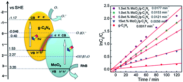
Novel Z-scheme type MoO3–g-C3N4 composites photocatalysts were prepared with a simple mixing–calcination method, and evaluated for their photodegradation activities of methyl orange (MO). The optimized MoO3–g-C3N4 photocatalyst shows a good activity with a kinetic constant of 0.0177 min−1, 10.4 times higher than that of g-C3N4. Controlling various factors (MoO3–g-C3N4 amount, initial MO concentration, and pH value of MO solution) can lead to the enhancement of the photocatalytic activity of the composite. Only MoO3 and g-C3N4 are detected with X-ray diffraction (XRD) and Fourier transform infrared spectroscopy (FT-IR) spectra. N2 adsorption and UV-vis diffuse reflectance spectroscopy (DRS) results suggest that the addition of MoO3 slightly affects the specific surface area and the photoabsorption performance. The transmission electron microscopy (TEM) image of MoO3–g-C3N4 indicates a close contact between MoO3 and g-C3N4, which is beneficial to interparticle electron transfer. The high photocatalytic activity of MoO3–g-C3N4 is mainly attributed to the synergetic effect of MoO3 and g-C3N4 in electron–hole pair separation via the charge migration between the two semiconductors. The charge transfer follows direct Z-scheme mechanism, which is proven by the reactive species trapping experiment and the ˙OH-trapping photoluminescence spectra.


1. Introduction

Organic pollutants are present in the water environment usually as a result of industrial effluents. With the development of industry, the volume of wastewater containing nonbiodegradable pollutants have increased rapidly. Dealing with the pollutants demands the development of new, effective, clean, and safe decontamination technologies. Photocatalysis represents a promising alternative technology for degradation of organic pollutants in water, and hence attracts much attention.1 Titanium oxide (TiO2) has been used to degrade many organic pollutants in water and air.2–6 It is considered as a promising photocatalyst since it is stable, insoluble, non-toxic, resistant to corrosion and relatively inexpensive. However, TiO2 photocatalyst is effective only under irradiation of UV light due to its wide band gap. Many strategies, such as metal or nonmetal element doping,7–9 dye sensitization10 and semiconductor coupling,11 have been applied to overcome the shortcoming. Cost-effective photocatalysts with high efficiencies under visible light are desired.

Currently, many scientists pay attention on the non-TiO2 based photocatalyst and developed a lot of novel photocatalysts, such as Bi2WO6,12 g-C3N4,13 Ag3PO4,14 AgBr,15 etc. Among them, g-C3N4 attracted a great deal of interests due to its good photoactivity, moderate band gap, and low cost. In addition, it is considered as a “sustainable” material since pure g-C3N4 is a metal-free semiconductor and can be synthesized by the simple calcination of urea and melamine at 500–600 °C.16,17 Three approaches have been applied to promote its photocatalytic activity in degrading organics more efficiently: increasing the surface area,18,19 doping with metal or nonmetal elements,20,21 coupling with another semiconductor.22,23 The last method was considered as the most feasible one, which has been applied to develop Pt/CdS/PdS composite with the highest quantum efficiency achieved so far.24 Since Wang et al. first reported the photocatalytic activity of g-C3N4,13 a varity of g-C3N4 based composite photocatalysts have been reported. A number of semiconductors, such as WO3,25 ZnO,26 SmVO4,27 Bi2WO6,28 AgBr29 etc., have been used as a doper to strengthen the photocatalytic activity of g-C3N4. The promotion effect was usually explained by three different mechanisms. The first one is sensitization in which electrons generated by visible-light irradiation in g-C3N4 migrate to a wider bandgap semiconductor (such as ZnO, TiO2, YVO4),26,30,31 while the photogenerated holes stay in the g-C3N4. This would facilitate electron–hole separation, suppress charge recombination, and hence improve the photocatalytic activity. The second one is often-associated with heterojunction composite photocatalyst in which both semiconductors can be excited to generate electron–hole pairs.27,28 The photogenerated electrons from g-C3N4 with a higher conduction band could transport to another semiconductor with a lower conduction band. Meanwhile, the photogenerated holes from the semiconductor with a lower valence band transport to the g-C3N4 with a higher valence band. The double charge transfer may also lead to the separation of electrons and holes and enhance the photo-catalytic efficiency. The third one could be called as direct Z-scheme type mechanism. Taking g-C3N4/TiO2−xSx photocatalyst as an example, the photogenerated electrons from the TiO2−xSx semiconductor with a lower conduction band recombine with photogenerated holes from the g-C3N4 with a higher valance band.32 By this way, the electrons on g-C3N4 and holes on S–TiO2 are separated efficiently. The g-C3N4/TiO2−xSx composite showed four times higher quantum efficiency than S doped TiO2 in the photodegrdation of acetaldehyde.32 Although all the three mechanisms have been widely used by scientists to explain their works, there are still some questions about how to choose the mechanism suitable to a specific research. The band edge potential of semiconductor is generally considered as the criterion which works well in the differentiation of the first two mechanisms. However, it is not enough to distinguish the double-charge-transfer mechanism and direct Z-scheme mechanism. More detailed works need to be done for resolving the issue.

A new Z-scheme type photocatalyst, MoO3–g-C3N4 composite in spite of its similarity to g-C3N4 doped MoO3 composite,33 was developed in this paper. Li et al.33 think that the g-C3N4–MoO3 composite follows the double-charge-transfer mechanism. However, this research proved that the real mechanism is Z-scheme type mechanism based on the active species trapping experiment. In addition, in comparison to Li's catalyst with 93.0 wt% MoO3, a very low content of MoO3 (1.5 wt%) was used in this work, while the latter exhibits excellent photocatalytic activity in dyes photodegradation under visible light irradiation. The high activity and low cost indicates that the MoO3–g-C3N4 is promising photocatalyst.

2. Experimental section

2.1 Catalysts preparation

(NH4)6Mo7O24·4H2O (>99.0%), melamine (99.0%), urea (99.0%), tetrabutyl titanate (>99.0%), benzoquinone (>98.0%), KI (>99.0%), isopropanol (>99.7%), coumarin (>99.0%), and ethanol (>99.5%) were purchased and used without further purification. N-doped TiO2 was prepared by a modified sol–gel method.34 Pure MoO3 was prepared by directly calcining at 500 °C for 4 h. Pure g-C3N4 powders were prepared by directly calcining melamine in a muffle furnace. In a typical synthesis run, 6 g of melamine was placed in an alumina crucible with a cover. The crucible was heated to 520 °C for 4 h at a heating rate of 10 °C min−1. After cooling to room temperature, yellow g-C3N4 was obtained in a powder form.

The MoO3–g-C3N4 composites were prepared according to the following procedure. 0.03 g of MoO3 and 1.97 g of g-C3N4 were mixed and ground in an agate mortar for 20 min. Then, the mixture was calcined at 400 °C for 2 h to obtain the 1.5 wt% MoO3–g-C3N4 catalyst. Other MoO3–g-C3N4 catalysts were prepared by a same method except the g-C3N4 concentration.

2.2 Photocatalytic reaction

The photocatalytic activities of the synthesized powders were evaluated by degrading methyl orange (MO) under visible-light irradiation. The light source for photocatalysis was a spherical Xe lamp (350 W). Two optical filters were used to eliminate the UV light and infrared light (800 nm > λ > 420 nm). The power density at the position of reactor is about 16 mW cm−2. The volume of initial MO solution is 100 mL. All the powder contents in the MO aqueous solution are 0.10 g 100 mL−1. Prior to irradiation, the mixture was agitated for an hour to ensure adsorption–desorption equilibrium at room temperature. At regular intervals, samples were withdrawn and centrifuged to remove photocatalyst for analysis. The concentration of aqueous MO was determined by measuring its absorbance at the range of 400 nm to 700 nm. The MO degradation was calculated by Lambert–Beer equation. Photoactivity for MO under visible-light irradiation in the absence of the photocatalyst was also evaluated. The photodegradation of other dyes, rodamine B (RhB) and methylene blue (MB), was also carried out by the similar procedure.

The examination experiment process of reactive species was similar to the photodegradation experiment. A quantity of scavengers was introduced into the MO solution prior to addition of the catalyst. The concentration of scavengers was controlled to be 0.01 mol L−1 according to the previous studies35,36 with the exception of benzoquinone (0.0001 mol L−1).

The formation rate of ˙OH at photo-illuminated sample/water interface was detected by the photoluminescence (PL) technique using coumarin (COU) as a probe molecule. The measurement was carried out in the photocatalytic testing system. In a typical run, 0.2 g of the samples was added to an aqueous solution (100 mL) containing 10 mM COU in a 250 mL beaker. After irradiation for a given time, 8 mL aliquots were sampled and centrifuged for analysis. The ˙OH formed in the system can react with COU and generate 7-hydroxycoumarin (7HC), the fluorescence intensity of which is directly proportional to the generated ˙OH.37

2.3 Characterization

Thermogravimetry analysis (TG-DTA; Netzsch STA449) was carried out in a flow of air (10 mL min−1) at a heating rate of 10 °C min−1. The specific surface areas were measured on Autosorb-1 (Quantachrome Instruments) with Brunauer–Emmett–Teller (BET) method. The XRD characterization of catalysts was carried out on Philips PW3040/60 X-ray diffractometer, using Cu Kα radiation (40 kV/40 mA). The FT-IR spectra of the catalysts were recorded on Nicolet NEXUS670 with a resolution of 4 cm−1. The X-ray photoelectron spectroscopy (XPS) measurements were performed with a Quantum 2000 Scanning ESCA Microprobe instrument using Al Kα. The C 1s signal was set to a position of 284.6 eV. The scanning electron microscopy (SEM) pictures were taken on a field emission scanning electron microscope (LEO-1530). The TEM images were collected with a JEM-2010F transmission electron microscope at an accelerating voltage of 200 kV. The DRS spectra of catalysts were recorded on a UV-vis spectrometer (PerkinElmer Lambda900) equipped with an integrating sphere. The PL spectra were collected on FLS-920 spectrometer (Edinburgh Instrument), using a Xe lamp (excitation at 365 nm and 332 nm for photocatalyst and 7HC, respectively) as light source.

Photocurrent was measured on an electrochemical analyzer (CHI660B) in a two-electrode system under zero bias. The prepared sample and a Pt wire are used as the working electrode and the counter electrode, respectively. A 350 W Xe arc lamp through a UV-cutoff filter (λ > 420 nm) served as a light source. Na2SO4 (0.5 mol L−1) aqueous solution was used as the electrolyte. Working electrodes were prepared based on the literature reported.27

3. Results and discussion

3.1 Characterizations of g-C3N4, MoO3 and MoO3–g-C3N4 composites

Thermo-gravimetric analysis was performed from room temperature to 800 °C under air conditions to determine the real content of MoO3 in MoO3–g-C3N4 composite. The results are shown in Fig. 1. For clarity, only the TG profiles of pure g-C3N4 and three MoO3–g-C3N4 composites are presented. It can be observed that the weight of pure g-C3N4 decreased rapidly in the temperature range of 600–750 °C, indicating that the decomposition of g-C3N4 occurred in this temperature range. For MoO3–g-C3N4 composite, the weight-loss range shift forwards to 480–570 °C, which suggests that the existence of MoO3 promotes the combustion of g-C3N4, as observed in the SmVO4/g-C3N4 photocatalyst.27 The real concentration of MoO3 can be easily calculated from the residuals after the samples were heated over 600 °C. As shown in Table 1, the real MoO3 content is slightly larger than the theoretical values. Actually, the difference between the real and theoretical value of g-C3N4 is usually large in the reported g-C3N4 based photocatalysts.27,38,39 The small difference in the MoO3–g-C3N4 might be ascribed to the low preparation temperature (400 °C). Besides the weight loss of g-C3N4, another weight loss between 750 and 800 °C in the MoO3–g-C3N4 composites, could be ascribed to the vaporization of MoO3. This result is consistent with the phase composition. The BET surface areas of MoO3–g-C3N4 composites are also shown in Table 1. Pure MoO3 exhibits much lower surface area than pure g-C3N4. However, the addition of a small amount of MoO3 to g-C3N4 led to the increase in the BET value of photocatalyst, which might be due to the interaction between the two semiconductors. With the further increase of the MoO3 concentration, the BET value decreased from 14.7 m2 g−1 to 6.9 m2 g−1 15 wt% MoO3–g-C3N4 sample presents the lowest specific surface area.
image file: c4ra00693c-f1.tif
Fig. 1 TG profiles of MoO3–g-C3N4 composites.
Table 1 Specific surface area and the real MoO3 concentrations of MoO3–g-C3N4 composites
Catalysts S m−2 g−1 MoO3 content per wt%
g-C3N4 13 0
MoO3 1.7 100
1.0 wt% MoO3–g-C3N4 14.7 1.2
1.5 wt% MoO3–g-C3N4 14.4 1.9
2.0 wt% MoO3–g-C3N4 11.6 2.4
5.0 wt% MoO3–g-C3N4 7.1 5.7
15 wt% MoO3–g-C3N4 6.9 15.8


Fig. 2 shows the TEM pictures of g-C3N4, MoO3, and 1.5 wt% MoO3–g-C3N4 composite. As shown in Fig. 2a, the morphology of pure g-C3N4 seems to be smooth, which is also confirmed by SEM observation (Fig. S2a). The layer structure might be the origin of the special morphology of g-C3N4. Different from g-C3N4, MoO3 sample had a particle size of 200–600 nm, and a grain-like morphology with polygonal grain shapes (Fig. 2b and S2b). For the MoO3–g-C3N4 composite, several MoO3 particles were sparsely observed on the g-C3N4 surface (Fig. S2c), and almost all particles were in direct contact with the g-C3N4. The TEM image of MoO3–g-C3N4 gives detailed information on the contact of the two semiconductors. As can be seen in Fig. 2c, the big black particles could be assigned to MoO3 grain, while g-C3N4 closely adhered to the surface of MoO3. The close contact can be observed more clearly in the high-resolution TEM image. As shown in Fig. 2d, two different phases were observed. The dark and big phase which exhibits fringes with an interplanar spacing of about 0.3501 nm can be indexed into the (040) plane of MoO3, whereas another phase without fringes can be assigned to g-C3N4. Additionally, because the MoO3–g-C3N4 hybrids were ultrasonicated for 20 min before TEM analysis, the result in Fig. 2c and d indicates that the interaction between the MoO3 particles and g-C3N4 is very strong, which is beneficial to the formation of hetero-junction of MoO3 and g-C3N4.


image file: c4ra00693c-f2.tif
Fig. 2 TEM images of g-C3N4 (a), MoO3 (b), and 1.5 wt% MoO3–g-C3N4 (c and d) photocatalysts.

The structure of MoO3–g-C3N4 composites was characterized by XRD and FT-IR. Fig. 3 shows the XRD patterns of MoO3, g-C3N4, and MoO3–g-C3N4 composites with different MoO3 concentration. Pure g-C3N4 shows two broad peaks at 2θ = 13.0° and 27.4°, which are the (001) and (002) diffraction planes of the graphite-like carbon nitride, respectively.13 Pure MoO3 is in its orthorhombic phase (JCPDF 35-0609) and exhibits several strong diffraction peaks at 2θ = 12.8°, 23.4°, 25.7°, 27.3°, 33.8°, 39.0°.40 In the MoO3–g-C3N4 composite, although the content of MoO3 is very low, MoO3 phase could still be observed due to its strong XRD signal. As MoO3 concentration increases from 1.0 wt% to 15 wt%, the diffraction peaks of MoO3 are gradually intensified, whereas the peaks of g-C3N4 are weakened. This result accords well with that of TG experiment. With the exception of MoO3 and g-C3N4, no other phase was observed. The same result was also obtained in the FT-IR experiment. As shown in Fig. 4, pure g-C3N4 shows strong IR signal in the range of 1200–1600 cm−1, which could be assigned to the C[double bond, length as m-dash]N and aromatic C–N stretching vibration modes.41 In addition, the signal of out–of plane bending modes of C–N heterocycle was also observed at 808 cm−1.41 For MoO3, only three strong IR peaks were observed. The peak at 996 cm−1 could be assigned to the Mo[double bond, length as m-dash]O stretching mode, while the peaks at 562 cm−1 and 859 cm−1 originate from the stretching mode of oxygen linked with three metal atoms and in the Mo–O–Mo units, respectively.42 In the case of MoO3–g-C3N4 composite, the position of these characteristic peaks is as same as that of pure phase. The peak intensity of MoO3 increases with the MoO3 content, which is consistent with the results obtained with XRD.


image file: c4ra00693c-f3.tif
Fig. 3 XRD patterns of MoO3–g-C3N4 composites with different MoO3 concentration.

image file: c4ra00693c-f4.tif
Fig. 4 FT-IR spectra of MoO3–g-C3N4 composites with different MoO3 concentration.

XPS measurements were performed to explicate the valence states of various species. Fig. 5 shows the C1s high resolution XPS spectra of MoO3, g-C3N4 and several MoO3–g-C3N4 composites. Pure g-C3N4 has two C1s peak located at 284.6 eV and 288.0 eV, which could be attributed to C–C coordination of carbon-containing contaminations and N–C–N coordination in graphitic carbon nitride, respectively.41 For MoO3, only the contaminated carbon was observed. Hence, the C1s peak at 288.0 eV could be used to verify the existence of g-C3N4 in the MoO3–g-C3N4 photocatalyst. Generally, the N1s peak can also be the standard. The peak at 398.5 eV and 400.6 eV could be separately assigned to the nitrogen atoms in C–N–C and –NHx groups.41 No shift was observed in the C1s or N1s peak after the addition of MoO3 except a slight red shift of N1s in 15 wt% MoO3–g-C3N4 sample. Li et al. observed the shift of N1s peak in 7 wt% g-C3N4–MoO3 composite and attributed it to the interaction between Mo and N atoms.33 However, it should be noted that the binding energy (BE) of Mo 3p3/2 is very close to that of N1s. We think the contribution of Mo 3p3/2 might be the reason of the N1s peak shift. Fig. 5c shows the high resolution XPS peak of O1s. MoO3 has a strong O1s peak at 530.8 eV which could be attributed to the O2− in molybdenum oxide,43 while g-C3N4 has a small O1s peak originated from adsorbed H2O.41 The O1s peak of MoO3–g-C3N4 could be seen as the overlap of the two oxygen species. With the increase of the MoO3 content, the position of O1s shifts to low binding energy end. The high-resolution XPS spectrum of Mo 3d exhibits the Mo (3d5/2, 3d3/2) doublet at 232.9 eV and 236.0 eV (Fig. 5d), the typical binding energies of Mo6+.44,45 In the case of MoO3–g-C3N4, the BEs of Mo 3d5/2 and Mo3d3/2 are 232.3 and 235.4 eV, respectively. Earlier studies show that the Mo 3d5/2 BE of MoO2 is 229.6 eV, and the BE of Mo5+ 3d5/2 is 232.0 eV.44,45 So, both Mo5+ and Mo6+ exist in the surface of MoO3–g-C3N4 samples, although the XRD experiment indicates that molybdenum exists in the forms of crystalline MoO3. The Mo5+ might originate from the catalytic oxidation of g-C3N4 by MoO3 during the preparation of MoO3–g-C3N4, which has been proven by the TG-DTA experiment (see Fig. S1). Some Mo6+ ions were reduced to Mo5+ during the heating process. The reaction between g-C3N4 and MoO3 consumed some g-C3N4. Meanwhile, it promotes the interaction between the two semiconductors, which benefits the decrease in interface energy and the charge transfer between them. In addition, it could be observed that the change in MoO3 concentration did not affect the BE of Mo3d5/2, which suggests that the surface Mo5+ concentration is stable in the MoO3–g-C3N4 composites. The surface phase composition of the 1.5 wt% MoO3–g-C3N4 sample was obtained based on the XPS spectra. The MoO3 concentration of the sample (Table S1) is 1.0 mol%, which is close to 1.2 mol% (obtained with TG). This result indicates well dispersion of MoO3 in composite, benefiting the formation of heterojunction structure between MoO3 and g-C3N4. The valence band (VB) XPS spectra of g-C3N4 and MoO3 are shown in Fig. 6. The position of the valence band edges of g-C3N4 and MoO3 are 1.53 eV and 3.20 eV, respectively.


image file: c4ra00693c-f5.tif
Fig. 5 XPS spectra of MoO3, g-C3N4 and MoO3–g-C3N4 composites, (a) C1s; (b) N1s; (c) O1s and (d) Mo3d.

image file: c4ra00693c-f6.tif
Fig. 6 Valence band XPS of MoO3 and g-C3N4.

Fig. 7 shows the UV-vis DRS spectra of MoO3–g-C3N4 composites. Only the spectra of 1.5 wt% MoO3–g-C3N4 and 15 wt% MoO3–g-C3N4 photocatalysts are presented. For all samples, the optical absorption edge was estimated to be at around 470 nm. This is consistent with their pale yellow color. Pure g-C3N4 has the best photoabsorption performance. The band gap was estimated to be 2.70 eV based on the K–M equation.46 The band gap of MoO3 is 2.80 eV. Both accord well with the reported values.13,45 The optical properties of MoO3–g-C3N4 fall in between those of MoO3 and g-C3N4. However, due to the low concentration of MoO3 and the similar band gap of the two semiconductors, the composites with different MoO3 contents exhibit nearly same photoabsorption performance.


image file: c4ra00693c-f7.tif
Fig. 7 UV-vis spectra of MoO3–g-C3N4 (a) composites and the estimated band gaps of g-C3N4 and MoO3 (b).

3.2 Photocatalytic activities of g-C3N4, MoO3, and MoO3–g-C3N4

To evaluate the photocatalytic activity of as-prepared MoO3–g-C3N4 composites, the degradation of MO was carried out. Fig. 8a displays the changes of the MO concentration versus the reaction time over MoO3–g-C3N4 photocatalysts prepared with different MoO3 concentrations. The blank test shows that MO is stable under visible light irradiation, indicating that the photolysis of MO is negligible. MoO3 barely shows activity for MO photodegradation, while g-C3N4 demonstrates good photocatalytic activity with a degradation rate of 0.0017 min−1 (Fig. 8b). The rate was estimated based on
 
ln(C0/Ct) = kt (1)
where C0 is the equilibrium concentration of MO after 60 min dark adsorption, Ct is the MO concentration remaining in the solution at irradiation time t (min), and k is the observed rate constant.28,29 The addition of MoO3 to g-C3N4 promotes the degradation of MO effectively. With the increase in MoO3 concentration, the photocatalytic activity of MoO3–g-C3N4 increases and then decreases. 1.5 wt% MoO3–g-C3N4 exhibits the highest photocatalytic efficiency. The k value is 0.0177 min−1 which is 10.4 times higher than that of pure g-C3N4. Obviously, the coupling of MoO3 and g-C3N4 creates an excellent composite photocatalyst. Considering that the MoO3–g-C3N4 composites have similar specific surface area with g-C3N4, the enhanced photoactivity may be ascribed to the improvement in charge transmission between the semiconductors g-C3N4 and MoO3 prolonged the lifetime of charge carriers. Moreover, the photoactivity of the coupled semiconductors was also affected by the surface contact between particles. 1.5 wt% MoO3–g-C3N4 might have the highest contact interfaces between MoO3 and g-C3N4, and thus the highest photocatalytic efficiency.

image file: c4ra00693c-f8.tif
Fig. 8 Photocatalytic activities of MoO3–g-C3N4 composites on photodegradation of MO under visible-light irradiation (a) and the corresponding kinetic studies (b). (Catalyst's content: 1.0g L−1; MO content: 20 ppm).

Reaction conditions also have great effects on the photocatalytic reaction. The optimal reaction condition could accelerate the photocatalytic oxidation of dye. Therefore, in order to degrade MO more efficiently, the 1.5 wt% MoO3–g-C3N4 sample was employed for the following investigations. It can be seen from Fig. 9a that the degradation rate of MO increased from 0.0145 min−1 to 0.0210 min−1 with catalyst amount, and then decreased slightly. Higher catalyst amount means higher availability of total surface area and active sites of the catalyst, and thus better adsorption of MO and degradation rate. However, when the catalyst is overdosed, the reductions in light penetration and light scattering would result in a reduction of degradation rate. An optimal amount (2.0 g L−1) was chosen for the further study. Fig. 9b shows the effect of the initial concentration of MO on the photocatalytic degradation of MO over 1.5 wt% MoO3–g-C3N4 composite. The increase in MO concentration reduces the degradation efficiency, which might be due to the light attenuation in MO solution or the visible light screening effect of the dye. The higher the MO concentration is, the fewer the light could pass through the MO solution and reach the photocatalyst, and lead to lower photoactivity.


image file: c4ra00693c-f9.tif
Fig. 9 Effect of catalyst content (a) and initial dye concentration (b) on the degradation of MO.

The influence of the pH on the photocatalytic degradation of MO was also investigated and the results are shown in Fig. 10a. When the pH value was lower than 9, pH had little effect on the photocatalytic oxidation of MO. The 1.5 wt% MoO3–g-C3N4 photocatalyst exhibited good photoactivity in both neutral and acid solution. However, when pH was higher than 9, the photocatalytic activity decreased considerably. The loss of MoO3 might result in the decrease in photoactivity, which was also observed in cyclic experiments. As shown in Fig. 10b, the degradation efficiency of 1.5 wt% MoO3–g-C3N4 composite decreased gradually with the increase in the number of cyclic test. In the fourth run, only 55% of the initial activity was obtained. According to the results of XRD experiments, the disappearance of MoO3 phase might lead to the inactivation of the photocatalyst (Fig. S2). Unlike WO3, MoO3 has very low solubility in water (4.9 g L−1, 28 °C). Although the strong interaction between g-C3N4 and MoO3 might slow down the dissolution process, the inactivation of the MoO3–g-C3N4 is observed. It should be noted that the inactivation of MoO3-containing photocatalyst has not been reported before. All researchers claimed that the synthesized MoO3-containing photocatalyst is stable and can be recycled with water purification.32,33,47,48 The observation might result from the high concentration of MoO3 in those photocatalysts. MoO3 dissolution did not affect photocatalytic activity during the several cyclic tests. However, it is no doubt that the inactivation of MoO3-containing photocatalyst during water purification is unavoidable due to the solubility of MoO3. The MoO3-containing composites are more suitable for air purification such as the photodegradation of acetone with SO42−–MoO3–MgF2 composite.41


image file: c4ra00693c-f10.tif
Fig. 10 Effect of the pH value of MO solution on the degradation of MO (a) and the cycling run of 1.5 wt% MoO3–g-C3N4 (b). (Content of catalyst: 2.0g L−1; content of MO: 20 ppm).

Fig. 11a shows the photocatalytic activity of MO, MB, and RhB in the presence of 1.5 wt% MoO3–g-C3N4 composite. It can be seen that the MB and RhB dyes can also be photodegraded efficiently over the MoO3–g-C3N4 photocatalyst besides MO. Indeed, many photocatalysts could only degrade one type of dye. For example, it was reported that CaBi6O10/Bi2O3 photocatalyst is only effective in RhB photodegradation.34 Yan et al. found that pure g-C3N4 exhibited much higher photocatalytic activity in RhB degradation than that in MO degradation.49 The result in Fig. 11a indicates the general applicability of the synthesized MoO3–g-C3N4 photocatalyst since RhB and MO are two different type of dyes. Fig. 11b displays the photocatalytic activities of different photocatalysts in photodegradation of MO. Actually, the g-C3N4–MoO3 and WO3–g-C3N4 composite photocatalysts were also prepared based on the reported literature.25,33 However, the synthesized photocatalysts display different photocatalytic behavior from the literature, which might be due to the inconsistence in preparation techniques and experimental conditions. Hence, in order to eliminate their effects, only the N–TiO2,34 DyVO4/g-C3N4,38 and YVO4/g-C3N4 (ref. 31) photocatalysts which we have previously reported were chosen as the reference photocatalysts. The result shown in Fig. 11b demonstrated the superiority of MoO3–g-C3N4 composite. The MoO3–g-C3N4 photocatalyst had better performance on photocatalytic oxidation of MO than the other g-C3N4 based photocatalysts.


image file: c4ra00693c-f11.tif
Fig. 11 The photodegradation of different organics over 1.5wt% MoO3–g-C3N4 sample (a) and the photodegradation of MO over different photocatalysts (b). (Content of catalyst: 2.0g L−1; pH = 6.0).

Therefore, the synthesized MoO3–g-C3N4 composite is an excellent photocatalyst for the photodegradation of MO, RhB, and MB. The addition of a small amount of MoO3 could greatly promote the photocatalytic activity of g-C3N4. The highest photocatalytic activity could be obtained by optimizing the influencing factors (MoO3 concentration, catalyst amount, initial MO concentration, pH of MO solution). However, MoO3–g-C3N4 composite is not stable in water. In order to make it applicable for water purification, the modification should be done, which includes coating the exposed MoO3 surface with a nanocarbon layer to prevent the dissolution of MoO3.

3.3 Possible mechanism of enhanced photocatalytic activity of MoO3–g-C3N4 composite

Although the surface area of a photocatalyst has been considered as an important factor in determining the photocatalytic activity, the separation efficiency of electron–hole pairs of the composite photocatalyst including MoO3–g-C3N4 used in this research is always the key factor. The excellent photocatalytic activity is associated with the enhanced separation efficiency resulting from the addition of MoO3. XPS experiment shows that some Mo5+ species formed on the surface of the composite might contribute to the separation of electron–hole pairs because Mo5+ has been reported to be the low trap of hole.7,45 However, due to the inconsistency of surface Mo5+ content and photoactivity, the coupling effect between MoO3 and g-C3N4 should be the main reason, suppressing the recombination of electron–hole pairs, and subsequently improving the photocatalytic efficiency. The PL experiment was carried out to confirm the assumption. Fig. 12 shows the PL spectra of pure g-C3N4, 1.5 wt% MoO3–g-C3N4 composite, and 1.9 wt% MoO3–g-C3N4 mixture. The mixture containing the same amount of MoO3 as the 1.5 wt% MoO3–g-C3N4 sample was prepared as a reference catalyst. G-C3N4 has a strong PL emission at about 460 nm, an indication of rapid recombination of electrons and holes.50 The other two samples have a PL peak at the same position. The peak intensities of three materials are in the order of g-C3N4 >1.9 wt% MoO3–g-C3N4 mixture > 1.5 wt% MoO3–g-C3N4 composite. In general, the surface energies of MoO3 and g-C3N4 in the mixture should be high due to the lack of calcination, which retards the interparticle electron transfer. Hence, the weak PL intensity of 1.9 wt% MoO3–g-C3N4 mixture could be mainly ascribed to the decrease in g-C3N4 concentration. The fact that PL peak of the 1.5 wt% MoO3–g-C3N4 composite is weaker than that of the physical mixture verifies the existence of the charge transfer between MoO3 and g-C3N4 particles.
image file: c4ra00693c-f12.tif
Fig. 12 Photoluminescence spectra of pure g-C3N4, 1.5 wt% MoO3–g-C3N4 composite, and 1.9 wt% MoO3–g-C3N4 physical mixture.

The PT experiments were also carried out to reveal dynamics of the charge transfer between the interfacial surface of MoO3 and g-C3N4 semiconductors. As shown from Fig. 13, the photocurrent of 1.5 wt% MoO3–g-C3N4 is much higher than that of g-C3N4 or MoO3, thus holds stronger ability in generating and transferring the photoexcited charge carrier under visible light irradiation.51,52 The results in Fig. 13 are well in agreement with those from the PL experiments.


image file: c4ra00693c-f13.tif
Fig. 13 Transient photocurrent responses for g-C3N4 and 1.5 wt% MoO3–g-C3N4 photocatalysts.

Therefore, it is obvious that the efficient charge separation is the origin of the high photoactivity of MoO3–g-C3N4. However, the associated mechanism is unclear. Based on

 
Ecb = EgEvb (2)
and the results of VB XPS and DRS, the band edge potentials of MoO3 and g-C3N4 could be determined. The sensitization mechanism should be excluded since both MoO3 and g-C3N4 have visible light absorption ability. However, the final determination of the working mechanism of the MoO3–g-C3N4 photocatalyst is still difficult because the band potentials of MoO3 and g-C3N4 meet the requirements of both mechanisms (Fig. 14). Li et al. reported that g-C3N4–MoO3 composite follows the double charge transfer mechanism (Fig. 14a).33 This mechanism was also thought to work in another similar photocatalyst, WO3/g-C3N4 composite.25,53 But these authors did not consider the possibility of Z-scheme type mechanism. Meanwhile, some studies showed that the photogenerated electrons migrate by the Z-scheme route in the MoO3-containing composite photocatalysts (MoO3/TiO2−xSx,32 Pt/MoO3/TiO2,54 MoO3–TiO2 (ref. 55)). The theory is based on the assumption that the photocatalytic efficiency of the Z-scheme photocatalysis system is limited by the semiconductor with a wider band gap.56 However, due to the similarity band gaps of MoO3 and g-C3N4, a new method is needed to determine the function mechanism of MoO3–g-C3N4.


image file: c4ra00693c-f14.tif
Fig. 14 Possible schemes for electron–hole separation and transport at the visible-light-driven MoO3–g-C3N4 composite interface: (a) double charge transfer mechanism, (b) Z-Scheme mechanism.

The activity of a photocatalyst originates from the redox ability of electrons and holes.1,2 The band potentials of a semiconductor greatly affect the photocatalytic reaction. Actually, band potential difference was considered as an important reason of the higher photoactivities of BiOCl and Zn2GeO4 than that of P25.57,58 However, for composite photocatalysts, the positive effect of charge separation is great enough to control the photocatalytic reaction. The effect of the change in redox ability resulting from the charge migration is usually neglected. Migration does influence the process of the photocatalytic oxidation of organics. For example, although the photocatalytic activity of g-C3N4 was greatly improved due to the addition of GdVO4, the ˙OH concentration in the presence of GdVO4/g-C3N4 composite is lower than that of pure g-C3N4 and GdVO4.59 This change in the content of reactive species could be attributed to the decrease in redox potentials of electron and holes. Hence, in the case of MoO3–g-C3N4 composite, the change in redox ability can affect the reactive species. If the MoO3–g-C3N4 photocatalyst follows the same mechanism as GdVO4/g-C3N4 composite (double charge transfer mechanism, Fig. 14 a), the decrease in ˙OH concentration would be observed over MoO3–g-C3N4 composite. In addition, due to the conduction band potential of MoO3 is lower than the standard reduction potential of ˙O2/O2, the electron transfer from the CB of g-C3N4 to that of MoO3 would lead to the significant decrease in ˙O2 concentration. It would be impossible that the MoO3–g-C3N4 composite and pure g-C3N4 have the same dominant reactive species ˙O2. On the contrary, if the MoO3–g-C3N4 follows the Z-scheme type mechanism, the opposite result would be obtained.

Fig. 15 shows the kinetic constants of g-C3N4 and 1.5 wt% MoO3–g-C3N4 photocatalyst in the presence of different quenchers. The addition of benzoquinone (BQ, ˙O2 scavenger) greatly decreased the photocatalytic activity of g-C3N4, indicating that ˙O2 is the dominant active species,35,36 which is consistent with the Ji's work.60 A small decrease in k was also observed after the addition of KI (KI, ˙OH and h+ scavenger),35,36 whereas isopropanol (IPA, a quencher of ˙OH) has nearly no effect on the degradation of MO in the presence of g-C3N4 catalyst.35,36 For the MoO3–g-C3N4 composite, similar results were obtained. The active trapping experiments verified that ˙O2 and h+ are the reactive species during the photocatalytic oxidation of MO. Thus, MoO3–g-C3N4 composite photocatalyst follows the direct Z-scheme type mechanism. Besides, it could be noted that the isopropanol scavenger displayed a stronger effect on the photoactivity of MoO3–g-C3N4 than that of g-C3N4. This phenomenon suggests the increase in ˙OH concentration in the MoO3–g-C3N4 composite, which is in a good agreement with the expectation of the Z-scheme mechanism.


image file: c4ra00693c-f15.tif
Fig. 15 Kinetic constants of g-C3N4 and 1.5 wt% MoO3–g-C3N4 photocatalyst with different quenchers.

The increased ˙OH concentration was verified via the experiments with the trapping of hydroxyl radicals (˙OH) by COU. Fig. 16a displays the ˙OH-trapping photoluminescence spectra of 1.5 wt% MoO3–g-C3N4 composite under visible-light irradiation. PL emission peak at approximately 456 nm was observed and gradually increased with prolonged irradiation time, which indicates that the ˙OH was photogenerated.37 The PL spectra of MoO3, g-C3N4 and 1.5 wt% MoO3–g-C3N4 samples under visible light irradiation for 30 min are shown in Fig. 16b. The 1.5 wt% MoO3–g-C3N4 sample has the strongest PL peak, indicating that ˙OH concentration was higher than that of pure g-C3N4. Obviously, due to the high concentrations of the electron on the CB of g-C3N4 and holes on the VB of MoO3, and thus ease in the generation of ˙OH species. The data in Fig. 15 and 16 clearly verifies that Z-scheme type mechanism is applicable to the MoO3–g-C3N4 composite photocatalyst.


image file: c4ra00693c-f16.tif
Fig. 16 ˙OH-trapping photoluminescence spectra of photocatalyst under visible-light irradiation in a solution of COU at room temperature (a) 1.5 wt% MoO3–g-C3N4. (b) MoO3, g-C3N4, and 1.5 wt% MoO3–g-C3N4 samples under light irradiation for 30 min.

4. Conclusion

The prepared MoO3–g-C3N4 powders exhibited excellent performance in the degradation of MO, RhB and MB, and displayed much higher photocatalytic activity than single g-C3N4 or MoO3 under visible-light irradiation (>420 nm). The synergic effect of g-C3N4 and MoO3 was considered as the origin of the high activity of MoO3–g-C3N4 composite. However, due to the very low solubility of MoO3, the deactivation of the photocatalyst in water is unavoidable. The photocatalyst containing MoO3 might be more suitable for the air purification. In addition, the reactive species trapping experiment and the coumarin photoluminescence probing technique were used for verification of the Z-scheme mechanism of MoO3–g-C3N4 composite. The method employed in this research could help researchers study the photocatalytic mechanism of new composite photocatalyst.

Acknowledgements

We acknowledge Dr Xiaodong Yi in Xiamen University for his help in XPS analysis. This work was financially supported by the National Natural Science Foundation of China (21003109, 51108424), the Opening-foundation of State Key Laboratory Physical Chemistry and Solid Surfaces, Xiamen University, China (201311), and the School of Energy Resources at University of Wyoming.

Notes and references

  1. S. Malato, J. Blanco, A. Vidal and C. Richter, Appl. Catal., B, 2002, 37, 1–15 CrossRef CAS.
  2. A. Fujishima, T. N. Rao and D. A. Tryk, J. Photochem. Photobiol., C, 2000, 1, 1–21 CrossRef CAS.
  3. S. Kwon, M. Fan, A. T. Cooper and H. Yang, Crit. Rev. Environ. Sci. Technol., 2008, 38, 197–226 CrossRef CAS.
  4. J. Qu and M. Fan, Crit. Rev. Environ. Sci. Technol., 2010, 40, 519–560 CrossRef CAS.
  5. H. Hu, Q. Yang, X. Lu, W. Wang, S. Wang and M. Fan, Crit. Rev. Environ. Sci. Technol., 2010, 40, 452–518 CrossRef.
  6. T. Li, Y. Chen, P. Wan, M. Fan and X. Yang, J. Am. Chem. Soc., 2009, 132, 2500–2501 CrossRef PubMed.
  7. W. Choi, A. Termin and M. R. Hoffmann, J. Phys. Chem., 1994, 98, 13669–13679 CrossRef.
  8. J. Zhang, C. X. Pan, P. F. Fang, J. H. Wei and R. Xiong, ACS Appl. Mater. Interfaces, 2010, 2, 1173–1176 CAS.
  9. R. Ashi, T. Morikawa, T. Ohwaki, K. Aoki and Y. Taga, Science, 2001, 293, 269–271 CrossRef PubMed.
  10. G. H. Qin, Z. Sun, Q. P. Wu, L. Lin, M. Liang and S. Xue, J. Hazard. Mater., 2011, 192, 599–604 CrossRef CAS PubMed.
  11. J. B. Mu, B. Chen, M. Y. Zhang, Z. C. Guo, P. Zhang, Z. Y. Zhang, Y. Y. Sun, C. L. Shao and Y. C. Liu, ACS Appl. Mater. Interfaces, 2012, 4, 424–430 CAS.
  12. A. K. P. Mann, E. M. P. Steinmiller and S. E. Skrabalak, Dalton Trans., 2012, 41, 7939–7945 RSC.
  13. X. C. Wang, K. Maeda, A. Thomas, K. Takanabe, G. Xin, J. M. Carlsson, K. Domen and M. Antonietti, Nat. Mater., 2009, 8, 76–80 CrossRef CAS PubMed.
  14. Y. P. Bi, S. X. Ouyang, N. Umezawa, J. Y. Cao and J. H. Ye, J. Am. Chem. Soc., 2011, 133, 6490–6492 CrossRef CAS PubMed.
  15. P. Wang, B. B. Huang, X. Y. Zhang, X. Y. Qin, H. Jin, Y. Dai, Z. Y. Wang, J. Y. Wei, J. Zhan, S. Y. Wang, J. P. Wang and M. H. Whangbo, Chem.–Eur. J., 2009, 15, 1821–1824 CrossRef CAS PubMed.
  16. S. C. Yan, Z. S. Li and Z. G. Zou, Langmuir, 2009, 25, 10397–10401 CrossRef CAS PubMed.
  17. G. Q. Li, N. Yang, W. L. Wang and W. F. Zhang, J. Phys. Chem. C, 2009, 113, 14829–14833 CAS.
  18. Y. J. Cui, J. H. Huang, X. Z. Fu and X. C. Wang, Catal. Sci. Technol., 2012, 2, 1396–1402 CAS.
  19. S. C. Lee, H. O. Lintang and L. Yuliati, Chem. – Asian J., 2012, 7, 2139–2144 CrossRef CAS PubMed.
  20. G. Liu, P. Niu, C. H. Sun, S. C. Smith, Z. G. Chen, G. Q. Lu and H. M. Cheng, J. Am. Chem. Soc., 2010, 132, 11642–11648 CrossRef CAS PubMed.
  21. X. C. Wang, X. F. Chen, A. Thomas, X. Z. Fu and M. Antonietti, Adv. Mater., 2009, 21, 1609–1612 CrossRef CAS.
  22. S. C. Yan, S. B. Lv, Z. S. Li and Z. G. Zou, Dalton Trans., 2010, 39, 1488–1491 RSC.
  23. Y. J. Wang, Z. X. Wang, S. Muhammad and J. He, CrystEngComm, 2012, 4, 5065–5070 RSC.
  24. J. H. Yang, H. J. Yan, X. L. Wang, F. Y. Wen, Z. J. Wang, D. Y. Fan, J. Y. Shi and C. Li, J. Catal., 2012, 290, 151–157 CrossRef CAS PubMed.
  25. L. Y. Huang, H. Xu, Y. P. Li, H. M. Li, X. N. Cheng, J. X. Xia, Y. G. Xu and G. B. Cai, Dalton Trans., 2013, 42, 8606–8616 RSC.
  26. J. X. Sun, Y. P. Yuan, L. G. Qiu, X. Jiang, A. J. Xie, Y. H. Shen and J. F. Zhu, Dalton Trans., 2012, 41, 6756–6763 RSC.
  27. T. T. Li, L. H. Zhao, Y. M. He, J. Cai, M. F. Luo and J. J. Lin, Appl. Catal., B, 2013, 129, 255–263 CrossRef CAS PubMed.
  28. L. Ge, C. C. Han and J. Liu, Appl. Catal., B, 2011, 108, 100–107 CrossRef PubMed.
  29. H. Xu, J. Yan, Y. G. Xu, Y. H. Song, H. M. Li, J. X. Xia, C. J. Huang and H. L. Wan, Appl. Catal., B, 2013, 129, 182–193 CrossRef CAS PubMed.
  30. H. J. Yan and H. X. Yang, J. Alloys Compd., 2011, 509, L26–L29 CrossRef CAS PubMed.
  31. J. Cai, Y. M. He, X. X. Wang, L. H. Zhang, L. Z. Dong, H. J. Lin, L. H. Zhao, X. D. Yi, W. Z. Weng and H. L. Wan, RSC Adv., 2013, 3, 20862–20868 RSC.
  32. K. Kondo, N. Murakami, C. Ye, T. Tsubota and T. Ohno, Appl. Catal., B, 2013, 142–143, 362–367 CrossRef CAS PubMed.
  33. L. Y. Huang, H. Xu, R. X. Zhang, X. N. Cheng, J. X. Xia, Y. G. Xu and H. M. Li, Appl. Surf. Sci., 2013, 283, 25–32 CrossRef CAS PubMed.
  34. Y. J. Wang, Y. M. He, T. T. Li, J. Cai, M. F. Luo and L. H. Zhao, Chem. Eng. J., 2012, 189–190, 473–481 CrossRef CAS PubMed.
  35. G. T. Li, K. H. Wong, X. W. Zhang, C. Hu, J. C. Yu, R. C. Y. Chan and P. K. Wong, Chemosphere, 2009, 76, 1185–1191 CrossRef CAS PubMed.
  36. J. Cao, B. Y. Xu, B. D. Luo, H. L. Lin and S. F. Chen, Appl. Surf. Sci., 2011, 257, 7083–7089 CrossRef CAS PubMed.
  37. Q. J. Xiang, J. G. Yu and P. K. Wong, J. Colloid Interface Sci., 2011, 357, 163–167 CrossRef CAS PubMed.
  38. Y. M. He, J. Cai, T. T. Li, Y. Wu, Y. M. Yi, L. H. Zhao and M. F. Luo, Ind. Eng. Chem. Res., 2012, 51, 14729–14737 CrossRef CAS.
  39. Y. M. He, J. Cai, T. T. Li, Y. Wu, H. J. Lin, L. H. Zhao and M. F. Luo, Chem. Eng. J., 2013, 215–216, 721–730 CrossRef CAS PubMed.
  40. T. H. Chiang and H. C. Yeh, Materials, 2013, 6, 4609–4625 CrossRef.
  41. H. J. Yan, Y. Chen and S. M. Xu, Int. J. Hydrogen Energy, 2012, 37, 125–133 CrossRef CAS PubMed.
  42. F. Prinetto, G. Cerrato, G. Ghiotti, A. Chiorino, M. C. Campa, D. Gazzoli and V. Indovina, J. Phys. Chem., 1995, 99, 5556–5567 CrossRef CAS.
  43. B. M. Reddy, B. Chowdhury, E. P. Reddya and A. Fernández, Appl. Catal., A, 2001, 213, 279–288 CrossRef CAS.
  44. K. Asakura, K. Nakatani, T. Kubota and Y. Iwasawa, J. Catal., 2000, 194, 309–317 CrossRef CAS.
  45. Y. M. He, L. H. Zhao, Y. J. Wang, T. T. Li, T. H. Wu, X. T. Wu and Y. Wu, Ind. Eng. Chem. Res., 2011, 50, 7109–7119 CrossRef CAS.
  46. H. J. Dong, G. Chen, J. X. Sun, C. M. Li, Y. G. Yu and D. H. Chen, Appl. Catal., B, 2013, 134–135, 46–54 CrossRef CAS PubMed.
  47. S. M. Lam, J. C. Sin, A. Z. Abdullah and A. R. Mohamedt, J. Mol. Catal. A: Chem., 2013, 370, 123–131 CrossRef CAS PubMed.
  48. A. Chithambararaj, N. S. Sanjini, S. Velmathi and A. C. Bose, Phys. Chem. Chem. Phys., 2013, 15, 14761–14769 RSC.
  49. S. C. Yan, Z. S. Li and Z. G. Zou, Langmuir, 2010, 26, 3894–3901 CrossRef CAS PubMed.
  50. D. Z. Li, Z. X. Chen, Y. L. Chen, W. J. Li, H. J. Huang, Y. H. He and X. Z. Fu, Environ. Sci. Technol., 2008, 42, 2130–2135 CrossRef CAS.
  51. M. Kong, Y. Z. Li, X. Chen, T. T. Tian, P. F. Fang, F. Zheng and X. J. Zhao, J. Am. Chem. Soc., 2011, 113, 16414–16417 CrossRef PubMed.
  52. Q. J. Xiang, J. G. Yu and M. Jaroniec, J. Phys. Chem. C, 2011, 115, 7355–7363 CAS.
  53. L. Y. Huang, H. Xu, Y. P. Li, H. M. Li, X. N. Cheng, J. X. Xia, Y. G. Xu and G. B. Cai, Dalton Trans., 2013, 42, 8606–8616 RSC.
  54. B. J. Ma, J. S. Kim, C. H. Choi and S. I. Woo, Int. J. Hydrogen Energy, 2013, 38, 3582–3587 CrossRef CAS PubMed.
  55. M. Navgire, A. Yelwande, D. Tayde, B. Arbad and M. Lande, Chin. J. Catal., 2012, 33, 261–266 CrossRef CAS.
  56. Y. Sasaki, H. Nemoto, K. Saito and A. Kudo, J. Phys. Chem. C, 2009, 113, 17536–17542 CAS.
  57. P. Ye, J. J. Xie, Y. M. He, L. Zhang, T. H. Wu and Y. Wu, Mater. Lett., 2013, 108, 168–171 CrossRef CAS PubMed.
  58. J. Liang, Y. Q. Cao, H. Lin, Z. Z. Zhang, C. C. Huang and X. X. Wang, Inorg. Chem., 2013, 52, 6916–6922 CrossRef CAS PubMed.
  59. Y. M. He, J. Cai, T. T. Li, Y. Wu, H. J. Lin, L. H. Zhao and M. F. Luo, Chem. Eng. J., 2013, 215–216, 721–730 CrossRef CAS PubMed.
  60. H. H. Ji, F. Chang, X. F. Hu, W. Qin and J. W. Shen, Chem. Eng. J., 2013, 218, 183–190 CrossRef CAS PubMed.

Footnote

Electronic supplementary information (ESI) available: TG-DTA profiles of pure g-C3N4 and 1.5 wt% MoO3–g-C3N4 composite (Fig. S1), SEM pictures of pure g-C3N4, MoO3, and 1.5 wt% MoO3–g-C3N4 composite (Fig. S2). XRD patterns of 1.5 wt% MoO3–g-C3N4 before and after reaction (Fig. S3). The elemental concentration in 1.5 wt% MoO3–g-C3N4 photocatalyst (Table S1). See DOI: 10.1039/c4ra00693c

This journal is © The Royal Society of Chemistry 2014