Two-dimensional assemblies of ultrathin titanate nanosheets for lithium ion battery anodes

Seung-Ho Yu abc, Mihyun Parkab, Hyun Sik Kimab, Aihua Jinab, Mohammadreza Shokouhimehrab, Tae-Young Ahnd, Young-Woon Kimd, Taeghwan Hyeon*ab and Yung-Eun Sung*ab
aCenter for Nanoparticle Research, Institute for Basic Science (IBS), Seoul National University, Seoul 151-742, Korea. E-mail: thyeon@snu.ac.kr; ysung@snu.ac.kr; Fax: +82-2-888-1604; Tel: +82-2-880-1889
bSchool of Chemical and Biological Engineering, Seoul National University, Seoul 151-742, Korea
cResearch Institute of Advanced Materials (RIAM), Seoul National University, Seoul 151-742, Korea
dDepartment of Materials Science & Engineering, Seoul National University, Seoul 151-742, Korea

Received 22nd January 2014 , Accepted 17th February 2014

First published on 18th February 2014


Abstract

Ultrathin titanate nanosheets of 0.5 nm thickness were successfully synthesized from the non-hydrolytic sol–gel reaction of tetraoctadecyl orthotitanate via a heat-up method. The synthesized nanosheets were easily assembled to be layer structures by being reacted with hydroxide ions in basic solutions such as LiOH, NaOH, and KOH. The hydrophobic surface of the titanate nanosheets was also modified to be a hydrophilic surface through an assembly process. The layer structured nanosheets were employed as anode materials for lithium ion batteries to visualize fast charging and discharging effects utilizing 2D structured electrodes, and stable cycling induced the mechanically stable layered structure. The ultrathin morphology of the 2D titanate electrodes affected not only the diffusion path of Li ions but also the reaction mechanism from the insertion reaction of the crystal interior to the surface reaction. Furthermore, the electrodes of the layer structured nanosheets had superior cycling and rate performances.

Introduction

During the last decade, the production of nanostructured titanium dioxide has gradually increased to include 0.7% of the overall TiO2 market and is predicted to continue increasing.1 Nanostructured titanium oxides are found in a wide variety of applications such as printing inks and glass, solar cells, photocatalysis, water treatment agents, and lithium ion batteries (LIBs). So far, variously shaped titanium oxide nanostructures2–7 such as nanoparticles, mesoporous materials, and nanotubes have been synthesized and investigated as well as their potential applications in various fields.

The emergence of two-dimensional (2D) synthetic chemistry as a new field of nanomaterial sciences8–10 has been accompanied by the development of colloidal chemistry processes to prepare 2D nanostructured metal oxide materials.11–16 However, relatively few reports exist on the general synthesis and characterization of ultrathin 2D structured anatase nanocrystals.17–19 Crystal structure characterization of ultrathin titanium oxide nanostructures including nanotubes and nanosheets determined that the crystal structure was a lepidocrocite hydrogen titanate structure, not an anatase structure.20–23 During the 2D nanocrystals synthetic process, the surface reconstruction would occur by incorporation of protonated surfactants in order to stabilize the ultrathin 2D nanocrystals.23 The lepidocrocite hydrogen titanate crystals are known to easily undergo topological transition to anatase crystals, since particular crystallographic features of lepidocrocite hydrogen titanate are very similar to those of anatase.22

A layered structure is observed in various situations arising both in nature and in artificial products. Recently, layer-structured materials were studied as a sources of monolayer sheets by delamination using a bulky cation.24 Layered structures show excellent mechanical properties that are related to its hierarchical structure.25,26 Between the layers, there are usually strong interactions induced by the infilling matrix, where pressures arising from the upper and lower layers provide a constrictive force.

LIBs have been rapidly developed as major energy storage devices for portable electronics and electric vehicles.27,28 Various materials such as metal oxides, as well as Si, and Sn based materials have been examined as potential anode materials for high energy density LIBs.29–33 However, they often suffer from complicated fabrication processes and slow charging rates. Titanium oxide nanomaterials such as lithium titanate, and titanium dioxide have attracted considerable attention for their applications to high rate LIB anodes,34,35 because they can accommodate small ions such as Li+ and H+ through insertion mechanism.37 Herein, we report the synthesis of ultrathin titanate nanosheets using colloidal synthetic methods and the method in which the nanosheets are restacked to a layered structure in an alkaline solution is also described. The titanate structures are easily assembled into a layered structure because of the inherent nature of the layered crystal structure.38 Finally, the resulting powder was employed as an anode material for LIBs by utilizing its nanostructured 2D nature, which in discussed in detail.

Experimental

Materials

Tetraoctadecyl orthotitanate and tri-n-octylamine (TOA) were purchased from Tokyo Chemical Instrument. Titanium(IV) isopropoxide (Ti(Oi-Pr)4) was purchased from Sigma-Aldrich. Oleyl amine and LiOH were purchased from Acros Organics.

Synthesis of titanate nanosheets

The syntheses of 2D nanocrystals were carried out under an argon atmosphere using standard Schlenk line techniques. In a typical synthesis of 30 nm sized titanate nanosheets, 10 mmol (11.26 g) of tetraoctadecyl orthotitanate was added in 80 g of TOA. The mixture was degassed at 60 °C for 1 h. The solution was then heated to 280 °C at a heating rate of 2 °C min−1. The reaction mixture was maintained at this temperature for 2 h. After cooling the solution to room temperature, a mixture of CHCl3 (20 mL) and ethanol (100 mL) was added to the solution. The solution was then centrifuged at 1700 rpm for 10 min to precipitate the nanosheets. The separated nanosheets were washed using CHCl3 (50 mL) and ethanol (150 mL) several times. For the synthesis of 20 nm sized titanate nanosheets, the same procedures were employed except that 160 g of TOA was used.

Layer assembly process

The nanosheets (1.5 g) were dispersed in THF (50 mL). Next, 10 mL of an aqueous 1 M LiOH solution was added into the nanosheet dispersion. The mixed solution was then heated to 70 °C and then stirred for 1 day. After cooling to room temperature, the solution was then centrifuged at 1700 rpm for 20 min to precipitate the nanosheets. The separated nanosheets were washed twice using water and THF and centrifuged at 1700 rpm for 30 min. The 2D nanocrystals were heated to 375 °C in the tubular furnace under air condition and then kept that temperature for 5 h.

Characterization

The nanosheets were characterized by transmission electron microscopy (TEM), X-ray diffraction (XRD), and FT-IR spectroscopy, Raman spectroscopy, atomic force microscopy (AFM). X-ray absorption near edge structure (XANES) spectroscopy, and X-ray photoelectron spectroscopy (XPS). The TEM and STEM images were obtained on a JEOL EM-2010 microscope and a Tecnai F-20 microscope. The powder X-ray diffraction patterns were recorded from a Rigaku D/Max-3C diffractometer equipped with a rotating anode and a Cu Kα radiation source (λ = 0.15418 nm). The FT-IR spectra were obtained with a Jasco Model FT-IR 200, and Raman spectrum was acquired from a LabRam ARAMIS equipped with a diode laser (785 nm) as an excitation beam source. AFM images were acquired from Bruker Dimension FlatScan. XANES spectra were recorded on the 10C beamline at the Pohang Light Source II (PLS II). X-Ray photoelectron spectroscopy (XPS, SIGMA PROBE, ThermoFisher Scientific, UK) was performed with Al Kα (1486.6 eV) as the X-ray source.

Electrochemical characterization

The slurry prepared for working electrode was composed of 70 wt% active material, 15 wt% super P and 15 wt% polyvinylidenefluoride by weight in n-methyl-2-pyrrolidinone. The prepared slurry was casted onto an aluminum foil which acts as a current collector, and dried in an oven. This was followed by pressing to enhance both particle to particle contact and particle to current collector contact. Before the electrochemical cells (2016 type coin cell) were assembled in an argon-filled glove box, the working electrode was dried again at 120 °C in a vacuum oven overnight. Li metal was used as counter and reference electrode. The electrolyte was 1.0 M LiPF6 dissolved in ethylene carbonate (EC) and diethyl carbonate (DEC) (1[thin space (1/6-em)]:[thin space (1/6-em)]1 volume ratio). The voltage window was between 1.0 and 3.0 V vs. Li/Li+. The electrochemical cells were galvanostatically charged and discharged with a WBCS3000 cycler (WanA Tech, Korea) at room temperature.

Result and discussion

Titanate nanosheets were synthesized from non-hydrolytic sol–gel reaction of tetraoctadecyl orthotitanate via a heat-up method (Fig. 1). In the current synthesis, tertiary amine was used as a surfactant and template to yield 2D titanate nanosheets. When tetraoctadecyl orthotitanate in tri-n-octylamine was heated at 280 °C for 2 h, elliptically shaped nanosheets with dimensions of ca. 30 nm × 20 nm were produced (Fig. 1a and c). The morphology of the single nanosheet was examined by atomic force microscopy (AFM). The AFM image obtained in tapping mode revealed a single nanosheet dispersed on the substrate with a lateral dimension of 30 nm (Fig. 2). The height profile showed that the thickness of the nanosheet was about 0.7 nm. Considering the thickness was influenced by the organic layer, it matched well with the high-resolution transmission electron microscopy (HRTEM) image, which indicated the nanosheet thicknesses were ca. 0.5 nm (Fig. 1c). The long alkyl chains of the titanium precursor are observed to play a critical role in inducing the 2D shape. When the titanium precursor was changed from tetraoctadecyl orthotitanate to titanium(IV) isopropoxide, agglomerated titanium oxide nanoparticles were synthesized (Fig. S1, ESI). It was inferred that the remaining 1-octadecanol induced the protonation of tri-n-octylamine under the high temperature reaction conditions. It should be noted that protonated amines play a critical role in stabilizing the ultrathin morphology.23
image file: c4ra00624k-f1.tif
Fig. 1 STEM images of (a) 30 nm sized nanosheets and (b) 20 nm sized nanosheets. Insets show corresponding TEM images. (c) HR-TEM image of 30 nm sized nanosheets.

image file: c4ra00624k-f2.tif
Fig. 2 AFM images of the nanosheets. (a and b) 3D, 2D mapping images, respectively. Inset of (b) shows height profiles performed along the white line.

Varying the concentration of tetraoctadecyl orthotitanate also controlled the dimensions of the nanosheets. Decreasing the concentration of the titanium reagent to 50 mM allowed for the successful synthesis of smaller sized nanosheets with dimensions of ca. 20 nm × 15 nm (Fig. 1b). It is worthy to note that the nanosheet thickness remained consistent regardless of experimental conditions. The high-resolution transmission electron microscopy (HRTEM) image showed that the nanosheets are ∼0.5 nm thick with clear crystal lattice measured to be 0.34 nm (Fig. 1c).

The Raman spectrum of the as-synthesized nanosheets (λexc = 785 nm) in the 150–1000 cm−1 region is shown in Fig. 3. Raman spectral analysis confirmed that the structures were indeed composed of titanate nanosheets, which displayed very broad bands near 185, 268, 285, 382, 445, 549, 674, and 701 cm−1, which was in good agreement with the previously reported titanate structure.20,22 When the reaction was performed using 20 mmol of titanium precursor, 1.8 g of titanate nanosheets were obtained in a single batch (Fig. S2), demonstrating that the current synthetic process can be easily scaled up.


image file: c4ra00624k-f3.tif
Fig. 3 Raman spectrum of titanate nanosheets.

As depicted in Scheme 1, assembly of individual titanate nanosheets to form layered structures was easily achieved by removal of the hydrophobic surfactants on the as-synthesized nanosheets by treatment with LiOH. This procedure promotes the reaction of hydroxide ion with the surface Ti atoms while the Li+ ion remains on the surface of titanate nanosheet. The 30 nm sized nanosheets was dispersed in THF and the solution was mixed with LiOH aqueous solution.39 After the reaction with LiOH, the titanate sheets were collected by centrifugation. HR-TEM images revealed that a few dozen of the titanate sheets were hierarchically assembled after reaction with LiOH (Fig. 4a and b). The distance between the two layers ranged from 0.6–1.2 nm, with the majority being in the region of 0.8–0.9 nm. The collected powder product was heated at 375 °C for 5 h in a tube furnace. An examination of the obtained materials showed that the structure, morphology, and individual thickness of the 2D structure was nearly unchanged after the thermal treatment (Fig. 4). FT-IR spectral analysis after treatment with LiOH, revealed that a large portion of surfactant was removed and a strong hydroxyl peak appeared (Fig. 5a). Small angle XRD patterns demonstrated that distances between two sheets were remarkably decreased after the assembly process (Fig. 5b). The peak at 2θ = 4.35° of as-synthesized nanosheets suggested that distances between two sheets are around 2.03 nm. On the other hand, after the assembly process, the broad peak across 6.50–12.15° was observed in the case of assembled nanosheets, indicating an inter-particle distance between 0.73 and 1.36 nm. The maximum peak point at around 9.75° demonstrated that the most dominant inter-particle distance was 0.91 nm. These results are in accordance with the HR-TEM study. Therefore, it was concluded that the nanosheets were closely stacked together by replacing most of the hydrophobic surfactants with Li ions through the assembly process.


image file: c4ra00624k-s1.tif
Scheme 1 Schematic illustration of layered assembly process.

image file: c4ra00624k-f4.tif
Fig. 4 TEM images of titanate nanosheets (a and b) after reaction with LiOH and (c and d) subsequently heat treated at 375 °C. Inset of (b) shows line profile measured at dotted white line in (b).

image file: c4ra00624k-f5.tif
Fig. 5 (a) FT-IR spectra of the nanosheets as synthesized, after LiOH and heat treatment. (b) Small angle XRD of titanate nanosheets before and after the assembly process.

The wide angle XRD patterns showed that the crystal structure was not changed after the heat treatment, but the XRD peaks sharpened slightly (Fig. S3). This observation suggests that the particle size slightly increased. Furthermore, layered assembly process could be achieved not only by using LiOH aqueous solution treatment but also by using NaOH or KOH solution through a similar process (Fig. S4), demonstrating that the current assembly process can be extended to various base solutions. In order to analyze the chemical composition and oxidation states of titanium on the surface, X-ray photoelectron spectroscopy (XPS) was performed (Fig. 6a and b). In the survey spectrum, only Ti, O, C were observed. Li was almost negligible due to low sensitivity of lithium. No other impurity elements were observed to exist on the surface. The high resolution XPS spectrum in the Ti 2p region indicated that oxidation states of Ti are mainly 4+ with minor amounts of 3+. Oxygen vacancies or Li ion chemisorptions on the surface would induce the reduction of Ti ion on the surface.40 In order to further understand the local structure and bulk oxidation state of titanate nanosheets, Ti K-edge X-ray absorption near-edge structure (XANES) analysis of the titanate nanosheets was performed (Fig. 6c). In the pre-edge region, anatase TiO2 and titanate nanosheets display clear three peaks, which correspond to the transitions from core 1s level to unoccupied 3d states.41 The total oxidation state of titanate nanosheets is estimated from the interpolation of the main-edge position of three standard samples (TiO, Ti2O3, anatase TiO2) versus Ti oxidation state linear fit (inset of Fig. 6c). The oxidation state of titanate nanosheets is confirmed to be +3.87. This is in good agreement with the XPS results. The lower average oxidation state of titanate nanosheets than that of titanium dioxide can increase the electrical conductivity.


image file: c4ra00624k-f6.tif
Fig. 6 XPS spectra of titanate nanosheets after assembly process: (a) survey spectrum and (b) high resolution spectrum of Ti 2p region. (c) Ti K-edge XANES spectra of titanate nanosheets after assembly process and various standard titanium oxide samples. Inset of (c) shows calibration of edge position of standard samples and determination of the Ti valence of titanate nanosheets.

Electrochemical performances of the layer assembled titanate nanosheets were evaluated using coin-type half cells. Charge and discharge voltage profiles are shown in Fig. 7a. The Li insertion/extraction was carried out in the voltage range of 1.0 to 3.0 V (vs. Li/Li+) with a rate of 1 C (=168 mA h g−1). Previous reports on the examinations of TiO2 anatase anode material charging profiles showed a large plateau at ca. 1.7 V, indicating the insertion reaction of Li ions into vacant sites.37 However, no significant plateau was observed in the current 2D titanate nanosheets, similar to previously reported titanate nanomaterials.42,43 When nanosheets were applied as anode, the first discharge and charge capacities were 245 mA h g−1 and 198 mA h g−1, respectively. The irreversible capacity loss was significantly reduced by repeating the cycles. The irreversible capacity loss in the first cycle could possibly be because of a variety of reasons such as dangling bonds, defects, or the existence of water on the surface area.40 Although these conditions normally exist, the wider surface area of the 2D nanosheet electrode increases the influence of these conditions.


image file: c4ra00624k-f7.tif
Fig. 7 (a) Charge–discharge profiles of the assembled nanosheets at a rate of 1 C. (b) Cycling performances of the assembled nanosheets at rate of 1, 5 and 10 C. (c) Rate performance of the assembled nanosheets.

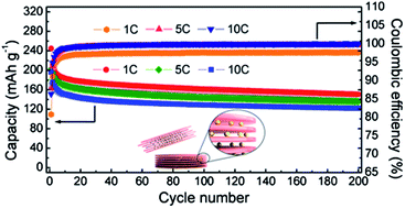
The cycling performances of the nanosheet anodes were tested over 200 cycles (Fig. 7b). The stable cycling behavior was observed at charging–discharging rates of 1, 5 and 10 C. The capacity decreases for initial several cycles, and then stabilized, which can be observed in previous titanium oxide anodes.44–46 The charge capacity at a high rate of 10 C was 122 mA h g−1 after 200 cycles, which is very high considering that the capacity at 1 C is 168 mA h g−1. Therefore, the layer assembled structure of the nanosheets enabled stable cycling performance. The rate performances of the 2D electrodes were also evaluated over a gradual increase in current rates at 0.1, 0.5, 1, 5, 10 and 50 C (Fig. 7c). The specific capacities, which were obtained by selecting the fifth charge capacity at each step, were 192, 175, 166, 141, 123, and 70 mA h g−1, respectively. The capacities cycled at high rates without cycling at low rates are higher than those cycled at various rates from low to high. It is reported that this might to be related to fast kinetics of pseudocapacitive interfacial storage process.3 In addition, the capacity recovered to 165 mA h g−1 when the rate was reduced to 1 C after cycling at high rates, indicating the good structural stability and tolerance for high rate cycling of the electrode materials. These results indicated that 2D titanate anodes have a higher capacity and superior capacity retention than typical TiO2 nanoparticles.36 The enhancement of the cyclic stability and rate capability seems to be a result of reduction of the particle dimensions.

Conclusions

In conclusion, ultrathin 2D titanate nanosheets with a thickness of 0.5 nm were successfully synthesized. Through layer assembly processes, few dozen titanate sheets were hierarchically assembled without deformation of the 2D shape of individual titanate nanosheets, accompanied by removal of most of the hydrophobic surfactants, i.e., the surface of the 2D titanate nanocrystals was transformed to a hydrophilic layer for easy accessibility of Li ions. The layer-assembled 2D nanocrystals were applied as anode materials for LIBs and showed superior electrochemical performances under high rates. The structure of nanocrystals leads to a fast charging reaction of Li ion due to shortened Li ion diffusion path. The ultrathin 2D titanate can be possibly applied to other areas such as catalysis, energy conversion, and development of optoelectronic devices.

Acknowledgements

This work was supported by the Institute for Basic Science (IBS) in Korea.

Notes and references

  1. C. O. Robichaud, A. E. Uyar, M. R. Darby, L. G. Zucker and M. R. Wiesner, Environ. Sci. Technol., 2009, 43, 4227 CrossRef CAS.
  2. T. J. Trentler, T. E. Denler, J. F. Bertone, A. Agrawal and V. L. Colvin, J. Am. Chem. Soc., 1999, 121, 1613 CrossRef CAS.
  3. J.-Y. Shin, D. Samuelis and J. Maier, Adv. Funct. Mater., 2011, 21, 3464 CrossRef CAS.
  4. G. K. Mor, K. Shankar, M. Paulose, O. K. Varghese and C. A. Grimes, Nano Lett., 2006, 6, 215 CrossRef CAS PubMed.
  5. J. Ye, W. Liu, J. Cai, S. Chen, X. Zhao, H. Zhou and L. Qi, J. Am. Chem. Soc., 2011, 133, 933 CrossRef CAS PubMed.
  6. X. W. Lou and L. A. Archer, Adv. Mater., 2008, 20, 1853 CrossRef CAS.
  7. F.-F. Cao, X.-L. Wu, S. Xin, Y.-G. Guo and L.-J. Wan, J. Phys. Chem. C, 2010, 114, 10308 CAS.
  8. Z. Tang, Z. Zhang, Y. Wang, S. C. Glotzer and N. A. Kotov, Science, 2006, 314, 274 CrossRef CAS PubMed.
  9. V. F. Puntes, D. Zanchet, C. K. Erdonmez and P. Alivisatos, J. Am. Chem. Soc., 2002, 124, 12874 CrossRef CAS PubMed.
  10. J. S. Son, X. D. Wen, J. Joo, J. Chae, S.-I. Baek, K. Park, J. H. Kim, K. An, J. H. Yu, S. G. Kwon, S.-H. Choi, Z. Wang, Y.-W. Kim, Y. Kuk, R. Hoffmann and T. Hyeon, Angew. Chem., Int. Ed., 2009, 48, 6861 CrossRef CAS PubMed.
  11. D. Wang, Y. Kang, V. Doan-Nguyen, J. Chen, R. Kngas, N. L. Wieder, K. Bakhmutsky, R. J. Gorte and C. B. Murray, Angew. Chem., Int. Ed., 2011, 50, 4378 CrossRef CAS PubMed.
  12. J.-W. Seo, Y.-W. Jun, S.-W. Park, H. Nah, T. Moon, B. Park, J.-G. Kim, Y. J. Kim and J. Cheon, Angew. Chem., Int. Ed., 2007, 46, 8828 CrossRef CAS PubMed.
  13. M. Park, N. Lee, S. H. Choi, K. An, S.-H. Yu, J. H. Kim, S.-H. Kwon, D. Kim, H. Kim, S.-I. Back, T.-A. Ahn, O. K. Park, J. S. Son, Y.-E. Sung, Y.-W. Kim, Z. Wang, N. Pinna and T. Hyeon, Chem. Mater., 2011, 23, 3318 CrossRef CAS.
  14. H. Wang, H. S. Casalongue, Y. Liang and H. Dai, J. Am. Chem. Soc., 2010, 132, 7472 CrossRef CAS PubMed.
  15. C. Zhang and Y. Zhu, Chem. Mater., 2005, 17, 3537 CrossRef CAS.
  16. J. Jang, S. H. Jeong, S. H. J. Seo, M.-C. Kim, E. Sim, Y. Oh, S. Nam, B. Park and J. Cheon, J. Am. Chem. Soc., 2011, 133, 7636 CrossRef CAS PubMed.
  17. H. G. Yang, C. H. Sun, S. Z. Qiao, J. Zou, G. Liu, S. C. Smith, H. M. Cheng and G. Q. Lu, Nature, 2008, 435, 638 CrossRef PubMed.
  18. T. R. Gordon, M. Cargnello, T. Paik, F. Mangolini, R. T. Weber, P. Fornasiero and C. B. Murray, J. Am. Chem. Soc., 2012, 134, 6751 CrossRef CAS PubMed.
  19. A. M. Ferrari, D. Szieberth and Y. Noel, J. Mater. Chem., 2011, 21, 4568 RSC.
  20. R. Z. Ma, K. Fukuda, T. Sasaki, M. Osada and Y. Bando, J. Phys. Chem. B, 2005, 109, 6210 CrossRef CAS PubMed.
  21. W. Zhou, H. Liu, R. I. Boughton, G. Du, J. Lin, J. Wang and D. Liu, J. Mater. Chem., 2010, 20, 5993 RSC.
  22. Y. Mao and S. S. Wong, J. Am. Chem. Soc., 2006, 128, 8217 CrossRef CAS PubMed.
  23. C.-T. Dinh, Y. Seo, T.-D. Nguyen, F. Kleitz and T.-O. Do, Angew. Chem., Int. Ed., 2012, 51, 6608 CrossRef CAS PubMed.
  24. Y. Omomo, T. Sasaki, L. Wang and M. Watanabe, J. Am. Chem. Soc., 2003, 125, 3568 CrossRef CAS PubMed.
  25. G. Hunt, R. Butler and C. Budd, Philos. Trans. R. Soc., A, 2012, 370, 1723 CrossRef PubMed.
  26. J. Wang, Q. Cheng and Z. Tang, Chem. Soc. Rev., 2012, 41, 1111 RSC.
  27. M. H. Oh, T. Yu, S.-H. Yu, B. Lim, K.-T. Ko, M.-G. Willinger, D.-H. Seo, B. H. Kim, M. G. Cho, J.-H. Park, K. Kang, Y.-E. Sung, N. Pinna and T. Hyeon, Science, 2013, 340, 964 CrossRef CAS PubMed.
  28. S. H. Lee, S.-H. Yu, J. E. Lee, A. Jin, D. J. Lee, N. Lee, H. Jo, K. Shin, T.-Y. Ahn, Y.-W. Kim, H. Choe, Y.-E. Sung and T. Hyeon, Nano Lett., 2013, 13, 4249 CrossRef CAS PubMed.
  29. S. Ding, D. Luan, F. Y. C. Boey, J. S. Chen and X. W. Lou, Chem. Commun., 2011, 47, 7155 RSC.
  30. C. K. Chan, H. Peng, G. Liu, K. Mcilwrath, X. F. Zhang, R. A. Huggins and Y. Cui, Nat. Nanotechnol., 2008, 3, 31 CrossRef CAS PubMed.
  31. J. E. Lee, S.-H. Yu, D. J. Lee, D.-C. Lee, S. I. Han, Y.-E. Sung and T. Hyeon, Energy Environ. Sci., 2012, 5, 9528 CAS.
  32. H. Kim and J. Cho, Nano Lett., 2008, 8, 3688 CrossRef CAS PubMed.
  33. S. Ding, Z. Wang, S. Madhavi and X. W. Lou, J. Mater. Chem., 2011, 21, 13860 RSC.
  34. Y. Q. Wang, L. Gu, Y.-G. Guo, H. Li, X.-Q. He, S. Tsukimoto, Y. Ikuhara and L.-J. Wan, J. Am. Chem. Soc., 2012, 134, 7874 CrossRef CAS PubMed.
  35. S.-H. Yu, A. Pucci, T. Herntrich, M.-G. Willinger, S.-H. Baek, Y.-E. Sung and N. Pinna, J. Mater. Chem., 2011, 21, 806 RSC.
  36. J. Liu, J. S. Chen, X. Wei, X. W. Lou and X.-W. Liu, Adv. Mater., 2011, 23, 998 CrossRef CAS PubMed.
  37. M. Wagemaker, W. J. H. Borghols and F. M. Mulder, J. Am. Chem. Soc., 2007, 129, 4323 CrossRef CAS PubMed.
  38. T. Gao, H. Fjellvåg and P. Norby, Inorg. Chem., 2009, 48, 1423 CrossRef CAS PubMed.
  39. A. Nag, M. V. Kovalenko, J.-S. Lee, W. Liu, B. Spokoyny and D. V. Talapin, J. Am. Chem. Soc., 2011, 133, 10612 CrossRef CAS PubMed.
  40. M. Wagemaker, D. Lützenkirchen-Hecht, A. A. van Well and R. Frahm, J. Phys. Chem. B, 2004, 108, 12456 CrossRef CAS.
  41. S. I. Shin, A. Go, I. Y. Kim, J. M. Lee, Y. Lee and S.-J. Hwang, Energy Environ. Sci., 2013, 6, 608 CAS.
  42. H. Zhang, G. R. Li, L. P. An, T. Y. Yan, X. P. Gao and H. Y. Zhu, J. Phys. Chem. C, 2007, 111, 6143 CAS.
  43. J. Li, Z. Tang and Z. Zhang, Electrochem. Commun., 2005, 7, 62 CrossRef CAS PubMed.
  44. Y.-G. Guo, Y.-S. Hu, W. Sigle and J. Maier, Adv. Mater., 2007, 19, 2087 CrossRef CAS.
  45. Y. Lan, X. Gao, H. Zhu, Z. Zheng, T. Yan, F. Wu, S. P. Ringer and D. Song, Adv. Funct. Mater., 2005, 15, 1310 CrossRef CAS.
  46. L. Ren, Y. Liu, X. Qi, K. S. Hui, K. N. Hui, Z. Huang, J. Li, K. Hunag and J. Zhong, J. Mater. Chem., 2012, 22, 21513 RSC.

Footnotes

Electronic supplementary information (ESI) available: TEM images, IR spectra, XRD patterns and electrochemical impedance spectra. See DOI: 10.1039/c4ra00624k
These authors contributed equally to this work.

This journal is © The Royal Society of Chemistry 2014