Pramod Kumar,
Anuradha and
Indrajit Roy*
Department of Chemistry, University of Delhi, Delhi-110007, India. E-mail: indrajitroy11@gmail.com; Tel: +91-9560721851
First published on 14th February 2014
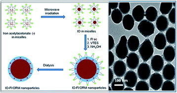
In this manuscript, we report the fabrication of a dual-modality nanoprobe involving the co-encapsulation of fluorophores and iron oxide nanoparticles within ormosil nanoparticles, and their applications in in vitro bioimaging. The entire synthesis, including microwave-mediated precipitation of the iron oxide nanoparticles, was carried out within the non-aqueous core of an oil-in-water microemulsion. The nanoparticles are spherical and monodispersed, with an average diameter of around 150 nm. After synthesis, their composition, stability, crystallinity, as well as their magnetic and optical properties were evaluated. X-ray diffraction studies of the non-encapsulated iron oxide nanoparticles showed them to be crystalline, corresponding to an α-Fe2O3 phase. However, their crystallinity was found to diminish upon ormosil encapsulation. These doped nanoparticles displayed both optical and superparamagnetic properties, by virtue of which they have the potential to serve as dual diagnostic probes. The stability of the fluorophores was found to increase upon nanoencapsulation. In vitro studies have shown that the nanoparticles are non-toxic to cells in culture and undergo efficient cellular uptake, as shown by optical bioimaging. These observations demonstrate the promise of such nanoparticles for non-toxic bioimaging applications.
Nanoparticles, owing to their small size and unique size-dependent properties, present ideal platforms for the fabrication of multimodal agents.6–10 Such multimodal agents are expected to play a leading role in medical diagnostics in the future. Two kinds of nanoparticle have been categorized based on their applications, which are (a) functional nanoparticles with some unique physical parameters, such as iron oxide and metallic nanoparticles, quantum dots, etc., and (b) structural nanoparticles, such as silica, biodegradable polymers, etc., which provide a matrix for hosting one or multiple active agent(s), including smaller functional nanoparticles. Moreover, their surfaces can be aptly functionalized to enhance their circulation in the blood and targeting specificity.11–13
Among the various kinds of functional nanoparticles available, iron oxide nanoparticles perhaps have the most promising applications in the field of medicine.14–17 Their ultrasmall size, superparamagnetic properties, ease of synthesis, and biocompatibility makes them ideally suited for a variety of medical applications. Their most important applications are as contrast enhancement agents in magnetic resonance imaging (MRI) and in the provision of magnetically guided drug delivery.18,19 Their use in magnetic field assisted triggering of local hyperthermia for localized anticancer activity has also been demonstrated.20,21 Moreover, they have been found to be largely biocompatible and non-toxic. Despite these benefits, iron oxide nanoparticles have limited applicability for in vitro bioimaging studies.
It has been proposed that a hybrid diagnostic agent comprising of iron oxide nanoparticles and an optical probe will have the dual capability of MRI and high-resolution optical imaging.1 Dual magnetic and optical probes using organic fluorophores conjugated onto the surface of iron oxide nanoparticles have been reported.22 However, such a design leads to modification of the iron oxide surface with the fluorophores, thereby altering their biodistribution and other pharmacological parameters. Moreover, the exposed fluorophore may get degraded or quenched in the physiological milieu. Therefore, co-encapsulation of iron oxide nanoparticles and fluorophores within inert, mesoporous nanostructures is an alternative strategy for preserving the magnetic and optical properties for potential bimodal bioimaging.
One of the most simple yet versatile structural nanomaterials is made up of organically modified silica (ormosil).23 Highly monodispersed ormosil nanoparticles encapsulating poorly water-soluble molecules, can be easily synthesized in an oil-in-water microemulsion media.24 The encapsulation of active molecules within these nanoparticle matrixes ensures that (a) the encapsulants are protected from environmental insult, and (b) the nanoparticle surface can be further modified for a specific application. Therefore, these doped nanoparticles have found a variety of applications, such as drug delivery in photodynamic therapy (PDT), gene therapy, optical bioimaging, etc.25–27 Their non-toxicity and biocompatibility has also been demonstrated in cultured cells, small animals, and drosophila.28–30 Moreover, their surfaces have been functionalized to incorporate inert polymers and biorecognition agents for enhanced bioavailability and target specificity.27 Taken together, ormosil nanoparticles are well suited to providing robust platforms for the fabrication of multimodal nanoparticles.
In this work, we have used a one-pot method for the synthesis of ormosil nanoparticles, co-encapsulating iron oxide nanoparticle and an organic fluorophore, for providing combined MRI and optical imaging capabilities, respectively. The iron oxide nanoparticles were synthesized in an oil-in-water microemulsion media by the microwave-assisted decomposition of the oil-soluble precursor, ferric acetylacetonate. Next, these magnetic nanoparticles, along with a fluorophore [nile red or ruthenium-tris(2,2′-bipyridyl) dichloride], were co-encapsulated within the ormosil nanoparticles by alkaline hydrolysis of the corresponding organosilane precursor. The resulting nanoparticles were characterized for their size, composition, functionality, crystallinity, along with their magnetic and optical behaviour. The stability of the nanoencapsulated fluorophore vis-a-vis that of the free fluorophore has also been investigated. Following that, they were treated with cultured cells to ascertain their non-toxicity and biocompatibility properties. Concurrently, their uptake into cells in culture was studied by optical bioimaging. The purpose of the above structural, functional, and preliminary in vitro investigations was to explore their potential usefulness for advanced biomedical imaging in the future.
![]() | ||
| Scheme 1 Schematic of the one-pot synthesis of ormosil nanoparticles co-encapsulating iron oxide nanoparticles and fluorophores (IO-Fl–ORM; Fl = NR or RU). | ||
The sizes of the nanoparticles were further analyzed by dynamic light scattering (DLS) measurements. Here, aqueous-dispersed samples of the nanoparticles were taken in glass cuvettes, and analysed using a NANO-ZS series MALVERN ZETASIZER instrument. A He–Ne laser (wavelength 633 nm, power 4 mW) was used as the light source. The number average hydrodynamic size distribution of the nanoparticles was plotted.
The magnetic properties of the dried nanoparticles were investigated using a vibrating sample magnetometer (VSM), Model 3473-70 electromagnet amplifier (CREST Performance CPX 900 power amplifier Instrument). High resolution powder X-ray diffraction (XRD) was used to analyze the phase composition of the nanoparticles, using a Brukar D8 Discover X-ray spectrometer, over the 2θ° range from 20–65° at a rate of 2.58 min−1, using Cu-Kα radiation (λ = 1.54060 Å).
The optical properties (UV-visible absorption and fluorescence emission spectra) of the various aqueous nanoparticulate samples, both with and without NR or RU, were recorded using a Shimadzu UV-1601 spectrophotometer (Shimadzu, Kyoto, Japan) and a Cary Eclipse fluorescence spectrometer (Varian, Palo Alto, CA), respectively.
The stability of the fluorophore RU, both free and nanoencapsulated, was studied using a fluorescence quenching experiment. Here, fixed concentrations of free and nanoencapsulated fluorophores in aqueous medium were treated with various concentrations of the chemical quencher Cu2+ (copper sulfate).32 Fluorescence emission intensities in the absence (Io) and presence (I) of various quencher concentrations was then measured using the Cary Eclipse fluorescence spectrophotometer. A plot was made with the natural log of (I/Io) versus quencher concentration [Q]. The slope of the plot correlated directly with the magnitude of chemical quenching of the fluorophore.
In addition, the fluorescence decay spectra of the free and nanoencapsulated fluorophores were obtained using the time-correlated single photon counting (TCSPC) technique.33 The samples were excited with a NanoLED 460 pulsed diode (Horiba, Jovin Yvon), at an excitation wavelength of 460 nm and pulse duration FWHM of 1.4 ns. Single photon detection was carried out using the TBX-04 (Horiba, Jovin Yvon) picosecond photon-detection module, along with FluoroHub (Horiba) timing electronics. Decay analysis was done using DAS6 software.
For analyzing the cell viability upon treatment with various nanoparticles, one day prior to treatment, the cells were trypsinized and resuspended in fresh media. 100
000 cells per 1 mL fresh media were added to each well of a sterilized 24-well plate, and transferred back to the incubator for attachment and overnight growth. The next day, to the cells at a confluency of 70–80%, three different dosages of the various samples were added, mixed by swirling, and transferred back to the incubator. The added samples were the NR fluorophore, the iron oxide (IO) nanoparticles, as well as the IO and NR co-encapsulated ormosil nanoparticles (IO-NR–ORM). After three days of incubation, the plate was taken out, and the cells in each well were washed three times with sterile PBS, and treated with 100 μL of MTT reagent [3-(4,5-dimethylthiazol-2-yl)-2,5-diphenyltetrazolium bromide, (5 mg mL−1 in PBS)] for 2 hours.24 The resulting blue-coloured formazan crystals were dissolved in DMSO, and the optical density of this solution was recorded at 570 nm using UV-visible spectrophotometry. The optical density of each solution reflected the viability of the cells in each well. The viability percentage of the treated cells was calculated after comparing their optical density with that of non-treated cells (positive control), the latter being arbitrarily assigned 100% viability. The experiment was carried out in triplicate for statistical significance.
For monitoring the uptake of the fluorophore-doped nanoparticles in cells, one day prior to treatment, the cells were seeded in a sterilized 6-well plate (200
000 cells per 2 mL fresh media in each well) and returned to the incubator. Next day, to the cells at a confluency of 70–80%, three dosages of the various samples were added, mixed by swirling, and returned to the incubator for two hours. After incubation, the plate was taken out, the cells in each well washed twice with sterile PBS, and fixed by a mixture of methanol and glycerol. The fixed cells were visualized under a fluorescent Nikon TS-100 inverted microscope, and photographed using a Nikon DIGITAL SIGHT DS-Fi1 Camera (Nikon, Japan).
Their sizes were further determined by dynamic light scattering (DLS), which measures the hydrodynamic diameter. Fig. 1c and d present the DLS data of IO and IO-NR–ORM, respectively, with respective mean hydrodynamic diameters of 8 nm and 155 nm. The DLS data agrees well with the TEM results. Overall, the sizes of IO-NR–ORM nanoparticles are reasonably small (diameter around 150 nm) and monodispersed.
Fig. 2a represents the powder XRD spectrum of the IO nanoparticles, showing diffraction peaks of (012), (014), (110), (113), (024), (116), (214) and (300), which match the characteristic peaks of α-Fe2O3 (Hematite, JCPDS86-0550).34 The sharpness of XRD peaks indicates the crystalline nature of the nanostructures. In comparison, for the XRD spectrum of IO-NR–ORM nanoparticles (Fig. 2b), the peaks appeared highly diminished, demonstrating that the crystalline IO nanoparticles are being shielded by a layer of amorphous ormosil matrix. This crystalline pattern is also observed in the SAED data (ESI, Fig. S1†).
![]() | ||
| Fig. 2 Powder X-ray diffraction (XRD) spectra of (a) free iron oxide (IO) nanoparticles, and (b) ormosil nanoparticles co-encapsulating iron oxide nanoparticles and nile red (IO-NR–ORM). | ||
Fig. 3 shows the magnetization curves of the IO and IO-NR–ORM nanoparticles, measured at room temperature using vibrating sample magnetometry (VSM). Under a large external field, the magnetization of the particles aligns with the field direction and reaches its saturation value for both of these nanoparticles. The pattern is typical of superparamagnetic nanoparticles.35 The saturation magnetization (Ms) values of IO and IO-NR–ORM nanoparticles were 1.0 and 0.80 emu g−1, respectively. This result shows that co-encapsulation with a fluorophore within the ormosil nanoparticles did not significantly alter the magnetic behaviour of IO nanoparticles.
![]() | ||
| Fig. 3 Magnetization (VSM) curves of free iron oxide nanoparticles (IO; solid line), and ormosil nanoparticles co-encapsulating iron oxide nanoparticles and nile red (IO-NR–ORM; broken line). | ||
The optical properties of the various samples were studied by UV-visible and fluorescence spectroscopies. Fig. 4a shows the absorption spectrum of IO nanoparticles, formed after microwave irradiation of ferric acetylacetonate dissolved in micelles. For comparison, the spectra of ferric acetylacetonate dissolved in DMSO, as well as within micelles prior to microwave irradiation, are also provided. It can be seen that the characteristic absorption peaks of ferric acetylacetonate, which are observed around 360 nm and 434 nm, disappear after microwave irradiation, indicating the decomposition of this compound and the formation of nanoparticles. Fig. 4b shows the absorption spectrum of IO-NR–ORM nanoparticles. For comparison, the spectra of NR/micelles, and IO-NR/micelles (after microwave irradiation), are also provided. It can be seen that the characteristic absorption peak of NR (around 560 nm) is substantially diminished after co-encapsulation with IO within micelles, as well as within the ormosil nanoparticles. This can be partially attributed to the shielding of the nile red absorption by scattering from IO and the ormosil nanoparticles, as well as some optical quenching by IO. Nevertheless, the presence of nile red in the nanoparticulate samples is ascertained from their fluorescence emission spectra. Fig. 4c shows the fluorescence spectra of NR in micelles, IO-NR in micelles, and IO-NR–ORM nanoparticles, each excited at 560 nm. The spectra show the characteristic emission of NR in each case; although the peak intensity of NR is diminished and red-shifted upon nanoencapsulation. Similar optical data were observed in the case of IO-RU–ORM nanoparticles (ESI, Fig. S2†).
The motivation behind encapsulating fluorophores within transparent nanomaterials is to enhance the optical stability of the fluorophores. We investigated whether nanoencapsulation enhances the stability of fluorophores, using a chemically-induced fluorescence quenching experiment. From Fig. 5a, it is clear that the free fluorophore is more sensitive to chemical quenching than the nanoencapsulated fluorophore, as the free fluorophores have a steeper fluorescence quenching curve. This indicates the enhanced stability of nanoencapsulated fluorophores. The result of a fluorescence decay experiment is given in Fig. 5b, showing an exponential decay curve for both the free and nanoencapsulated fluorophore. However, the free fluorophore has a steeper fluorescence decay curve, and hence, a shorter fluorescence lifetime, when compared to that of the nanoencapsulated fluorophore. This result indicates that fluorescence lifetime is increased upon nanoencapsulation. These results demonstrate that although the fluorescence intensity diminished for the nanoencapsulated fluorophore, it became more photostable with an enhanced fluorescence lifetime. Overall, these optical data, along with the magnetism data from Fig. 3, show that the dually-doped ormosil nanoparticles show both magnetic and optical behaviour. Thus, they are promising bimodal contrast agents for efficient biomedical imaging.
Finally, in order to study the biocompatibility and non-cytotoxicity of the various nanoparticles, we treated live cells with these nanoparticles and analyzed the cell viability. It can be seen from Fig. 6a that after 72 hours of nanoparticle treatment at various dosages, the cells remained more than 90% viable, even at a dosage as high as 1200 μg mL−1. These data demonstrate that the iron oxide nanoparticles, whether in free form or co-encapsulated with the nile red within ormosil nanoparticles, exert a negligible toxic effect on the cells. This preliminary experiment indicates that these nanoparticles are non-toxic to cells, and can be used in biological applications.
We have also used fluorescence microscopy of cells treated with the IO-NR–ORM nanoparticles to investigate their cellular uptake. The fluorescence of the fluorophore nile red is used to optically track the doped nanoparticle within the cell. Fig. 6c shows a fluorescence image of the treated cells, showing a robust optical signal from the cells (pseudo-coloured in greyscale). The treated cells looked morphologically healthy, further indicating the non-toxicity of the particles. Control experiments using untreated cells did not result in any optical signal from the cells (Fig. 6b). It may be noted from the above result that efficient optical imaging is possible with a nanoparticle dosage of 300 μg mL−1, which was the lowest dosage used in the cell viability studies. These experiments show that the nanoparticles are efficiently taken up by the cells, thus further highlighting the promise of these nanoparticles as non-toxic optical labels in bioimaging studies.
The above observations of in vitro non-toxicity, as well as cellular uptake without the necessity of any active cell-entry strategies, of only fluorophore-doped ormosil nanoparticles have also been observed previously; although the size of the particles were much smaller in the previous reports (diameter below 50 nm).24–30 Their biocompatibility and non-immunogenicity can be attributed to the inertness of the ormosil matrix. The mechanism of their cellular uptake is yet to be determined, though fluid-phase endocytosis is suspected to be the possible route.
Footnote |
| † Electronic supplementary information (ESI) available: EDX and SAED data. See DOI: 10.1039/c4ra00331d |
| This journal is © The Royal Society of Chemistry 2014 |