Dopamine polymerization-induced surface colouration of various materials
Liang Heab,
Vicky Lai Lai Soab and
John H. Xin*ab
aInstitute of Textiles & Clothing, The Hong Kong Polytechnic University, Hung Hom, Hong Kong, P. R. China. E-mail: tcxinjh@polyu.edu.hk; Fax: +852 2773 1432; Tel: +852 2766 6474
bShenzhen Research Institute, The Hong Kong Polytechnic University, Shenzhen, P. R. China
First published on 10th April 2014
Abstract
Bio-inspired by melanins and adhesive of marine mussels, a novel method was developed for colouration of various material surfaces. Using dopamine polymerization to form an adhesive coating, the surface colouration of various materials was easily achieved, including metal, ceramic, polymers and even textile fabrics (resistant to colouration) through a simple dip-coating procedure. The colour appearance of the dyed materials could be tuned in a controllable way due to the reactivity of dopamine with nucleophiles such as amino acids and heterocycle components during its oxidization step. Commercially available colorants could also be used in this procedure to enrich the colour gamut. The surface compositions, morphology and wettability of the dyed surfaces were studied by X-ray photoelectron spectroscopy, scanning electron microscopy and water contact angle measurement, respectively. The obtained results showed that the material surfaces were successfully coloured, which was verified by the obvious changes in surface properties compared to the blank samples. For this colouration method, less energy consumption and dyeing auxiliaries were needed, indicating that it is an environmentally friendly approach. Moreover, it is a promising alternative to the traditional colouration techniques, especially for those materials, which are resistant to colouration.
Introduction
Through evolution, living organisms in nature are highly perfect. Because of their adaptation to specific environments, they often possess unique and fascinating characteristics. Moreover, with the gradual disclosure of their underlying mechanisms, we are able to exploit their unique properties and thus greatly benefit from them.As is well known, melanins are natural pigments formed in the hair and skin of humans and many other animals and plants. For example, melanins in human hair share a common biosynthetic pathway related to 3,4-dihydroxyphenylalanine (DOPA),1,2 which is an amino acid formed by post-translational hydroxylation of tyrosine residues. After enzymatic oxidation, DOPA cyclizes and polymerizes to form melanins. Moreover, in the tidal environment, marine mussels possess remarkable adhesive ability,3 which is rapid, permanent and versatile to virtually all types of solid surfaces, including organic and inorganic materials.4 In their adhesive plaques, it was found that the adhesive has a higher DOPA content, which is believed to play a vital role in the mussel adhesion.5–7
In the chemical structure of DOPA, there are two main functional groups, i.e., catechol and ethylamine, which are also the main functional groups of dopamine. Similar to DOPA, dopamine is also easily oxidized and subsequently self-polymerizes when exposed to air, weak lye and oxidants.8–10 As a result, it is seen as an ideal biomimetic molecule of DOPA. Inspired by this, the use of dopamine was widely reported for preparation of various adhesive ad-layers,10,11 adhesive polymers,12,13 and surface modifications of inorganic materials and nano capsules.14,15 However, to the best of our knowledge, few studies on dopamine polymerization-induced surface colouration of versatile material surfaces have been reported.
Material colouration is also an important functionalization field in modern chemical, biological and material sciences, including decoration,16,17 optical application,18,19 anti-corrosion,20,21 fluorescent labelling and fluorescence switch.22,23 The traditional colouration method can work well on some materials to some extent by strictly complying with the material-dependence rule, but it still lacks efficacy on a broad range of materials.
Inspired by the environmentally friendly nature and versatility of melanins and adhesive of mussels in nature, we achieved the functionalization of carbon nanotubes with polydopamine.24 Based on this successful experience, herein, we advance the study of dopamine polymerization-induced surface colouration of various materials in a simple way, as indicated in Scheme 1. The materials, including metal, ceramic, cellulose, protein fibres and materials resistant to colouration, were studied in detail.
 | ||
| Scheme 1 A description of dopamine polymerization-induced surface colouration. | ||
Experimental
Materials
Cotton, wool and polybenzimidazole (PBI) fabrics were selected as representatives of cellulose, protein fibres and materials resistant to colouration. They were commercially available and were washed in 5‰ sodium dodecyl sulfonate solution for 10 min before use. Then, they were dried in air. Glass and aluminium slides were selected as representatives of ceramic and metal materials. They were commercially available and ultrasonically cleaned first in ethanol for 5 min, and then in deionized water for another 5 min before use. In addition to dopamine (1), 2,4-dihydroxybenzophenone (2), aniline (3), N-(2-hydroxyethyl)aniline (4), L-histidine (5), trans-4-hydroxy-L-proline (6) and 1-(4-sulfophenyl)-3-methyl-5-pyrazolone (7) were also investigated as representatives of phenols, amines, amino acids and heterocycles. Rhodamine B was used as a model of a commercially available colorant. All chemical reagents were obtained from Sigma-Aldrich and used as received.Colouration procedure
Wool fabrics were immersed in 10 wt% dopamine solution overnight at room temperature. Subsequently, they were taken out of the bath and sprayed with 1 wt% oxidant solution (potassium periodate, except as indicated) and air dried. In order to tune the colour appearance of dyed fabrics, a second component of 2–7 was also added to the dopamine solution with the mass ratio of 1![[thin space (1/6-em)]](https://www.rsc.org/images/entities/char_2009.gif) :5. The total concentration of the second component and dopamine was maintained at 10 wt% The dyed fabrics were soaped with 5‰ sodium dodecyl sulfonate solution at 60 °C for 10 min. After drying, the obtained samples were used for colorimetric analysis and tests.
:5. The total concentration of the second component and dopamine was maintained at 10 wt% The dyed fabrics were soaped with 5‰ sodium dodecyl sulfonate solution at 60 °C for 10 min. After drying, the obtained samples were used for colorimetric analysis and tests.
Other materials were immersed in a solution containing 5 mg mL−1 rhodamine B and 5 mg mL−1 dopamine hydrochloride at room temperature overnight. The solution was buffered to pH 8.5 with Tris. After a predetermined time, the resulting samples were taken out, and after washing in deionized water for 10 min, the obtained samples were air dried for measurements.
Colouration characterization
From the reflectance values at the λmax of the coloured materials, the colour yield (K/S) was calculated using the Kubelka–Munk equation.25 Wash fastness was assessed using grey scales according to AATCC Test Method 61-2010 3A at 71 °C.26 Crocking fastness was measured using the crockmeter method according to AATCC Test Method 8-2007. Light fastness was obtained according to the standard method: ISO 105 B02-2013 (xenon-arc lamp). The UV-vis absorption of the coloured materials was tested using a Perkin Elmer Lambda 18 UV-vis spectrometer with a scanning speed of 240 nm min−1.The chemical composition of the material surface was determined by X-ray photoelectron spectroscopy (XPS). XPS measurements were carried out on a SKL-12 X-ray photoelectron spectrometer (Shenyang, China) equipped with a VG CLAM 4MCD electron energy analyzer. XPS is configured with a dual anode source from VG (type XR3E2) and non-monochromatic Mg Kα radiation (1253.6 eV) at a current of 15 mA with an ultrahigh vacuum (<8 × 10⁻¹⁰ Torr). To compensate for surface charging effects, all binding energies were referenced to the C1s hydrocarbon peak at 284.6 eV. The morphology of the samples was investigated by scanning electron microscopy (SEM) on a TEM 3000 Tabletop Microscope (Hitachi, Japan). The wettability of the samples was investigated by water contact angle measurement using a Model CAM-Micro Contact Angle Meter (Tantec, USA).
Results and discussion
Colouration in protein fabrics
Under room temperature, the fresh dopamine solution is colourless and there is no absorption in the visible region, as shown in Fig. 1a. After the addition of oxidant, the solution gradually changed to yellowish brown and an absorption peak centred about 350 nm appeared because of dopamine polymerization induced by oxidation. Polydopamine has an adhesive ability; thus, it was used to prepare adhesive coatings and polymers. This feature could be utilized to increase its fixation on the fibres. Thus, dopamine is used as a precursor molecule for surface colouration. | ||
| Fig. 1 (a) UV-vis absorption spectra of dopamine before and after oxidation; (b) UV-vis absorption spectra of the coloured wool fabrics by using dopamine and various components; (c) images of the coloured wool fabrics; (d) UV-vis absorption spectra and their images of the coloured wool fabrics using dopamine and component 7 in the presence of different oxidants. | ||
Firstly, dopamine was used to dye protein fibres (wool), which showed a natural yellow colour. It was reported that dopaquinone, formed during the oxidation of DOPA, has the ability to react with various nucleophilic species.2 Based on this understanding, during the oxidation process, we attempted to tune the obtained colour appearances by adding different kinds of nucleophiles, such as UV absorbers (2,4-dihydroxybenzophenone), amino acids (L-histidine and trans-4-hydroxy-L-proline), heterocyclic components (1-(4-sulfophenyl)-3-methyl-5-pyrazolone) and aromatic amines (aniline and N-(2-hydroxyethyl)aniline). After colouration, their maximum UV absorptions were centred about 350 nm, but there were differences in their visible absorptions (Fig. 1b). As shown in Fig. 1c, there were noticeable variations in the colour appearance of the dyed fabrics. This tuning of the obtained colour appearance was also seen in their colorimetric parameters, as listed in Table 1. Upon the incorporation of different components into the dopamine polymerization process, the L*, a* and b* values of the obtained colours were obviously changed, and the coloured fabrics had good colour yields with K/S values of 12–18 (Table 1). In addition, their colour appearances could also be tuned by using oxidants with different oxidizing abilities, as shown in Fig. 1d. By using hydrogen peroxide, appearance of a pink colour could be obtained.
| Compt. | K/S | L* | a* | b* | Light fastness | Crocking fastness | |
|---|---|---|---|---|---|---|---|
| Dry | Wet | ||||||
| 1 | 14.9 | 45.8 | 7.4 | 21.3 | 3 | 4–5 | 4–5 |
| 2 | 12.5 | 52.5 | 5.9 | 21.4 | 2–3 | 4–5 | 3–4 |
| 3 | 17.9 | 37.7 | 9.0 | 19.1 | 2–3 | 4 | 3–4 |
| 4 | 15.0 | 40.8 | 12.0 | 18.5 | 2–3 | 4–5 | 4–5 |
| 5 | 15.4 | 42.0 | 8.0 | 19.7 | 3 | 4–5 | 3 |
| 6 | 15.8 | 42.9 | 7.8 | 20.8 | 2–3 | 4–5 | 3–4 |
| 7 | 12.2 | 59.0 | 9.5 | 37.8 | 3–4 | 5 | 4–5 |
The fastness of the dyed fabrics was also investigated, including light fastness, crocking fastness and wash fastness, as listed in Tables 1 and 2. Light fastness of the fabrics dyed by histidine and 1-(4-sulfophenyl)-3-methyl-5-pyrazolone was 3 and 3–4, which was better than that of fabrics dyed by other components. This showed that the incorporation of a heterocyclic component was favourable to improve the light fastness, which is a useful guide for future studies. Moreover, their dry crocking fastness was in the range of 4 to 5, which was a little better than their wet crocking fastness. Concerning the wash fastness listed in Table 2, the ratings of the colour change was in the range from 4 to 4–5, except the sample dyed by 1-(4-sulfophenyl)-3-methyl-5-pyrazolone in the presence of dopamine. This was possibly due to the fact that its incorporation reduced the polymerization degree of dopamine, leading to higher water solubility. Upon the addition of N-(2-hydroxyethyl)aniline, the dyed fabric showed lower colour staining ratings, indicating that N-(2-hydroxyethyl)aniline-modified polydopamine derivatives had an adhesive tendency to wool, polyester and nylon fibres. All other samples had colour staining ratings of 5, showing low colour staining to their adjacent multi-fibres.
It was observed that the wash fastness and crocking fastness of the dyed fabrics were better than their light fastness. This was consistent with the fact that underwater mussels' adhesive proteins suffer less direct light radiation and their photostability is possibly not superior to their adhesion. This possibly resulted in the poor photostability of the colorants biomimicked from underwater mussels' adhesive. However, the photostability of dyed fabrics could be improved through the incorporation of photostable heterocyclic compounds according to the light fastness results.
By expanding the application ranges, it was found that the colouration approach also works well on silk fabrics. However, the results were not so satisfactory on the other textile materials, such as cotton, PET and nylon, which was as expected. Note that wool and silk are protein fibres; thus, they belong to the same type as human hair and skin, in which natural melanin forms. Other fibres cannot provide a similar medium of human hair and skin; thus, their colouration only depended on the adhesive ability of polydopamine derivatives to fibres. Due to the limited understanding of the adhesive mechanism of mussels, the biomimetic adhesion was not very versatile. Based on the fact that mussels can adhere to the anti-adhesive Teflon, the developed colouration method would also work well on other fibres, once the adhesive mechanism of mussels is completely discovered.
In addition, the colour spectrum was still not rich, although several methods were used, including changes in the nucleophilic species, oxidant types and ratio of dopamine to nucleophilic species. Therefore, we propose to utilize commercially available colorants rather than nucleophilic species to produce a large colour gamut.
Colouration in natural and synthetic fabrics
Polydopamine has absorption at about 350 nm, perhaps showing some influence on the colour appearance. Thus, a red colorant of rhodamine B was preliminarily chosen as an example to investigate the colouration method. In order to avoid the influence of oxidants on the chromophore, dopamine polymerization was induced by a typical seawater pH of 8.5 buffered with Tris.The influence of dopamine polymerization on the colour appearance of rhodamine B was first investigated using UV-vis spectroscopy at room temperature. Fig. 2a shows the absorption spectra of rhodamine B in pH 8.5 Tris solution with and without dopamine. Under the experimental conditions, the presence of dopamine did not cause obvious changes in the λmax region of rhodamine B solution, although the absorption intensity at other bands had some increase due to dopamine polymerization. Moreover, there were no obvious naked-eye-visible changes in the solution colour. Furthermore, there was no precipitation observed in the solution even after centrifugation at a speed of 14000 rpm for 10 min. This indicated that the presence of polydopamine did not obviously affect the application of rhodamine B.
 | ||
| Fig. 2 (a) UV-vis absorption spectra of rhodamine B with and without dopamine; (b) visible absorption spectra of fabrics coloured with rhodamine B in the presence of dopamine; (c) water contact angles of PBI before and after colouration with rhodamine B; (d) images showing the wetting properties of water droplets on a 45° slope before and after colouration. | ||
In the presence of dopamine, cotton and PBI fabrics could also be dyed with red colour by rhodamine B. As shown in Fig. 2b, their visible absorptions were centred at 550 nm, which was similar to that of rhodamine B solution. Their colour measurements were also investigated, as listed in Table 3. Compared with the case in the absence of dopamine, the presence of dopamine caused small colour changes in woollen fabrics, but larger colour changes in cotton and PBI fabrics. However, the K/S values of the cotton and PBI fabrics were obviously better than those in the absence of dopamine, which showed the improved dyeability of the fabric.
| Fabric | Presence of dopamine | Absence of dopamine | ||||||
|---|---|---|---|---|---|---|---|---|
| K/S | L* | a* | b* | K/S | L* | a* | b* | |
| Wool | 20.1 | 33.9 | 49.4 | −16.9 | 20.0 | 38.8 | 57.9 | −20.5 |
| Cotton | 14.2 | 32.6 | 33.5 | −3.1 | 4.2 | 58.0 | 47.3 | −28.3 |
| PBI | 14.3 | 28.2 | 22.6 | −0.9 | 5.2 | 50.2 | 33.6 | −2.0 |
Moreover, after colouration, the surface wettability of PBI was obviously changed. Its water contact angle decreased from about 130° to 83° (Fig. 2c). Furthermore, the slope test further verified these changes, as shown in Fig. 2d. The water droplets on the un-dyed PBI surface rolled rapidly down a 45° slope under gravity because of no adsorption on this superhydrophobic surface, while the water droplet on the dyed fabric surface did not roll down the slope. These results clearly showed the different interfacial interactions of the PBI surfaces before and after colouration, demonstrating the successful colouration on the hydrophobic surface in the presence of dopamine.
Surface morphology of the cotton and PBI fabrics before and after colouration was further investigated by SEM. In the case of cotton fibres, it can be seen that the pristine fibre surface is smoother (Fig. 3a, left), while the surface roughness of the dyed fibre increased, showing the obviously contrasting results, as shown in Fig. 3a (right). In addition, the dyed layer was composed of the lamellar ad-layer and aggregated particles. The surface morphology of PBI showed similar results (Fig. 3b). From these results, obvious changes observed in their surface morphology before and after colouration indicated the successful colouration.
 | ||
| Fig. 3 SEM images of the surface morphology of the fibres before (left) and after (right) colouration; (a) cotton, (b) PBI. Bar = 5 μm. | ||
Colouration in inorganic materials
Using this approach, other kinds of materials, such as metal (Al slide) and ceramic (glass), could also be successfully dyed with red colour by using rhodamine B in the presence of dopamine. Their coloured surfaces were characterized with XPS, SEM and water contact angle measurement.XPS analysis was employed to quantitatively determine the chemical composition of the material surface. For the blank materials, the material signals of Si2s and Si2p as well as Al2s and Al2p were observed, while no nitrogen (N1s) was found in the XPS spectra (Fig. 4, left). However, the XPS spectra of the coloured materials showed no obvious substrate signal. On the contrary, peaks from the atomic composition of colouration components were clearly observed, especially the nitrogen signal (N1s) (Fig. 4, right).
 | ||
| Fig. 4 XPS spectra of glass (a) and Al (b) slides before (left) and after (right) colouration. | ||
Compared to the initial surface, the C1s and O1s XPS spectra signals were evidently altered after colouration (Fig. 5). In the case of glass, two peak species at the binding energy of about 284.6 eV and 287.7 eV, attributable to C–C and C![[double bond, length as m-dash]](https://www.rsc.org/images/entities/char_e001.gif) O species, respectively, were observed in the C1s core-level spectrum of pristine glass. After colouration, the C1s core-level spectrum can be curve-fitted into four peak species at binding energies of about 284.6 eV, 285.5 eV, 286.5 eV and 287.7 eV, attributable to the carbon in C–C/CC, C–O, C–N, and CO species, respectively.27 The colouration of a glass surface by the rhodamine B/polydopamine system resulted in two additional peak species for C–N and C–O. Both the C–O and CO signals existed, which confirmed the carboxyl group in rhodamine B and the catechol and quinone groups in polydopamine. The colouration of glass was further confirmed by the O1s core-level spectra of pristine glass and the dyed glass. In the O1s spectrum of pristine glass, a species at the binding energy of about 532 eV was observed. The O1s spectra of the dyed glass can be curve-fitted into two species at binding energies of about 532 eV and 533 eV, attributable to CO and C–O, respectively.10 The C1s and O1s XPS spectra for the coloured Al surface showed similar results.
O species, respectively, were observed in the C1s core-level spectrum of pristine glass. After colouration, the C1s core-level spectrum can be curve-fitted into four peak species at binding energies of about 284.6 eV, 285.5 eV, 286.5 eV and 287.7 eV, attributable to the carbon in C–C/CC, C–O, C–N, and CO species, respectively.27 The colouration of a glass surface by the rhodamine B/polydopamine system resulted in two additional peak species for C–N and C–O. Both the C–O and CO signals existed, which confirmed the carboxyl group in rhodamine B and the catechol and quinone groups in polydopamine. The colouration of glass was further confirmed by the O1s core-level spectra of pristine glass and the dyed glass. In the O1s spectrum of pristine glass, a species at the binding energy of about 532 eV was observed. The O1s spectra of the dyed glass can be curve-fitted into two species at binding energies of about 532 eV and 533 eV, attributable to CO and C–O, respectively.10 The C1s and O1s XPS spectra for the coloured Al surface showed similar results.
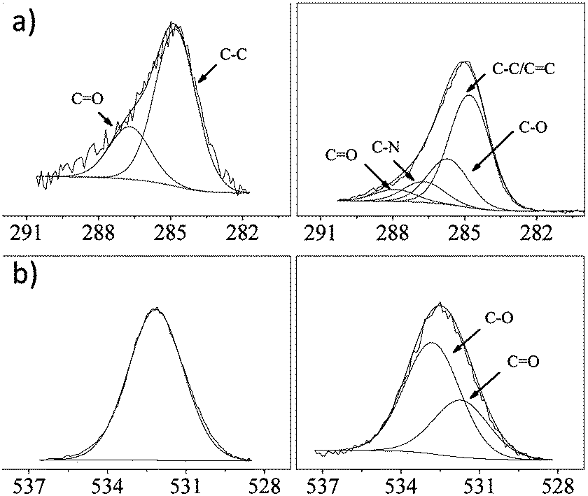 | ||
| Fig. 5 XPS C1s (a) and O1s (b) core-level spectra for glass before (left) and after (right) colouration. | ||
The changes in their surface morphology before and after colouration were further confirmed by the successful colouration, as shown in Fig. 6. In the case of the pristine surfaces, it can be seen that they are much smoother. After colouration, the blank surfaces were completely covered by the lamellar ad-layers and aggregated nano particles. Thus, the coloured surfaces had a higher roughness, showing obviously contrasting results to the blank surfaces.
 | ||
| Fig. 6 SEM images of the surface morphology of glass (a) and Al (b) slides before (left) and after (right) colouration. Bar = 5 μm. | ||
The wettability of the coloured glass and Al slides was also confirmed using water contact angle measurements. For the pristine surfaces, their water contact angles were about 100°, while after colouration, the contact angles decreased to about 80° (Fig. 7). All these results indicated that the material surfaces were successfully coloured by rhodamine B in the presence of dopamine.
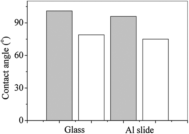 | ||
| Fig. 7 Water contact angles of glass and Al slides before (grey) and after (white) colouration. | ||
Conclusions
In conclusion, we have developed a simple method for surface colouration of various materials via dopamine polymerization in aqueous media. This method could successfully colour different kinds of materials, including textile materials, organic and inorganic materials, and even the materials resistant to colouration. This method showed a material-independence and could be achieved at room temperature, which was completely different from the conventional colouration process. This method expands the application of dopamine and proposes a novel colouration idea; moreover, it is a promising alternative to the traditional colouration process.Acknowledgements
The authors acknowledge funding from the GRF project (no. PolyU 5316/10E) by the Research Grants Council of the Hong Kong SAR Government and the National Natural Science Foundation of China (no. 21376197).Notes and references
- G. Prota, Med. Res. Rev., 1988, 8, 525 CrossRef CAS.
- K. C. Brown, E. Marlowe, G. Prota and G. Wenke, J. Soc. Cosmet. Chem., 1997, 48, 133 CAS.
- J. H. Waite and M. L. Tanzer, Science, 1981, 212, 1038 CAS.
- J. H. Waite, Integr. Comp. Biol., 2002, 42, 1172 CrossRef CAS PubMed.
- M. J. Sever, J. T. Weisser, J. Monahan, S. Srinivasan and J. J. Wilker, Angew. Chem., Int. Ed., 2004, 43, 448 CrossRef CAS PubMed.
- H. Lee, N. F. Scherer and P. B. Messersmith, Proc. Natl. Acad. Sci. U. S. A., 2006, 103, 12999 CrossRef CAS PubMed.
- Q. Lin, D. Gourdon, C. Sun, N. Holten-Andersen, T. H. Anderson, J. H. Waite and J. N. Isradlachvili, Proc. Natl. Acad. Sci. U. S. A., 2007, 104, 3782 CrossRef CAS PubMed.
- H. Lee, S. M. Dellatore, W. M. Miller and P. B. Messersmith, Science, 2007, 318, 426 CrossRef CAS PubMed.
- S. M. Kang, J. Rho, I. S. Choi, P. B. Messersmith and H. Lee, J. Am. Chem. Soc., 2009, 131, 13224 CrossRef CAS PubMed.
- Q. Wei, F. Zhang, J. Li, B. Li and C. Zhao, Polym. Chem., 2010, 1, 1430 RSC.
- H. Lee, Y. Lee, A. R. Statz, J. Rho, T. G. Park and P. B. Messersmith, Adv. Mater., 2008, 20, 1619 CrossRef CAS.
- H. Lee, J. Rho and P. B. Messersmith, Adv. Mater., 2009, 21, 431 CrossRef CAS PubMed.
- G. Westwood, T. N. Horton and J. J. Wilker, Macromolecules, 2007, 40, 3960 CrossRef CAS.
- B. Yu, D. A. Wang, Q. Ye, F. Zhou and W. M. Lin, Chem. Commun., 2009, 6789 RSC.
- J. K. Ryu, S. H. Ku, H. Lee and C. B. Park, Adv. Funct. Mater., 2010, 20, 2132 CrossRef CAS.
- O. J. X. Morel and R. M. Christie, Chem. Rev., 2011, 111, 2537 CrossRef CAS PubMed.
- S. M. Burkinshaw and D. S. Jeong, Dyes Pigm., 2012, 92, 1025 CrossRef CAS PubMed.
- B. Kahr and R. W. Gurney, Chem. Rev., 2001, 101, 893 CrossRef CAS PubMed.
- T. Bullard, K. L. Wustholz, E. D. Bott, M. Robertson, P. J. Reid and B. Kahr, Cryst. Growth Des., 2009, 9, 982 CAS.
- E. E. Oguzie, G. N. Onuoha and A. I. Onuchukwu, Mater. Chem. Phys., 2005, 89, 305 CrossRef CAS PubMed.
- S. Deng, X. Li and H. Fu, Corros. Sci., 2011, 53, 760 CrossRef CAS PubMed.
- M. Sameiro and T. Goncalves, Chem. Rev., 2009, 109, 190 CrossRef PubMed.
- S. Pu, G. Liu, R. Wang and B. Chen, Dyes Pigm., 2013, 98, 238 CrossRef CAS PubMed.
- B. Fei, B. Qian, Z. Yang, R. Wang, W. C. Liu, C. L. Mak and J. H. Xin, Carbon, 2008, 46, 1795 CrossRef CAS PubMed.
- R. McDonald, J. Soc. Dyers Colour., 1980, 96, 486 CrossRef.
- AATCC Test Method 61-2010 Colorfastness to Laundering: Accelerated, Test Condition 3A, AATCC Technical Manual, The American Association of Textile Chemists and Colorists, US, 2011 Search PubMed.
- W. Wang, A. Zhang, L. Liu, M. Tian and L. Zhang, J. Electrochem. Soc., 2011, 158, D228 CrossRef CAS PubMed.
| This journal is © The Royal Society of Chemistry 2014 |