Iqbal M. I. Ismailab,
M. Aslama,
T. Almeelbiac,
S. Chandrasekarana and
A. Hameed*ad
aCentre of Excellence in Environmental Studies (CEES), King Abdulaziz University, Jeddah 21589, Saudi Arabia. E-mail: afmuhammad@kau.edu.sa
bChemistry Department, Faculty of Science, King Abdulaziz University, P.O. Box 80203, Jeddah 21589, Saudi Arabia
cDepartment of Environmental Sciences, King Abdulaziz University, P.O. Box 80203, Jeddah 21589, Saudi Arabia
dNational Centre for Physics, Quaid-e-Azam University, Islamabad 44000, Pakistan
First published on 13th March 2014
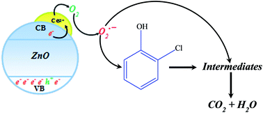
The surface of pre-synthesized hexagonal ZnO was tailored by Ce3+ states. The modified catalyst inveterate enhanced spectral response in the visible region and substantially quenched the luminescence without altering the morphology of the ZnO support. Compared to bare ZnO, the synthesized catalyst exhibited significant high activity both for degradation and mineralization of 2-chlorophenol (2-CP) in sunlight exposure.
ZnO, with a bandgap of 3.2 eV, is an important substitute for TiO2 and sometimes preferred due to its higher photon absorption cross section.10–12 However, ZnO suffers the problems of fast recombination rate and low stability under illumination. It is well established that the photocatalytic activity and the stability of ZnO can be enhanced by modifying its surface.2,13,14 Being renewable, the use of sunlight as an excitation source for the generation of highly energetic reactive radicals is a conspicuous option and can expand the scope of this technology. However, the non-availability of sunlight active photocatalysts limits the use of this cheap natural photon source in photocatalysis. Therefore, for the future and widespread commercial use of this technology, it is highly desirable to develop either new sunlight responsive active photocatalysts or modify the existing active photocatalysts for better sunlight response and activity. Various strategies are reported in the literature to make ZnO responsive in the visible region.2 These strategies include metal ion doping, composite formation, surface modification by metal impregnation and insertion of non-metals such as S and N. Several reports are available in the literature to enhance the spectral response and photocatalytic activity of ZnO by using Ce3+ and Ce4+ ions. However, these approaches are either based on composite formation or on inserting (doping) the Ce3+ or Ce4+ ions (CexZn1−xO) in the lattice of ZnO.15
In this communication, considering photocatalysis as a surface phenomenon, we adopted an innovative approach for suppressing the undesired e−–h+ pair recombination, improved absorption of photons in the visible region and enhanced photocatalytic activity without disturbing the lattice of ZnO. The impregnated Ce3+ ions, instead of being inserted into the lattice, reside at the surface of ZnO by sharing the singly charged surface oxygen to form Zn2+–O–Ce3+ type structures.
The comparison of the solid-state absorption spectra of bare and 10% Ce3+ impregnated ZnO powders is presented in Fig. 1(a), where, compared to pure ZnO, an increased absorption in the visible region (400–510 nm) can be observed. The appearance of two absorption edges in the graphical evaluation of bandgap (Fig. 1(b)), the major absorption edge at ∼3.1 eV that represents the ZnO support and the additional edge at ∼2.4 eV, predicts that Ce3+ entities deposit layer-by-layer at the surface of ZnO to form surface bound Ce2O3. As presented in Fig. 1(c), the presence of surface Ce3+ entities significantly suppresses the luminescence of pure ZnO, which indicates the enhanced lifetime of excited states. The intensity of the characteristic bands of ZnO at 382 nm, 429 nm and 505 nm16 is decreased by ∼68%, ∼100% and ∼50%, respectively, which specifies that the presence of Ce3+ significantly affects the bandgap, Zn interstices and surface oxygen (Zn–O−) vacancies. The depletion of surface oxygen (the band at 505 nm) for Ce3+ impregnated ZnO, as evidenced by PL spectra, supports the sharing of surface oxygen by the impregnating Ce3+ with Zn2+.
![]() | ||
Fig. 1 Comparison of (a) solid state absorption spectra, (b) graphical evaluation of bandgaps, (c) PL spectra of bare and Ce3+ impregnated hexagonal ZnO. |
The SEM analysis (Fig. S1, ESI†) revealed that the surface deposition of Ce3+ does not alter the morphology of the hexagonal ZnO support and the incoming Ce3+ ions are evenly distributed on the ZnO surface. Noticeably, the diminishing of sharp edges of the hexagonal ZnO particles in Ce3+ impregnated powder further verifies the homogeneous distribution of Ce3+ entities. The XRD pattern of Ce3+ impregnated ZnO is presented in Fig. S2 (ESI†). The major reflections at 2θ values of 31.957, 34.603, 36.437, 47.711, 56.758, 63.012, 68.098, 69.236, 72.714 and 77.102 were matched with hexagonal ZnO (JCPDS-36-1451). The appearance of reflection at 2θ = 28.85° represented the hexagonal Ce2O3 (002) phase (JCPDS-23-1048). The shifting of the observed reflection at 2θ = 28.85° by 0.6 degrees compared to 2θ = 29.45° (JCPDS-23-1048) confirms the existence of Ce2O3 as a surface bonded entity. The other reflections due to surface Ce2O3 are entrapped in high intensity ZnO peaks. The calculated crystallite size of the surface Ce2O3, as evaluated by the extrapolation of reflections at 2θ = 28.85°, was ∼6.6 nm. The change in the oxidation state of Ce3+ was not evidenced.
The photocatalytic activity of the synthesized Ce3+ impregnated ZnO, in comparison to that of bare ZnO, was evaluated for the degradation and mineralization of 2-chlorophenol (2-CP). The optimized amount (100 mg) was suspended in 100 cm3 of 30 ppm solution. The catalyst/2-CP suspension was exposed to sunlight in a glass reactor and the progress of the reaction was monitored by analyzing the samples by HPLC and total organic carbon (TOC) for degradation (loss of aromaticity) and mineralization (conversion to CO2 and H2O) progress respectively, at regular intervals of time. Along with monitoring of the decrease in 2-CP concentration, HPLC analysis was also used for the estimation of intermediates formed during the degradation process. The comparison of HPLC degradation profiles of 2-CP in the sunlight exposure over bare and Ce3+ impregnated ZnO is presented in Fig. S3 (ESI†). The Ce3+ loaded catalyst showed higher activity for the degradation of 2-CP compared to bare ZnO. For bare ZnO, 2-CP is degraded with the formation of a high concentration of intermediates compared to impregnated catalyst, where the simultaneous degradation of 2-CP substrate as well as intermediates was observed.
The percentage degradation of 2-CP as a function of sunlight exposure time is presented in Fig. 2(a). In the initial 20 minutes of sunlight exposure, compared to ∼40% 2-CP degradation for bare ZnO, ∼86% of 2-CP substrate was removed in the presence of Ce3+ loaded ZnO. The impregnated catalyst completely (≥99.5%) degraded 2-CP substrate in 90 minutes of exposure while for bare ZnO ∼95% of 2-CP was removed in 150 minutes. Compared to impregnated catalysts, a low TOC removal, as presented in Fig. 2(b), was observed for bare ZnO. Compared to ∼98% TOC removal for Ce3+ loaded catalyst in 150 minutes of exposure, 49% organic carbon removal was observed for bare ZnO, which was even lower than that observed for Ce3+ impregnated catalyst in 20 minutes of exposure.
![]() | ||
Fig. 2 Comparison of (a) HPLC based percentage degradation (b) TOC removal of 2-NP over bare and Ce3+ impregnated hexagonal ZnO. |
The comparison of degradation and TOC removal profiles of 2-CP (Fig. 2(a) and (b)) clearly elaborates that although ZnO is capable of degrading 2-CP in natural sunlight, it fails to carry out efficient mineralization (TOC removal). On the other hand, Ce3+ impregnated catalyst showed an excellent ability of simultaneous degradation and mineralization of 2-CP substrate. ZnO is a wide bandgap (3.1 eV) photocatalyst and can harvest only 3–5% of total incident photons (UV region) of the sunlight. Another negative aspect associated with ZnO is the high recombination of charge carriers. Therefore, it fails to deliver the photo-excited electrons to reductants, and thus a significant number of photons are lost in the recombination process. The significantly high simultaneous removal of 2-CP substrate and intermediates led to the conclusion that a “trap” and “transfer” synergy exists between ZnO and impregnated Ce3+ states.
The rapid degradation and mineralization of 2-CP substrates suggest the involvement of superoxide anion radicals, both in the degradation and in the mineralization process; however, further investigation is needed in this regard. These investigations are underway in our laboratories and the results will be reported in detail. The anticipated plausible mechanism of 2-CP mineralization is presented in Scheme 1.
The photocatalytic activity of the synthesized catalyst was also evaluated in the visible portion of sunlight by using Pyrex glass as the UV filter. A sustained activity of the catalyst was observed for Ce3+ impregnated catalyst compared to ZnO where a substantial decrease in the degradation of 2-CP was observed.
The solid-state absorption and diffuse reflectance spectra (DRS) of bare and Ce3+ impregnated ZnO catalysts were recorded by a Perkin Elmer UV-visible diffuse reflectance spectrophotometer in the 190–900 nm range. DRS were further manipulated to evaluate the absorption edges by plotting (F(R) × hν)1/2 versus hν (photon energy, eV). HORIBA Scientific (Jobin Yvon Fluoro Log 3), France, macro PL system was used to record the photoluminescence spectra of bare and Ce3+ modified ZnO powders at 325 nm excitation wavelength. The emissions were recorded from 350 to 650 nm. The powder XRD patterns of bare and Ce3+ impregnated powders were recorded by an Xpert X-ray powder diffractometer (Philips PW1398) with a Cu Kα radiation source from 20° to 80° (2θ) with a step time of 3 seconds and step size of 0.05°. Scherer's equation was applied on the main reflections to evaluate the crystallite size of various phases. The changes in the morphology of the ZnO base and the surface presence of Ce3+ species were examined by a field emission scanning electron microscope (JEOL JSM 6490-A).
Footnote |
† Electronic supplementary information (ESI) available. See DOI: 10.1039/c4ra00097h |
This journal is © The Royal Society of Chemistry 2014 |