Development of novel alginate lyase cross-linked aggregates for the oral treatment of cystic fibrosis

G. A. Islan a, Y. N. Martineza, A. Illanesb and G. R. Castro*a
aNanobiomaterials Laboratory, Institute of Applied Biotechnology (CINDEFI, UNLP-CONICET-CCT La Plata), Department of Chemistry, School of Sciences, Universidad Nacional de La Plata, Calle 47 y115, C.P. 1900, La Plata, Argentina. E-mail: grcastro@gmail.com; Fax: +54 221 483 37 94 ext. 132/103; Tel: +54 221 483 37 94 ext. 132/103
bEscuela de Ingeniería Bioquímica, Universidad Católica de Valparaíso, Avenida Brasil 2147, Valparaíso, Chile

Received 20th December 2013 , Accepted 13th February 2014

First published on 13th February 2014


Abstract

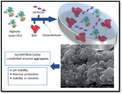
Alginate lyase (AL) from Sphingobacterium multivorum is unsuitable for oral delivery because of its rapid inactivation under acidic conditions. The synthesis of a novel crosslinking enzyme aggregate (CLEA) of AL (AL-CLEA) is proposed. AL precipitation with 95% ammonium sulfate and combined with low methoxylated pectin (LMP), showed 100% precipitation yield. Crosslinking with glutaraldehyde reduced the AL-CLEA activity to less than 1%, but addition of bovine serum albumin (BSA) and LMP during AL-CLEA synthesis increased the activity yield to 14.7%. AL-CLEA exposed to simulated gastric conditions (pH 1.2 to 3.0) showed more than 70% retention of enzymatic activity. Moreover, AL-CLEA showed thermal stability at temperatures over 37 °C. Stability against chemical denaturants (ethanol, acetone and propylene glycol) showed that AL-CLEA was 14 times more stable than free AL in all cases. Finally, a 25% viscosity reduction of alginate solution was achieved with AL-CLEA. This is the first report of AL-CLEA synthesis and evaluation.

Introduction

Over the last few years, alginate lyase (AL) has gained attention due to its ability to hydrolyze bacterial alginate produced by opportunistic pathogens.1 AL can be extracted from different microbial sources and it is classified as an endohydrolase enzyme acting over β-1,4-glycosidic linkages via a β-elimination reaction to produce mono-, di-, and tri-oligosaccharides.2 Recently, the use of AL in cystic fibrosis (CF) treatments has become an interesting alternative, since it is effective in the reduction of the viscosity of alginate produced by the mucoid Pseudomonas aeruginosa strains present in the lung and intestine of patients. It has been demonstrated that AL promotes diffusion of aminoglycosides through the extracellular polysaccharide of mucoid strains.1 Cystic fibrosis (CF) is an inherited autosomal disease produced by defective chloride transport and is the most common lethal genetic disease amongst Caucasians.3 The main colonizing bacterium in CF pathologies is Pseudomonas aeruginosa, producing considerable amounts of the exopolysaccharide alginate. Alginate is a linear anionic copolymer of (1,4)-linked β-D-mannuronate and α-L-guluronate residues, which is undegradable by mammalian cells.4 Alginate accumulates and increases viscoelasticity of pulmonary and intestinal secretions causing severe problems of gas transport (O2 and CO2) and food adsorption problems in CF. Mucoid strains are capable of surviving aggressive antibiotic therapies and once infection is established it becomes extremely difficult to eradicate.5 Microorganisms first colonize lung surface and then spread out colonizing the intestinal tract. As a consequence, pancreas pathways are clogged and proper digestion is impaired, increasing mortality due to malnutrition.

The co-immobilization of AL with Ciprofloxacin encapsulated into biopolymeric microspheres was recently reported by our laboratory as a promising system to reduce the visco-elasticity of CF secretions, allowing the antibiotic diffusion into the biofilm where pathogens are embedded.6 However, three major obstacles hamper the use of AL in oral and/or pulmonary delivery: (1) enzyme sensitivity to environmental denaturants such as salts and solvents commonly used in formulations, (2) the loss of enzyme activity at pH values below 3.0,6 and (3) enzyme inactivation at temperatures over 37 °C. These adverse conditions are present in oral administration, because of the gastric acidic environment, or when temperature is increased over 40 °C in patients with persistent fever. Crosslinked enzyme aggregates (CLEAs) of AL (AL-CLEAs) have been proposed as an alternative to conventional surface immobilization in order to overcome inactivation. CLEAs have been developed with a wide variety of enzymes such as laccases, fitases, galactose oxidases, trypsin, glucose oxidases, β-galactosidases and alcohol dehydrogenases. In all cases, the enzyme activity was not only retained but also enhanced, highlighting CLEAs as versatile and reusable biocatalysts.7 CLEAs are produced by enzyme precipitation under non-denaturing conditions and crosslinking with bifunctional reagents. Precipitating agents and chemical crosslinkers have to be selected and conditions optimized to maximize enzyme activity in the protein aggregates form.

The use of anionic biopolymers to co-precipitate the AL represents an attractive alternative to get fully physically stable CLEAs.8 Alginate and pectins are interesting biopolymers currently used in biomedical applications, being non-toxic, non-immunogenic and highly biocompatible.9 Pectins are water-soluble polymers composed of linear polysaccharides of partially methoxylated poly α-(1,4)-D-galacturonic acids present in plant cell wall. Pectins are grouped according to their degree of esterification (DE) into low methoxylated (LMP) with DE below 40%, medium methoxylated (MMP) with DE between 40 and 60%, and high methoxylated (HMP) with DE higher than 60%.10

The aim of the present study was to produce AL-CLEAs by biopolymer co-precipitation with alginate and different pectins, followed by crosslinking with bifunctional reagents. The formulations were tested under different denaturing environments including extreme pHs, high temperatures, solvents and high ionic strength.

Results and discussion

Determination of the best conditions for AL-CLEA synthesis

AL precipitation was performed by several agents commonly used in protein fractionation.11 Methanol, ethanol and acetone (75%, v/v) completely precipitated AL. The properties of polar solvents, like dielectric constant and log[thin space (1/6-em)]P, make them suitable to displace the water molecules directly involved in the enzymatic hydrolysis of alginate, affecting the active site residues which participate in hydrogen bonding with the substrate.2 Besides, the polar organic solvents tested as precipitating agents were discarded because of full AL inactivation. Protein precipitation by salting out mechanism is based on the desolvation of polar residues of the protein due to the presence of high salt concentration in the medium and the increase of hydrophobic interactions in the protein core.12 Ammonium sulfate (AS) was used as salting out agent and showed no enzyme inactivation. The increase of ammonium sulfate concentration in solution led to an increase in AL precipitation without losing activity (Fig. 1).
image file: c3ra47850e-f1.tif
Fig. 1 Precipitation of AL at increasing concentrations of ammonium sulfate. image file: c3ra47850e-u1.tif, supernatant; image file: c3ra47850e-u2.tif, resuspended precipitate; image file: c3ra47850e-u3.tif, total.

However, enzyme precipitation was only around 60% with 95% (v/v) ammonium sulfate, probably because of the low concentration of enzyme present in solution.

In this scenario, a co-precipitant could be a feasible alternative in order to get full enzyme precipitation by combining salting out process with biopolymer flocculation. Alginate and pectin were selected because they do not inactivate AL (data not shown). AL co-precipitation yields were 30 and 70% with HMP and alginate respectively, meanwhile with LMP 100% AL precipitation was reached (Fig. 2a).


image file: c3ra47850e-f2.tif
Fig. 2 Precipitation of AL with combined precipitating agents at enzyme–biopolymer ratio of 1[thin space (1/6-em)]:[thin space (1/6-em)]10: (a) ammonium sulfate (95%) with different biopolymers (1.0%): alginate (ALG), high methoxyl pectin (HMP) and low methoxyl pectin (LMP). (b) Effect of increase in LMP concentrations on AL precipitation.

The difference in enzyme precipitation by LMP could be explained in terms of the molecular structure of the biopolymers.10

LMPs are more polar than HMPs because of the high availability of free carboxylate groups. Pectins usually precipitate in the presence of high concentrations of ammonium sulfate as ammonium pectinates.13 The free carboxylate groups of LMP produce higher salt precipitation yields, and are able to improve AL precipitation more than HMP. Also, LMP have hydrophobic regions because of some methoxylated zones on the biopolymeric chain which can interact with hydrophobic residues of AL (Scheme 1). In consequence, precipitation yield is higher using LMP than HMP and alginate because of the contribution of hydrophobic and ionic zones. Hydrophobic regions of HMP strongly interact with AL, but the biopolymer is not able to be fully precipitated by the ammonium salt. In the presence of HMP, AL precipitation yield is lower than only with ammonium sulfate because most of the enzyme remains soluble and bound to the biopolymer (Fig. 1).


image file: c3ra47850e-s1.tif
Scheme 1 (a) HMP and LMP structure. (b) Interactions of the hydrophobic and hydrophilic regions of HMP and LMP with AL.

In order to determine the minimum concentration of LMP required to completely precipitate AL, decreasing concentrations of LMP were tested. As shown in Fig. 2b, full AL precipitation was only obtained at 1.0% (w/v) at an enzyme–biopolymer ratio of 1[thin space (1/6-em)]:[thin space (1/6-em)]10, precipitation being reduced at lower LMP concentrations down to less than 60% at 0.1% (w/v).

The most common crosslinking agent used in CLEA development is glutaraldehyde. However, AL was almost fully inactivated in the presence of this agent (Table 1). In order to avoid inactivation, BSA was proposed as a protective and inert agent.14 In fact, glutaraldehyde would mostly interact with BSA residues because its molecular weight is at least two times higher than the one of AL (Scheme 2).

Table 1 Alginate lyase activity after incubation with crosslinker and protective agents for 1 hour
Effectors Relative activitya (%)
a Relative to AL activity at optimal conditions (see Materials and methods).
Cross-linker
Glutaraldehyde (Glut) 0.9 ± 0.3
 
Protective agent
BSA–AL (5[thin space (1/6-em)]:[thin space (1/6-em)]1) 100.0 ± 5.1
BSA–AL (10[thin space (1/6-em)]:[thin space (1/6-em)]1) 100.0 ± 3.0
 
Combinations of both agents
BSA–AL (5[thin space (1/6-em)]:[thin space (1/6-em)]1)/Glut 20.5 ± 5.6
BSA–AL (10[thin space (1/6-em)]:[thin space (1/6-em)]1)/Glut 32.7 ± 3.0


image file: c3ra47850e-s2.tif
Scheme 2 (A) Glutaraldehyde reaction with amine residues of AL. (B) Glutaraldehyde reaction with amine residues of BSA incubated with AL preserving AL active site.

BSA–AL ratio of 5[thin space (1/6-em)]:[thin space (1/6-em)]1 and 10[thin space (1/6-em)]:[thin space (1/6-em)]1 did not affect AL activity in aqueous solution (Table 1). The protective effect of BSA was then evidenced when soluble AL and BSA were incubated with the crosslinkers. Protection was higher at BSA–AL ratio of 10[thin space (1/6-em)]:[thin space (1/6-em)]1, being the residual activity of AL in the presence of BSA 36 times higher than in the presence of glutaraldehyde alone.

Considering the previous results, the effect of the different components of AL-CLEA was evaluated on four different formulations. Table 2 shows AL-CLEA yields in the four cases.

Table 2 AL-CLEA yield using different formulations. Abbreviations: HMP, high methoxylated pectin; LMP, low methoxylated pectin; BSA, bovine serum albumin
CLEA formulation of AL AL-CLEA yield (%)
AL/HMP 0.05 ± 0.00
AL/LMP 0.35 ± 0.04
AL/BSA 4.53 ± 0.36
AL/LMP/BSA 14.74 ± 1.72


Pectin based AL-CLEAs showed the lowest values with 0.05 and 0.35% yield for HMP and LMP respectively.

It is evident that AL inactivation by glutaraldehyde is produced during CLEA synthesis. According to AL crystalline structure,15 the active site is composed of His and Tyr residues. Carboxylate and amino groups may react with glutaraldehyde leading to rigidification and inactivation of the enzyme. In the presence of BSA, however, AL-CLEA yield was increased to 4.5%, confirming the BSA protective effect (Table 1). In the presence of BSA, the crosslinker reacts mainly with carboxylate and amino residues of BSA rather than with the active site of AL (Scheme 2). In this way, the use of both BSA and LMP was tested, obtaining a significant increase in AL-CLEA yield up to 14.7%. The protective synergistic effect on AL is explained in terms of biopolymers affecting the enzyme microenvironment. The presence of ionic polymers near to the proteins could have two effects: (i) the change in the microenvironment due to the ionic effect of the polymers and (ii) the reduced mobility of the enzyme structure caused by multipoint polymer–enzyme interaction and cross-linking.

Characterization of CLEAs

SEM images were obtained of the different CLEA formulations. In AL/HMP CLEA, size distribution covered the 300 to 700 nm diameter range with a low production of aggregates, as seen in Fig. 3a. Apparently, the biopolymer is not fully crosslinked forming an aggregate with the enzyme, remaining instead as a film. In the case of AL/LMP CLEA, a bimodal size distribution was found, with aggregates populations of 400 nm and 700 nm, and more aggregates were observed (Fig. 3b). These differences can be explained based on the results obtained during the precipitation experiments where AL precipitation yield was increased in the presence of LMP instead of HMP (Fig. 2).
image file: c3ra47850e-f3.tif
Fig. 3 SEM images of the pectin based CLEAs formulations: (a) AL/HMP; (b) AL/LMP; at 10[thin space (1/6-em)]000 magnification.

BSA based formulations showed a homogeneous size distribution of aggregates with an average of 135 ± 21 nm (Fig. 4a). Similar results were observed in a “combi-CLEA” of Pseudomonas cepacia lipase and penicillin acylase, where in the presence of BSA enzymatic activity was increased and more homogeneous size and morphology distribution was achieved.16 On the other hand, AL/BSA/LMP formulation showed gnocchi like shape aggregates of an average size around 3 μm, even though lower size aggregates were observed (Fig. 4b).


image file: c3ra47850e-f4.tif
Fig. 4 SEM images of different AL-CLEA formulations: (a) AL/BSA; (b) AL/LMP/BSA at 10[thin space (1/6-em)]000 magnification.

Stability of soluble AL and AL-CLEAs

AL/LMP/BSA CLEAs incubated under denaturing conditions for the soluble enzyme (pH = 1.2 for 30 and 120 min), retained 80 and 60% of initial activity respectively (Fig. 5).
image file: c3ra47850e-f5.tif
Fig. 5 Stability against pH of CLEA formulations incubated at pH = 1.2 and 37 °C for 30 (image file: c3ra47850e-u4.tif) and 120 (image file: c3ra47850e-u5.tif) minutes.

In order to infer which of the formulation components offered the protective effect, AL/LMP and AL/BSA CLEAs were also incubated at the same experimental conditions. Fig. 5 shows that the presence of LMP was required to obtain 3.5 and 2 times more residual activity than the BSA component after 30 and 120 min of incubation respectively. Moreover, the combination of both LMP and BSA produced a synergistic protective effect on AL-CLEA. The hydrophobic regions of the BSA and LMP may have precluded the free diffusion of H+ to sensitive regions of AL avoiding enzyme inactivation, as previously suggested.17 Based on the above results, AL/LMP/BSA CLEA was selected for further experiments.

Enzymatic stability of the selected biocatalysts and free AL was studied in the pH range from 1.2 to 8.2, by measuring residual activity after 1 hour incubation (Fig. 6). Soluble AL and AL-CLEA were stable in the pH range from 5.0 to 7.4. However, enzymatic activity of the soluble AL decreased by 60% at pH 4.0 and was completely inactivated below pH 3.0 after 1 hour incubation. On the contrary, AL-CLEA was completely active after incubation at pH 3.0 and activity decreased only by 30% after incubation at pH 1.2 and 2.0 under the same experimental conditions.


image file: c3ra47850e-f6.tif
Fig. 6 Effect of pH on the stability (residual activity after 1 hour of incubation) of soluble alginate lyase (image file: c3ra47850e-u6.tif) and AL/LMP/BSA CLEAs (image file: c3ra47850e-u7.tif) at 37 °C.

Thermal stability was studied in the range from 25 to 60 °C by measuring residual activity after 1 hour incubation at pH 7.4. Significant differences (p ≤ 0.05) were found for AL-CLEAs and soluble AL after incubation at temperatures higher than 40 °C. CLEA showed enzymatic residual activity of 50, 70 and 10% higher than free AL at 40, 45 and 60 °C respectively (Fig. 7).


image file: c3ra47850e-f7.tif
Fig. 7 Effect on AL stability (residual activity) after one hour incubation at different temperatures: (image file: c3ra47850e-u8.tif) soluble alginate lyase and (image file: c3ra47850e-u9.tif) AL/LMP/BSA CLEA at pH 7.4.

In addition, to obtain an approach to the physiological environment generated in a patient with fever, stability of soluble AL and AL-CLEAs was tested at pH 1.2 (gastric pH) under incubation temperatures of 40 and 45 °C per one hour. While soluble enzyme showed a complete inactivation under both experimental conditions, the AL-CLEAs showed 52.6 ± 5.0% and 43.0 ± 2.1% residual activity at 40 and 45 °C (pH = 1.2) respectively. The results enhance the AL-CLEAs formulation for potential use in CF patients suffering fever symptoms.

In order to study which of the components offered the thermal protective effect, BSA and LMP/CLEA were incubated at 60 °C, condition at which the free enzyme is fully inactivated (Table 3).

Table 3 Thermal stability (residual activity after 1 hour of incubation) of soluble AL and AL-CLEAs at 60 °C and pH 7.4
Biocatalyst Residual activity (%)
Soluble AL 0.0
AL/LMP CLEA 14.2 ± 4.1
AL/BSA CLEA 0.7 ± 0.2
AL/BSA/LMP CLEA 11.1 ± 1.2


It was observed that LMP is needed to provide thermal protection to AL, because no thermal protective effect was observed when only BSA was incorporated into the CLEA.

As mentioned before, soluble AL was unstable in the presence of organic solvents. Accordingly, the stability of CLEA formulation (AL/LMP/BSA) was tested in solvents commonly used in the pharmaceutical industry and relevant for drug delivery purposes since water availability at the tissue interface will be scarce. AL/LMP/BSA CLEA was incubated in ethanol, acetone and 1,2-propylene glycol (50%, v/v) showing AL residual activity of 6.4, 18.0 and 37.5% respectively. On the other hand, soluble AL showed enzyme activity lower than 2.0% for all the solvents tested (Fig. 8).


image file: c3ra47850e-f8.tif
Fig. 8 Effect on AL stability (residual activity) after one hour incubation at organic solvents at 50% (v/v) at 37 °C.

The effect of NaCl up to 1 M on the stability of soluble AL and AL/LMP/BSA-CLEA was determined by measuring residual activity after one hour of incubation at 37 °C and pH = 7.4. The activity of both biocatalysts remained unaffected after exposure for such period of time at the NaCl concentrations in the tested range, with no significant differences (p ≥ 0.05) between samples.

Reusability

Reuse of the selected biocatalyst (AL/LMP/BSA-CLEA) is relevant since the potential use of the enzyme in drug delivery devices is based on its performance under different and harsh environmental interface conditions involving the drug delivery device, the microbial biofilm, and the tissue.

The AL-CLEA activity was reduced to 40% after the first cycle, but remained constant from then on, at least up to the fifth cycle. This drastic reduction can be explained because the aggregates were not robust enough, and leaking of unbound enzyme was produced after the first cycle, and also, because the reaction products cannot diffuse out of the matrix and partially inhibit the enzyme as previously suggested.18

Viscosity reduction of alginate solution

Viscosity reduction of alginate aqueous solution is an important factor for the treatment of cystic fibrosis patients. This viscosity reduction leads to higher diffusion of antibiotics through the bacterial biofilm.1 In this way, rheological experiments were carried out showing that viscosity reduction of 25% was produced after incubation of AL/LMP/BSA-CLEA in 2% (w/v) alginate solution at 37 °C for 3 hours. Considering that the mucoid alginate present in the patients is less than 2% (w/v), this reduction is quite significant for treatment.6 These results suggest the advantage of using crosslinked enzyme aggregates as a stable and functional alternative for AL oral administration.

Experimental

Materials

AL from Sphingobacterium multivorum (E.C. 4.2.2.11, poly(alpha-L-guluronate)lyase) was supplied by Sigma-Aldrich (Buenos Aires, Argentina). Acetone, ethanol and 1,2-propylene glycol were of analytical grade provided by Anedra S.A. (Buenos Aires, Argentina). Ammonium sulfate of analytical grade was supplied by Merck (Darmstadt, Germany). Alginate was supplied by Monsanto (St. Louis, MO, USA) and LMP and HMP were gifts from CP Kelco (Buenos Aires, Argentina).

Enzyme assay

AL activity was determined by monitoring the cleavage of alginate to its monosaccharide products, which absorbed at 233 nm. For free AL, a volume of 20 μl (AL stock 4.0 EU ml−1, 100 μg ml−1 protein concentration) of enzyme solution was mixed with 480 μl of 2.0% alginate solution (in 25 mM phosphate buffer, pH 7.4) and incubated at 37 °C for 30 minutes. With immobilized AL, a defined mass of CLEAs of each formulation was weighted in order to have the same enzymatic units than the soluble AL tested and mixed with 500 μl of 2.0% alginate solution in 25 mM phosphate buffer, pH 7.4 and incubated at 37 °C for 1 hour. In both cases, reaction was stopped by the addition of 500 μl of NaOH (100 mM). The resulting solution was diluted 20 times and the absorbance was measured at 233 nm. One AL unit was defined as the amount of enzyme producing an increase of 1 unit of absorbance at 233 nm per minute at the above conditions.

Biopolymers solution preparation

Alginate, LMP and HMP solutions were prepared at 2.0% (w/v) in 25 mM phosphate buffer (pH = 7.4). In the case of pectins, pH was adjusted by the addition of 1.0 N NaOH. Bovine Serum Albumin (BSA) stock solutions of 20 mg ml−1 were prepared at neutral pH and kept at 5 °C to until use.

Alginate lyase precipitation

Cold methanol, ethanol, acetone or ammonium sulfate (76% (w/v) at 25 °C) was slowly added to 500 μl of AL (1.0 mg ml−1) to obtain precipitating agents concentrations of 75, 88 and 95% (v/v). The resulting suspensions were immediately centrifuged at 1000 × g for 10 minutes and then AL activity was determined in the supernatant. The pellet was dissolved in 500 μl of 25 mM phosphate buffer (pH = 7.4) and AL activity was measured.

In the case of co-precipitation of the enzyme with the biopolymers (alginate, HMP or LMP), 250 μl of AL (2.0 mg ml−1) was mixed with 250 μl of the biopolymer solution (2.0%) and cold ammonium sulfate was slowly added up to 71% (w/v), following then the same procedure as above.

Production of CLEAs

AL-CLEAs were prepared by precipitation of the enzyme in the presence of the selected biopolymers (either alginate, LMP or HMP) and crosslinking with glutaraldehyde. 10 mg of AL was added to 10 ml of a solution containing 100 mg of biopolymer and mixed under stirring at 600 rpm (50 mg of BSA was added in the case of the AL/BSA and AL/BSA/LMP CLEAs). Subsequently, 90 ml of cold ammonium sulfate was added slowly through the beaker walls and left under mild stirring for 30 minutes. Then, 8.65 ml of glutaraldehyde (25%) was added under agitation at 0 °C for 1 hour and then the suspension was centrifuged at 10[thin space (1/6-em)]000 × g for 10 minutes. The supernatant was discarded, and the precipitate was washed with 10 ml of 25 mM phosphate buffer (pH 7.4) and then centrifuged at 10[thin space (1/6-em)]000 × g for 10 minutes. The washing and centrifugation steps were then repeated with deionized water until no AL activity was detected in the supernatant. The resulting CLEA was assayed for AL activity and stored at 5 °C until use.

Alginate lyase activity in the presence of cross-linking and protective agents

50 μl of soluble AL (1.0 mg ml−1) were mixed with 50 μl of the crosslinking agent glutaraldehyde (25%, v/v) in a total volume of 500 μl and incubated for 1 hour at room temperature followed by AL activity assay.

BSA was proposed as a protective protein against AL inactivation. In this way 5.0 and 10.0 mg ml−1 of BSA were mixed with AL and glutaraldehyde and enzymatic activity was measured as described before.

CLEA yield

CLEA yield was determined as the ratio of the units of AL activity in the total weighed CLEAs after synthesis and the units of AL activity added (400 units).

SEM analysis of AL-CLEAs

SEM analysis was performed on freeze-dried AL-CLEAs. Samples were prepared by sputtering the surface with gold using Balzers SCD 030 metalizer obtaining layer thicknesses between 15 and 20 nm. CLEAs morphologies and distribution were observed using Philips SEM 505 model (Rochester, USA), and processed by an image digitalizer program (Soft Imaging System ADDA II (SIS)).

Stability of AL-CLEAs and soluble AL

Effect of pH. Soluble AL and AL-CLEAs were exposed to pH ranging from 1.2 to 8.2 at 37 °C for 1 hour, mimicking the residence time of the formulation in the physiological fluids. 50 mg of AL-CLEAs or 100 μg ml−1 of soluble AL were incubated with a volume of 50 mM KCl/HCl buffer (pH 1.2 and 2.0), and in 25 mM citrate buffer (pH 3.0), acetate buffer (pH 4.0 and 5.0) and phosphate buffer (pH 6.3, 6.8, 7.4 and 8.2). After incubation, AL activity was assayed as previously described.
Thermal stabilities. Thermal stability was determined by incubation of free AL and AL-CLEAs at pH 7.4 and 25, 37, 45 and 60 °C for 1 hour and then AL activity was assayed as previously described.
Effect of organic solvents on enzyme stabilities. AL-CLEAs and soluble AL were incubated in 50% (v/v) ethanol, acetone and 1,2-propylene glycol at 37 °C for 1 hour. Residual enzyme activity was assayed as mentioned before.
Effect of ionic strength on AL-CLEAs and soluble AL stabilities. Soluble AL and AL-CLEAs were incubated in 25 mM phosphate buffer (pH 7.4) containing NaCl in the range from 50 mM to 1.0 M at 37 °C for one hour, followed by enzymatic activity assays.

Reuse of the biocatalyst

In the first cycle, 50 mg of CLEAs was mixed with 500 μl of 2.0% alginate solution in 25 mM phosphate buffer (pH 7.4) and incubated at 37 °C for 1 hour. The suspension was centrifuged at 10[thin space (1/6-em)]000 × g for 5 minutes and then activity was measured in the supernatant. For the second cycle and the following ones, the pellet was washed twice with phosphate buffer (pH 7.4) and enzyme activity was measured again as described before.

Rheological assays

50 mg of CLEAs were placed in a solution containing 10 ml of sodium alginate (2.0%, w/v) and 25 mM phosphate buffer (pH 7.4) incubated at 37 °C for different time intervals. At specified times, aliquots of 500 μl were removed and diluted 20 times in phosphate buffer. The relative viscosity was evaluated at 37 °C in S100 Ubbelohde viscometer.

Statistical analysis

Experiments were carried out at least in triplicate. Comparisons of mean values were performed by the one way analysis of variance (ANOVA) with a significance level of 5.0% (p < 0.05) followed by Fisher's least significant difference test at p < 0.05.

Conclusions

The AL-CLEA technique was proposed for cystic fibrosis therapy. As far as we know, this is the first report of AL-CLEA synthesis. CLEA formulation based on AL/BSA/LMP was chosen because of high yield and improved stability under different conditions of pH, temperature, ionic strength, presence of crosslinking agents and organic co-solvents and compared to free AL. A synergistic protective effect was found between BSA and LMP as CLEA components.

In conclusion, one of the major limitations associated with the use of AL for oral delivery has been solved by its use in the form of CLEAs with significantly improved stability.

Acknowledgements

The present work was supported by Argentine grants from CONICET (National Council for Science and Technology, PIP 0214), The National Agency of Scientific and Technological Promotion (ANPCyT, PICT 2011-2116), Fundación Argentina de Nanotecnología and UNLP (National University of La Plata, 11/X545 and PRH 5.2).

Notes and references

  1. R. A. Hatch and N. L. Schiller, Antimicrob. Agents Chemother., 1998, 42, 974–977 CAS.
  2. T. Y. Wong, L. A. Preston and N. L. Schiller, Annu. Rev. Microbiol., 2000, 54, 289–340 CrossRef CAS PubMed.
  3. Cystic Fibrosis Foundation, http://www.cff.org/research/ClinicalResearch/PatientRegistryReport, September 2009.
  4. S. Bajpai and S. Sharma, React. Funct. Polym., 2004, 59, 129–140 CrossRef CAS PubMed.
  5. N. A. Hodges and C. A. Gordon, Antimicrob. Agents Chemother., 1991, 35, 2450–2452 CrossRef CAS.
  6. G. A. Islan, V. E. Bosio and G. R. Castro, Macromol. Biosci., 2013, 13, 1238–1248 CrossRef CAS PubMed.
  7. R. Schoevaart, M. Wolbers, M. Golubovic, M. Ottens, A. Kieboom, F. Van Rantwijk, L. Van der Wielen and R. Sheldon, Biotechnol. Bioeng., 2004, 87, 754–762 CrossRef CAS PubMed.
  8. L. Wilson, G. Fernández-Lorente, R. Fernández-Lafuente, A. Illanes, J. M. Guisán and J. M. Palomo, Enzyme Microb. Technol., 2006, 39, 750–755 CrossRef CAS PubMed.
  9. A. D. Augst, H. J. Kong and D. J. Mooney, Macromol. Biosci., 2006, 6, 623–633 CrossRef CAS PubMed.
  10. I. Braccini and S. Pérez, Biomacromolecules, 2001, 2, 1089–1096 CrossRef CAS PubMed.
  11. Enzyme Biocatalysis: Principles and Applications, ed. A. Illanes, Springer, Netherlands, 2008 Search PubMed.
  12. Current Protocols Protein Science, ed. P. Wingfield, John Wiley and Sons Inc., Germany, 2001, Appendix 3F Search PubMed.
  13. L. Fan, M. Cao, S. Gao, W. Wang, K. Peng, C. Tan, F. Wen, S. Tao and W. Xie, Carbohydr. Polym., 2012, 88, 707–712 CrossRef CAS PubMed.
  14. T. Dong, L. Zhao, Y. Huang and X. Tan, Bioresour. Technol., 2010, 101, 6569–6571 CrossRef CAS PubMed.
  15. http://www.brenda-enzymes.info/php/result_flat.php4?ecno=4.2.2.11%26organism.
  16. S. Shah, A. Sharma and M. N. Gupta, Anal. Biochem., 2006, 351, 207–213 CrossRef CAS PubMed.
  17. S. M. Krall and R. F. McFeeters, J. Agric. Food Chem., 1998, 46, 1311–1315 CrossRef CAS.
  18. S. Talekar, V. Ghodake, T. Ghotage, P. Rathod, P. Deshmukh, S. Nadar, M. Mulla and M. Ladole, Bioresour. Technol., 2012, 123, 542–547 CrossRef CAS PubMed.

Footnotes

Electronic supplementary information (ESI) available. See DOI: 10.1039/c3ra47850e
Both authors contribute equally to the work.

This journal is © The Royal Society of Chemistry 2014
Click here to see how this site uses Cookies. View our privacy policy here.