A water soluble FRET-based ratiometric chemosensor for Hg(II) and S2− applicable in living cell staining

Buddhadeb Sena, Manjira Mukherjeea, Siddhartha Pala, Koushik Dharab, Sushil Kumar Mandalc, Anisur Rahman Khuda-Bukhshc and Pabitra Chattopadhyay*a
aDepartment of Chemistry, The University of Burdwan, Golapbag, Burdwan 713104, India. E-mail: pabitracc@yahoo.com; Fax: +91-342-2530452; Tel: +91-342-2558554 ext. 424
bDepartment of Chemistry, Sambhu Nath College, Labpur, Birbhum 731303, W.B., India
cMolecular Biology and Genetics Laboratory, Department of Zoology, Kalyani University, India

Received 19th December 2013 , Accepted 30th January 2014

First published on 31st January 2014


Abstract

A new highly sensitive and selective Hg(II) probe, 2-(rhodamine-b-hydrazido)-N-(quinolin-8-yl)acetamide (L1) was developed and characterized. L1 specifically binds to Hg(II) in the presence of a large excess of other competing ions with visually observable changes in both electronic and fluorescence spectral behaviour to make possible the naked eye detection of Hg(II) at a very low level (up to 4.5 × 10⁻⁷ M) through a fluorescence resonance energy transfer (FRET) process in HEPES buffer (1 mM, pH 7.4; 2% EtOH) at 25 °C. The theoretical and experimental kinetic study also support the binding of Hg(II) ion to induce the opening of the spirolactam ring in L1 for enabling the FRET process. Further studies reveal that the selective dissociation of the L–Hg complex in the presence of sulphide anions to restore the native structure of L1 is also useful in the detection of sulfide anions with a detection limit of a submicromolar range in the same medium of HEPES buffer (1 mM, pH 7.4; 2% EtOH) at 25 °C. L1 could be employed as a FRET based time dependent reversible chemosensor for imaging Hg(II) in living cells and whole bodies, and also could be used as an imaging probe for the detection of sulfide anions in HeLa cells.

Introduction

Hg(II) is considered as one of the most hazardous and ubiquitous pollutants.1 It is widely distributed in the air, water, soil, and anthropogenic materials2 through different processes, such as volcanic emissions, mining, solid waste incineration, and the combustion of fossil fuels.3 Inorganic mercury and its compounds can be accumulated in the body, and easily passes through biological membranes,4 such as skin, gastrointestinal and respiratory tissues and shows a high affinity for thiol groups in proteins.5 Very low levels of mercury ions is known to cause neurological, reproductive, cardiovascular, and developmental disorders, DNA damage, Minamata disease,5 some kinds of autism and damage to the brain, kidneys, central nervous system, immune system and endocrine system.4–6

In general, several traditional methods7 for the detection of mercury ions in various samples have been developed, including atomic absorption spectroscopy (AAS),8 inductively coupled plasma mass spectroscopy (ICP-MS),9 inductively coupled plasma-atomic emission spectrometry (ICP-AES),10 capillary electrophoresis-ICP-MS11 and high performance liquid chromatography-ICP-MS.12 Although these methods are quantitative, most of these methods require expensive instruments and so are not well-suited for the quick in-field detection of Hg(II) or for in vivo studies of Hg(II) in biology and toxicology.

Fluorescence techniques have become powerful tools for sensing and imaging metal ions in trace amounts because of their simplicity, high sensitivity and real-time monitoring with a short response time.13 Generally, Hg(II) ions are known to produce fluorescence quenching when binding to fluorophore molecules via the spin–orbit coupling effect. In consequence, the turn-off is the usual response upon binding in most instances. But the sensors with fluorescence enhancement (turn-on response) through Fluorescence Resonance Energy Transfer (FRET) are of considerable interest as FRET is a distance dependent radiationless transfer of energy from an excited donor fluorophore to a suitable acceptor fluorophore to investigate the molecular level interactions. However to date, a lot of rhodamine-based fluorescent probes for Hg(II) have been reported.14–16 Of these sensors, most of them are performed in organic solvent or water with organic cosolvent,15 only a few of them work well in aqueous buffer solutions containing less than 20% organic cosolvent.16 As a result, there is an increasing demand to develop a rhodamine based probe for Hg(II) detection in a medium containing a much smaller amount of organic cosolvent with a proper molecular level interaction through FRET.

Anion recognition and sensing has received considerable interest because of the pivotal role it plays in the areas of biology, environmental hazards, medicine, catalysis, etc.17 Among the anions, sulfide is one of the most biologically and environmentally important anions, as sulfide anions are generated not only as a byproduct in industrial processes, but also in biosystems due to the microbial reduction of sulfate by anaerobic bacteria and the formation of sulfur-containing amino acids in meat proteins.18 Continuous exposure to sulfide anion can cause gradual and cumulative damage, such as loss of consciousness, irritation of mucous membranes, and suffocation.19 Once protonated, sulfide anions become HS or H2S which are more toxic and caustic than the sulfide itself. However, recent studies have demonstrated that protonated sulfide can cause a reduction in blood pressure, mediation of neurotransmission, inhibition of insulin signaling and regulation of inflammation.20 In addition, H2S levels are important in several diseases, such as Alzheimer's disease,21 Down's syndrome,22 diabetes,23 and liver cirrhosis.24 Therefore, the development of a quick and sensitive method for sulfide anion detection in aqueous media and in biological systems is very important for the treatments and helpful towards understanding the mechanisms of action and regulation.

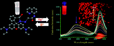
In this paper, we have designed and synthesized a new rhodamine-based chemosensor (L1) which behaves as a highly selective and ratiometric FRET-based fluorescent probe for Hg(II) in HEPES buffer (1 mM, pH 7.4; 2% EtOH) at 25 °C. The fluorescence spectrum of L1 excited at 330 nm exhibits a fluorescence maximum at 440 nm which decreases along with the gradual increase of a new peak at 575 nm upon the addition of Hg(II). This phenomenon due to the ring-opening of the spirolactam system of rhodamine gives rise to strong fluorescence emission and also a visual color change from colorless to violet to pink. Ratiometric responses are more attractive because the ratio between the two emission intensities can be used to measure the analyte concentration and sensor molecule concentration, providing a built-in correction for environmental effects and stability under illumination.13b,c,25 Interestingly, the presence of an excess of the biologically relevant (Na+, K+, Ca2+ etc.) and other metal (Cr3+, Mn2+, Fe3+ etc.) ions does not affect the “switch ON” behaviour of the receptor L1 observed in the presence of Hg(II) ions due to the formation of the L–Hg complex which is reversible in nature. But the reversibility of the L–Hg system to regenerate L1 is only in the presence of S2− anions and it results in “ON–OFF” behavior to sense sulphide anion in the same medium of HEPES buffer (1 mM, pH 7.4; 2% EtOH) at 25 °C. The kinetics study of the reaction of Hg(II) with L1 showed two consecutive steps and it clearly indicated that the opening of the spirolactam-ring was operated to produce xanthenes, formed after the chelation of L1 with Hg(II). Fluorescence microscopic studies confirmed that L1 could also be used as an imaging probe for detection of the uptake of both ions in HeLa cells and whole bodies.

Experimental section

Materials and methods

High-purity HEPES, 8-aminoquinoline, 2-chloroacetyl chloride and mercury(II) nitrate monohydrate and mercury(II) chloride were purchased from Sigma Aldrich (India) and rhodamine B from E. Merck. The solvents used were of spectroscopic grade. All the metal salts were used as either their nitrate or their chloride salts. Other chemicals were of analytical reagent grade and used without further purification except when specified. Milli-Q, 18.2 MΩ cm−1 water was used throughout the experiments. A Shimadzu (model UV-1800) spectrophotometer was used for recording the electronic spectra. FTIR spectra were recorded using a Perkin Elmer FTIR model RX1 spectrometer preparing KBr disk. The 1HNMR spectra were obtained on a Bruker Avance DPX 400 MHz spectrometer using DMSO-d6 solution and for 13C NMR it was 500 MHz. Electrospray ionization (ESI) mass spectra were recorded on a Qtof Micro YA263 mass spectrometer. A Systronics digital pH meter (model 335) was used to measure the pH of the solution and the adjustment of the pH was done using either 50 mM HCl or NaOH solution. Steady-state fluorescence emission and excitation spectra were recorded with a Perkin Elmer LS 55 spectrofluorimeter. Time-resolved fluorescence lifetime measurements were performed using a HORIBA JOBIN Yvon picosecond pulsed diode laser-based time-correlated single-photon counting (TCSPC) spectrometer from IBH (UK) at λex = 340 nm and MCP-PMT as a detector. Emission from the sample was collected at a right angle to the direction of the excitation beam maintaining magic angle polarization (54.71). The full width at half-maximum (FWHM) of the instrument response function was 250 ps, and the resolution was 28.6 ps per channel. The data was fitted to multiexponential functions after deconvolution of the instrument response function by an iterative reconvolution technique using IBH DAS 6.2 data analysis software in which reduced w2 and weighted residuals serve as parameters for the goodness of fit.

Synthesis of the probe (L1)

The probe L1 was synthesised by a 3 step reaction (Scheme 1).
image file: c3ra47800a-s1.tif
Scheme 1 Synthesis of L1.

At first, the rhodamine B-hydrazide (1) was prepared following a literature method.26 In brief, 85% hydrazine hydrate (4 mL) was added to a solution of rhodamine B (1 g, 2.09 mmol) in ethanol (40 mL). The solution was refluxed for 6 h. Then, the reaction mixture was evaporated under reduced pressure to give an orange oil, which was then recrystallized from methanol–water to afford rhodamine B-hydrazide as a light-orange crystal (77%).

Then 2-chloro-N-(quinol-8-yl)acetamide (2) was prepared from the reaction of 2-chloroacetyl chloride and 8-aminoquinoline as follows-

2-Chloroacetyl chloride (5.31 mL) was dissolved in chloroform (5 mL) and then added dropwise to a cooled stirred solution of 8-aminoquinoline (2.88 g, 20 mmol) and Et3N (3.0 mL) in chloroform (10 mL) within 1 h. After being stirred for 2 h at room temperature, the mixture was removed under reduced pressure to obtain a white solid, which was filtered out and extracted with dichloromethane to afford compound 2.27 Yield: 82%. mp (°C): 133 ± 2.

In the final step, rhodamine B-hydrazide (1) was taken in dry acetonitrile with anhydrous K2CO3 and 2-chloro-N-(quinolin-8-yl)acetamide (2) in dry acetonitrile and was added dropwise with stirring. The resulting reaction mixture was refluxed for 6 h. The volume of the solution was reduced to obtain a solid and then extracted with dichloromethane and finally purified by silica gel column chromatography using dichloromethane as the eluent. Yield: 70%, mp (°C): 152 ± 2; anal. found: C, 73.54; H, 6.76; N, 12.89%; calc.: C, 73.10; H, 6.29; N, 13.12%. IR (cm−1): νNH, 3327; νC[double bond, length as m-dash]C, 2971; νC[double bond, length as m-dash]O, 1693; νC[double bond, length as m-dash]N, 1614; 1H NMR (400 MHz, DMSO-d6): 8.93 (dd, 1H), 8.59 (d, 1H), 8.41 (dd, 1H), 7.78–7.71 (m, 2H), 7.66–7.58 (m, 2H), 7.52–7.46 (m, 2H), 6.99–6.94 (m, 1H), 6.42–6.30 (m, 6H), 4.56 (s, 2H, –CH2–CO), 3.29 (q, 8H, 4CH2), 1.06 (t, 12H, 4CH3); 13C NMR (125 MHz, DMSO-d6): 165.22, 164.92, 152.98, 152.61, 151.89, 149.13, 148.13, 138.07, 136.66, 133.66, 132.34, 129.56, 128.08, 127.82, 127.65, 126.89, 123.44, 122.65, 122.28, 122.12, 116.59, 107.85, 105.50, 97.43, 64.72, 43.61, 12.38; ESI-MS m/z 640.99 [M + H+, 10%], 663.00 [M + Na+, 23%].

Synthesis of the L–Hg complex as [Hg(L)Cl2]

To a 10 mL ethanolic solution of L1 (0.01 mmol), a solution of mercury(II) chloride was added dropwise and stirred for 4 h. The solvent was removed using a rotary evaporator, and a blood red solid was obtained (Scheme S1). [Hg(L)Cl2]: C39H40Cl2HgN6O3: anal. found: C, 52.17; H, 4.30; N, 9.09%; calc.: C, 51.35; H, 4.42; N, 9.21%. IR (cm−1): νNH, 3295; νC[double bond, length as m-dash]C, 2972; νC[double bond, length as m-dash]O, 1686; νC[double bond, length as m-dash]N, 1612; 1H NMR (400 MHz, DMSO-d6): 8.95 (dd, 1H), 8.62 (d, 1H), 8.44 (dd, 1H), 7.78–7.72 (m, 2H), 7.67–7.59 (m, 2H), 7.50–7.46 (m, 2H), 6.99–6.95 (m, 1H), 6.42–6.30 (m, 6H), 4.58 (s, 2H, –CH2–CO), 3.31 (q, 8H, 4CH2), 1.06 (t, 12H, 4CH3); ESI-MS in methanol: [M + Na]+, m/z, 935.11 (obsd with 24% abundance) calc.: m/z, 935.22; where M = [Hg(L)Cl2]; yield: 67–70%.

Emission study

Organic moiety (L1) shows a very weak emission at 575 nm in HEPES buffer (1 mM, pH 7.4; 2% EtOH) at 25 °C when excited at 550 nm considering the absorption at 550 nm. Fluorescence quantum yields (Φ) were estimated by integrating the area under the fluorescence curves with the equation:
image file: c3ra47800a-t1.tif
where A is the area under the fluorescence spectral curve and OD is the optical density of the compound at the excitation wavelength, 550 nm, η is the refractive index of the solvent used. The standard used for the measurement of the fluorescence quantum yield was rhodamine-B (Φ = 0.7 in ethanol).

General method of UV-vis and fluorescence titration

The path length of the cells used for the absorption and emission studies was 1 cm. For UV-vis and fluorescence titrations, a stock solution of L1 was prepared in HEPES buffer (1 mM, pH 7.4; 2% EtOH) at r.t. Working solutions of L1, Hg(II) and S2− were prepared from their respective stock solutions. Fluorescence measurements were performed using a 15 nm × 5 nm slit width. Except the time dependent spectra, all the fluorescence and absorbance spectra were taken after 30 minutes of mixing to acquire the optimised spectra.

Job's plot from the fluorescence experiments and UV-vis

A series of solutions containing L1 and Hg(NO3)2 were prepared such that the total concentration of L1 remains constant in all the sets. The mole fraction (X) of Hg(II) ions was varied from 0.1 to 0.75. The absorbance at 555 nm was plotted against the mole fraction of Hg(II) ions in solution and also the fluorescence intensity at 575 nm was plotted against the mole fraction of Hg(II) ions.

Calculation of the Förster distance (R0)

The Förster distance (R0) for the FRET process was calculated from the following simplified equation below:28,29
image file: c3ra47800a-t2.tif
where η is the refractive index (η = 1.33 in water);30 ΦD is the quantum yield of the donor; k denotes the average squared orientational part of a dipole–dipole interaction, typically k2 = 2/3; JDA expresses the degree of spectral overlap between the donor emission and the acceptor absorption; ID(λ) is the normalized fluorescence spectra of the donor; εA(λ) is the molar absorption coefficient of the acceptor.

Theoretical calculation

To clarify the understanding of the configurations and the mechanism of the process of the enhancement of fluorescence, DFT calculations of the excited state character of the probes L1 and its corresponding L–Hg complexes were performed using Gaussian-09 software over a Red Hat Linux IBM cluster. Molecular level interactions have also been studied using density functional theory (DFT) with the B3LYP/6-31G (d) functional model and basis set for the probe, L1 and LANL2DZ for the corresponding L–Hg complex.

Kinetic measurements

The kinetic studies were done on a Shimadzu UV 1601 PC spectrophotometer attached to a thermoelectric cell temperature controller (Model TCC 240A, accuracy ±0.1 °C). The absorption due to the ligand was subtracted by using an equi-molar ratio of ligand–HEPES buffer (1 mM, pH 7.4; 2% EtOH) mixture in the reference cell. The progress of the reaction was followed by monitoring the increase in absorbance at 550 nm. A conventional mixing technique was followed and pseudo first order conditions with respect to probe concentration were maintained throughout the course of the reaction.

Preparation of cell and in vitro cellular imaging with L1

Human cervical cancer cell, HeLa cell line was purchased from the National Center for Cell Science (NCCS), Pune, India and was used throughout the study. Cells were cultured in Dulbecco's modified Eagle's medium (DMEM, Gibco BRL) supplemented with 10% FBS (Gibco BRL), and 1% antibiotic mixture containing penicillin, streptomycin and neomycin (PSN, Gibco BRL), at 37 °C in a humidified incubator with 5% CO2. For experimental study, the cells were grown to 80–90% confluence, harvested with 0.025% trypsin (Gibco BRL) and 0.52 mM EDTA (Gibco BRL) in PBS (phosphate-buffered saline, Sigma Diagnostics) and plated at the desired cell concentration and allowed to re-equilibrate for 24 h before any treatment. Cells were rinsed with PBS and incubated with DMEM-containing L1 (10 μM, 1% DMSO) for 30 min at 37 °C. All the experiments were conducted in DMEM containing 10% FBS and 1% PSN antibiotic. The imaging system was composed of a fluorescence microscope (ZEISS Axioskop 2 plus) with an objective lens [10×].

To find out the applicability of this sensor (L1) for the visual detection of mercury in fish, an experiment was carried out with Chanda nama fish. The fish were treated with aqueous Hg(II) (10 μM) for 30 min, washed with PBS to remove any Hg2+ adhering to the surface, and were subsequently treated with a solution of L1 (10 μM in DMSO) for 20 min, and then washed with PBS. The visual images were taken with a digital camera.

Cell cytotoxicity assay

To test the cytotoxicity of L1, MTT [3-(4,5-dimethyl-thiazol-2-yl)-2,S-diphenyl tetrazolium bromide] assay was performed by the procedure described earlier.31 After treatment of the probes (5, 10, 20, 50, and 100 μM), 10 μL of the MTT solution (10 mg mL−1 PBS) was added in each well of a 96-well culture plate and incubated continuously at 37 °C for 6 h. All mediums were removed from wells and replaced with 100 μL of acidic isopropanol. The intracellular formazan crystals (blue-violet) formed were solubilized with 0.04 N acidic isopropanol and the absorbance of the solution was measured at 595 nm wavelength with a microplate reader. The values are means ± S.D. of three independent experiments. The cell cytotoxicity was calculated as percent cell cytotoxicity = 100% cell viability.

Results and discussion

Synthesis and characterisation

The synthesis of L1 first involves the conversion of rhodamine B to rhodamine B-hydrazide and then 2-chloro-N-(quinolin-8-yl)acetamide was prepared from the reaction of 2-chloroacetyl chloride and 8-aminoquinoline in chloroform medium in the presence of NEt3 (Scheme 1). Then the probe, L1 was isolated from the reaction of rhodamine B-hydrazide and 2-chloro-N-(quinolin-8-yl)acetamide in a dry acetonitrile solution in the presence of anhydrous K2CO3. The formulation of L1 was confirmed by physico-chemico and spectroscopic methods (Fig. S1A–G).

UV-vis spectroscopic studies of L1

UV-vis spectra of L1 was recorded in HEPES buffer (1 mM, pH 7.4; 2% EtOH) at 25 °C showing an absorption maximum at 314 nm which may possibly be attributed to the intramolecular π–π* charge transfer (CT) transition. The absorption of L1 at 314 nm gradually increased, accompanied by the formation of a new absorption peak at 555 nm (Fig. 1) as the Hg(II) concentration were increased stepwise (0–30 μM) and the solution turned from colorless to pink (Fig. S2) via an intermediate color change to blue violet with time (Fig. 2).
image file: c3ra47800a-f1.tif
Fig. 1 UV-vis titration spectra of L1 (10 μM) upon the incremental addition of Hg(II) concentrations (0–30 μM) in HEPES buffer (1 mM, pH 7.4; 2% EtOH) at 25 °C.

image file: c3ra47800a-f2.tif
Fig. 2 Color change detected by the naked eye with time, (A) probe only, (B) immediately and (C) 30 min after the addition of Hg(II) ions in HEPES buffer (1 mM, pH 7.4; 2% EtOH) at 25 °C.

On account of the complexity of the intracellular environment, an additional examination of the probe was performed to determine whether other ions were potential interferents or not. To establish this fact, metal ion selectivity assays were performed while keeping the other experimental conditions unchanged. No significant change in the UV-vis spectral pattern was observed upon the addition of 10 equivalents excess of relevant metal ions i.e. Na(I), K(I), Ca(II), Mg(II), Al(III), Cr(III), Mn(II), Fe(III), Co(II), Ni(II), Zn(II), Cd(II), and Pb(II).

Fluorescence spectroscopic studies of L1

In the absence of Hg(II) ion, L1 exhibited the fluorescence of weak intensity and showed the pH independency over the pH range 6.0 to 10.0 (Fig. S3). The emission spectrum of the L1 excited at 330 nm exhibits a fluorescence maximum at 440 nm in HEPES buffer (1 mM, pH 7.4; 2% EtOH) at 25 °C. The intensities at 440 nm were significantly decreased with a concomitant increase in intensities at 575 nm showing an isoemissive point at about 540 nm, when various concentrations of Hg(II) (0–30 μM) were added (Fig. 3).
image file: c3ra47800a-f3.tif
Fig. 3 (a) Emission spectra of L1 in the presence of Hg(II) ions (0–30 μM) at λex = 330 nm in HEPES buffer (1 mM, pH 7.4; 2% EtOH) at 25 °C. (b) The ratiometric signaling of the fluorescence output at two different wavelengths is plotted as a function of the concentration of Hg(II) ions in HEPES buffer (1 mM, pH 7.4; 2% EtOH) at 25 °C.

Ratiometric signaling of the fluorescence output at two different wavelengths plotted as a function of concentration of Hg(II) indicates that the fluorescence intensity ratio of the wavelengths 440 nm and 575 nm (I575/I440) gradually increases with increasing the concentration of Hg(II) ions (viz. Fig. 3).

L1 exhibited an increase of about 45-fold of its fluorescence intensity upon the addition of only 3.0 equivalent of Hg(II) ions. The detection limit was determined and was found to be 90 ppb (Fig. S4), which is significantly low. Interestingly, the introduction of other metal ions causes the fluorescence intensity to be either unchanged or weakened. A metal ion selectivity study was then performed for L1 to understand this phenomenon under identical experimental conditions. The fluorescence enhancement of L1 (10 μM) was not observed upon the addition of an excess of 50 equivalents of biologically relevant metal ions i.e. Na(I), K(I), Ca(II), and Mg(II) and 10 equivalents excess of several competitive metal ions [Al(III), Cr(III), Mn(II), Fe(III), Co(II), Ni(II), Cu(II), Zn(II), Cd(II), and Pb(II)] (Fig. S5), and also by naked eye (Fig. S6 and 7). In the presence of a 10 times excess of various tested ions together with L1 and Hg(II), almost no adverse effect on intensity was observed (Fig. S8). The time dependent fluorescence spectrum also shows that the emission band at 440 nm diminishes with the appearance of an intense emission band at 575 nm (Fig. 4).


image file: c3ra47800a-f4.tif
Fig. 4 Fluorimetric titration spectra of L1 (10 μM) with Hg(II) ions (10 μM) at λex = 330 nm with time in HEPES buffer (1 mM, pH 7.4; 2% EtOH) at 25 °C.

As the receptor L1 bears two different fluorophore units we consider it to be appropriate to study the metal binding event of L1 at two different excitation wavelengths corresponding to the excitation wavelength of the xanthene unit (550 nm) and quinoline unit (330 nm). Fig. 5 shows that the excitation of L1 at 550 nm in the absence of Hg(II) did not show any significant emission over the range from 550 to 700 nm initially with a quantum yield of only 0.03. This supports the fact that the receptor remains in the spirolactam form in the absence of metal ions, and the nonexistence of the highly conjugated xanthene form results in the suppression of emission in the above mentioned region. But the addition of Hg(II) to this chemosensor (L1) induces a significant switch ON fluorescence response near 575 nm, with a visual display of reddish fluorescence.


image file: c3ra47800a-f5.tif
Fig. 5 (a) Fluorimetric titration spectra of L1 with Hg(II) ions (0–30 μM) at λex = 550 nm in HEPES buffer (1 mM, pH 7.4; 2% EtOH) at 25 °C, (b) Fluorescence intensity as a function of Hg(II) concentration at λex = 550 nm in HEPES buffer (1 mM, pH 7.4; 2% EtOH) at 25 °C.

The switch ON response for the absorption spectral band at 555 nm and the emission band at ∼575 nm upon binding to Hg(II) suggests the opening of the spirolactam ring of L1 upon metal ion coordination (Scheme 2). It is observed that Hg binding with L1 to form L–Hg species induced the ring-opening of L1 and the generation of xanthene moiety that is selective towards Hg(II) ions and does not reveal any noticeable spectral change for the other tested metal ions.


image file: c3ra47800a-s2.tif
Scheme 2 Probable mechanism of the Hg(II) induced FRET process.

The binding of Hg(II) ion induces opening of the spirolactam ring of L1 with an associated switch on UV-vis spectral response in the range 450–600 nm, which has a significant spectral overlap with the emission spectrum of the N-(quinol-8-yl)-acetamide fragment (Fig. S9) and this fact unlocks a plausible route for the nonradiative transfer of excitation energy from donor quinoline to acceptor xanthene moiety within the Förster critical distance (R0) which was calculated to be 40.6 Å, and initiates an intramolecular FRET process (viz. Scheme 2). In the free state of L1 the FRET pathway is totally suppressed, and only an emission maximum near 440 nm is observed when excited at 330 nm. Binding of the receptor to Hg(II) induces the FRET process to produce an intense rhodamine-based reddish emission; i.e., the energy transfer from N-(quinol-8-yl)-acetamide moiety to xanthene is due to the ring-opening32 resulting in an increase of the overlap integral between the N-(quinol-8-yl)-acetamide and xanthene moiety. Thus, when titrated with increasing concentration of the Hg(II) the emission band with a λmax near 440 nm starts to decrease, along with a concomitant generation of a new fluorescence band at 575 nm. This change in fluorescence was also observed visually, and the color changed through violet to pink (viz. Fig. 2).

The complex formed between L1 and Hg(II) ions is found to be 1[thin space (1/6-em)]:[thin space (1/6-em)]1 in stoichiometry, which was established with the help of Job's plots (Fig. S10 and 11) by the fluorescence and absorbance study also. The apparent binding constant (K) determined by the Benesi–Hilderbrand method33 was estimated to be 6.73 × 10⁶ M−1 (Fig. S12). Further confirmation of the 1[thin space (1/6-em)]:[thin space (1/6-em)]1 stoichiometry was obtained by the physico-chemical and spectroscopic data of the L–Hg complex isolated in the solid form and the 1H NMR titration is also in support of the chelation of L1 with Hg2+ ions in the solution state (Fig. S13). The molecular-ion peak in the ESI-MS of L–Hg was observed at m/z 935.11 as evidence for the 1[thin space (1/6-em)]:[thin space (1/6-em)]1 stoichiometric species (Fig. S1F).

The proposed mechanism also correlates with the fluorescence lifetime data (Fig. 6, Tables 1 and 2). In the fluorescence life time experiment (λem = 440 nm), the average lifetime was found to be 1.75 ns. After 5 min of the addition of Hg(II) to the solution of L1, the average lifetime (λem = 440 nm) of the L–Hg species increased to 2.44 ns, which is ascribed to the chelation enhanced fluorescence (CHEF) process. After 30 min, the value reduces to 1.11 ns, corresponding to the decrease of the CHEF process of the L–Hg system with time, as proposed in the mechanism (Scheme 2). On the contrary, the average lifetime of the L–Hg system after 30 min (λem = 575 nm) increased from 1.72 ns to 2.10 ns, which resembles the Hg(II)-induced FRET process, (viz. Fig. 6).


image file: c3ra47800a-f6.tif
Fig. 6 Time resolved fluorescence decay of L1 (10 μM) only and in the presence of added Hg(II) (10 μM) in HEPES buffer (1 mM, pH 7.4; 2% EtOH) at 25 °C using a nano LED of 340 nm as the light source (a) at λem = 575 nm, (b) at λem = 440 nm.
Table 1 Fluorescence life time (ns) of the corresponding singlet excited states of L1 and L–Hg
System τav (ns) at λem = 440 nm χ2 τav (ns) at λem = 575 nm χ2
L1 1.75 1.13 1.72 1.07
L–Hg complex After 5 min 2.44 1.07 1.98 1.01
After 20 min 1.11 1.12 2.10 1.00


Table 2 Fluorescence quantum yield (Φ) and life time (ns) of L1 and L–Hg at λem = 575 nm
λem = 575 nm Φ τav (ns) kr (108 s−1) knr (109 s−1) χ2
L1 0.03 1.72 0.2 0.56 1.07
L–Hg 0.43 2.10 2.0 0.36 1.00


According to the equations:34 τ−1 = kr + knr and kr = Φf/τ, where kr = the radiative rate constant, and knr = total nonradiative rate constant, the values of kr and knr for the organic moiety, L1 and L–Hg species are listed in Table 2. The data suggests that kr slightly changed, but the factor that induces fluorescent enhancement is mainly ascribed to the decrease of knr.

Kinetics study

To verify the sequence of the reaction of Hg(II) with the probe (L1) in terms of chelation and ring opening, an experiment was performed to get the plot of ln(AAt) (where A is the absorbance at infinite time, and At is the absorbance at time t) against time (t) which was found to be nonlinear (Fig. S14). This is curved at the initial stage and then subsequently a constant slope, indicating that the reaction proceeds via two consecutive steps. The first step is dependent on [Hg(II)] and it is due to chelation. Using the Weyh and Hamm's method,35 the rate constant (k1) was calculated from the plot of ln[thin space (1/6-em)]Δ versus time (t) where time (t) is small. The second step is the ring opening step as it is independent of [Hg(II)]. At a particular temperature (40 °C) the slope of ln(AAt) versus time (t) plots for different [Hg(II)] were found to be constant in the linear region. From this limiting linear portion of the plot, the k2 values were obtained directly from the slope for different [Hg(II)]. The calculated values for k1 and k2 are tabulated in Table S1.

The overall pseudo first-order kinetic plot of the reaction of L1 (10 μM) with Hg(II) ((0.1–1.0 mM)) in HEPES buffer (1 mM, pH 7.4; 2% EtOH) at 25 °C was also done from the fluorescence emission intensity, and from the slope the overall rate constant, k′ was found to be 9.66 × 10⁻⁴ s−1 (Fig. S15).

Spectroscopic studies of the L–Hg complex in the presence of S2−

To gain insight into the reversibility of the ring-opening of the spirolactam form to the generation of the xanthene form, the rupture of the L–Hg species to regenerate L1 and the subsequent change of optical properties of the L–Hg system were also studied. UV-vis titration spectra of L–Hg (1[thin space (1/6-em)]:[thin space (1/6-em)]1 complex, 10 μM) upon incremental addition of S2− (0–30 μM) in HEPES buffer (1 mM, pH 7.4; 2% EtOH) at 25 °C showed the gradual decrease of absorption at 550 nm (Fig. S16). This fact is in support of the reclaiming of L1 from the L–Hg system in the presence of S2−. Here several anions (F, Cl, Br, I, CN, NO3, ClO4, H2PO4, HPO42−, OAc, SO42−, S2O32−, CrO42−, S2−, SCN, and PO43−) were used, but only S2− selectively quenches the fluorescence intensity of the L–Hg system (Fig. 7).
image file: c3ra47800a-f7.tif
Fig. 7 Changes in the fluorescence spectra of the L–Hg complex in the presence of different anions (λex = 550 nm).

This phenomenon proves that the regeneration of L1 occurred selectively by the rupture of L–Hg species and simultaneous lowering of the fluorescence intensity at 575 nm was observed. Hence the fluorescence “ON–OFF” switching property of the L–Hg complex could be used as a ratiometric sensor for the detection of S2− ions in physiological conditions (1 mM HEPES buffer, pH 7.4; 2% EtOH; at 25 °C) (Fig. 8).


image file: c3ra47800a-f8.tif
Fig. 8 Fluorescence titration spectra (λex = 330 nm) of L1 (10 μM) with 2 equiv. of Hg(II) ions upon the continuous addition of sodium sulphide (up to 30 μM) in HEPES buffer (1 mM, pH 7.4; 2% EtOH) at 25 °C.

The LOD value for sulphide ions for this method was found to be 1.7 × 10⁻⁷ M (Fig. S17). The intensities of the fluorescence were recorded within 30 min after sulfide anion addition and varied with time. As a result of this fact it may be concluded that the monitoring system is virtually real-time and stable, and the sensor L1 was recycled during the detection of sulfide anions.

Theoretical study

From theoretical calculation it is reflected that the HOMO and LUMO of L1 are less stabilized than the L–Hg complex (Fig. 9). From the energy optimization of the HOMO and LUMO of the L–Hg complex, it could be easily pointed out that more electronic charge density in HOMO over the rhodamine unit is pulled towards the quinoline unit in the LUMO. The energy gap between the HOMO and LUMO is less in the case of free ligand i.e. spirolactam ring form than the L–Hg complex i.e. xanthene form.
image file: c3ra47800a-f9.tif
Fig. 9 Optimized structures of the L1 and L–Hg complex.

Biological studies of L1 in the presence of Hg(II) and S2−

To examine the utility of the probe in biological systems, it was applied to the human cervical cancer HeLa cell and Chanda nama fish. In these experiments both the Hg(II) and L1 were allowed to be taken up by the cells of interest and the images of the cells were recorded by fluorescence microscopy following excitation at ∼550 nm. After incubation with L1 (10 μM) for 30 min, the cells displayed very faint intracellular fluorescence. However, the cells exhibited intensive fluorescence when exogenous Hg(II) was introduced into the cell via incubation with Hg-salt (Fig. 10). The fluorescence responses of the probe with various concentrations of added Hg(II) are clearly evident from the cellular imaging. Moreover, the intensive fluorescence was deeply suppressed by scavenging Hg(II) from the cell with the addition of Na2S (Fig. 11). This experiment proves that the binding of Hg(II) with this chemosensor, L1, is readily reversible inside the cellular environment. In addition, the in vitro study showed that 10 μM of L1 did not show any cytotoxic effect to cell up to 6 h (Fig. S18). These results indicate that the probe has huge potential for both in vitro and in vivo applications as a Hg(II) sensor, as well as imaging in different ways, as the same manner for live cell imaging can be followed instead of fixed cells.
image file: c3ra47800a-f10.tif
Fig. 10 (1) Fish; (2) phase contrast; fluorescence, and ratio image of HeLa cells after incubation with (3, 4) 0 μM; (5, 6) 3 μM; (7, 8) 5 μM; (9, 10) 8 μM; (11, 12) 10 μM Hg(II) with L1 for 30 min at 37 °C respectively and the samples were excited at ∼550 nm.

image file: c3ra47800a-f11.tif
Fig. 11 Fluorescence, and its corresponding ratio image of HeLa cells after incubation with (1, 2) 0 μM, (3, 4) 10 μM with Hg(II) and (5, 6) 15 μM of Na2S with L1 respectively for 30 min at 37 °C and the samples were excited at λ = 550 nm.

Conclusions

In summary, we may conclude that a newly designed fluorescent chemosensor (L1) behaves as a highly specific and selective FRET-based ratiometric fluorescence probe towards Hg(II) with a response that can be detected by the naked eye. On excitation at 330 nm the fluorescence spectrum of this probe exhibits a fluorescence maximum at 440 nm by CHEF which decreases with the gradual increase of a new peak at 575 nm on the addition of Hg(II) ions due to the ring-opening of the spirolactam system of rhodamine for FRET to enable this probe to act as a ratiometric sensor with greater advantages and this phenomenon also gives rise to a visual colour change from colourless to violet to pink. Dissociation of the L–Hg complex formed takes place only in the presence of sulfide anions, and this fact enables the L–Hg complex to be an efficient sensor for sulfide anions. With the incubation of cultured living cells (HeLa) with L1, intracellular Hg(II) and S2−could be monitored through fluorescence microscopy study.

Acknowledgements

Financial assistance from CSIR, New Delhi, India is gratefully acknowledged. B. Sen wishes to thank to UGC, New Delhi, India for offering him the fellowship. We sincerely acknowledge Prof. Samita Basu and Mr Ajay Das, Chemical Science Division, SINP, Kolkata for enabling the TCSPC instrument.

Notes and references

  1. (a) Q. Wang, D. Kim, D. D. Dionysiou, G. A. Sorial and D. Timberlake, Environ. Pollut., 2004, 131, 323 CrossRef CAS PubMed; (b) H. H. Harris, I. J. Pickering and G. N. George, Science, 2003, 301, 1203 CrossRef CAS PubMed.
  2. P. Grandjean, P. Weihe, R. F. White and F. Debes, Environ. Res., 1998, 77, 165 CrossRef CAS PubMed.
  3. (a) R. Von Burg and M. R. Greenwood, Metals and their Compounds in the Environment, VCH, Weinheim, 1991, p. 1045 Search PubMed; (b) A. Renzoni, F. Zino and E. Franchi, Environ. Res., 1998, 77, 68 CrossRef CAS PubMed.
  4. (a) X. Zhang, Y. Xiao and X. Qian, Angew. Chem., Int. Ed., 2008, 47, 8025 CrossRef CAS PubMed; (b) G. Guzzi and C. A. M. La Porta, Toxicology, 2008, 244, 1 CrossRef CAS PubMed.
  5. M. Harada, Crit. Rev. Toxicol., 1995, 25, 1 CrossRef CAS PubMed.
  6. (a) C. M. L. Carvalho, E.-H. Chew, S. I. Hashemy, J. Lu and A. Holmgren, J. Biol. Chem., 2008, 283, 11913 CrossRef CAS PubMed; (b) T. W. Clarkson, L. Magos and G. J. Myers, N. Engl. J. Med., 2003, 349, 1731 CrossRef CAS PubMed.
  7. K. Leopold, M. Foulkes and P. Worsfold, Anal. Chim. Acta, 2010, 663, 127 CrossRef CAS PubMed.
  8. (a) Y. Gao, Z. Shi, Z. Long, P. Wu, C. Zheng and X. Hou, Microchem. J., 2012, 103, 1 CrossRef CAS; (b) A. Q. Shah, T. G. Kazi, J. A. Baig, H. I. Afridi and M. B. Arain, Food Chem., 2012, 134, 2345 CrossRef CAS PubMed.
  9. (a) E. Kenduzler, M. Ates, Z. Arslan, M. McHenry and P. B. Tchounwou, Talanta, 2012, 93, 404 CrossRef CAS PubMed; (b) F. Moreno, T. Garcia-Barrera and J. L. Gomez-Ariza, Analyst, 2010, 135, 2700 RSC.
  10. X. Chai, X. Chang, Z. Hu, Q. He, Z. Tu and Z. Li, Talanta, 2010, 82, 1791 CrossRef CAS PubMed.
  11. Y. Zhao, J. Zheng, L. Fang, Q. Lin, Y. Wu, Z. Xue and F. Fu, Talanta, 2012, 89, 280 CrossRef CAS PubMed.
  12. (a) X. Jia, Y. Han, X. Liu, T. Duan and H. Chen, Spectrochim. Acta, Part B, 2011, 66, 88 CrossRef; (b) Y. Yin, M. Chen, J. Peng, J. Liu and G. Jiang, Talanta, 2010, 81, 1788 CrossRef CAS PubMed.
  13. (a) S. Sen, T. Mukherjee, B. Chattopadhyay, A. Moirangthem, A. Basu, J. Marek and P. Chattopadhyay, Analyst, 2012, 137, 3975 RSC; (b) S. Sen, S. Sarkar, B. Chattopadhyay, A. Moirangthem, A. Basu, K. Dhara and P. Chattopadhyay, Analyst, 2012, 137, 3335 RSC; (c) S. Sen, T. Mukherjee, S. Sarkar, S. K. Mukhopadhyay and P. Chattopadhyay, Analyst, 2011, 136, 4839 RSC; (d) U. C. Saha, K. Dhara, B. Chattopadhyay, S. K. Mandal, S. Mondal, S. Sen, M. Mukherjee, S. V. Smaalen and P. Chattopadhyay, Org. Lett., 2011, 13, 4510 CrossRef CAS PubMed; (e) U. C. Saha, B. Chattopadhyay, K. Dhara, S. K. Mandal, S. Sarkar, A. R. Khuda-Bukhsh, M. Mukherjee, M. Helliwell and P. Chattopadhyay, Inorg. Chem., 2011, 50, 1213 CrossRef CAS PubMed; (f) K. Dhara, U. C. Saha, A. Dan, M. Manassero, S. Sarkar and P. Chattopadhyay, Chem. Commun., 2010, 46, 1754 RSC.
  14. (a) C. Wang and K. M.-C. Wong, Inorg. Chem., 2013, 52, 13432 CrossRef CAS PubMed; (b) P. Mahato, S. Saha, E. Suresh, R. Di Liddo, P. P. Parnigotto, M. T. Conconi, M. K. Kesharwani, B. Ganguly and A. Das, Inorg. Chem., 2012, 51, 1769 CrossRef CAS PubMed; (c) H. N. Kim, S.-W. Nam, K. M. K. Swamy, Y. Jin, X. Q. Chen, Y. Kim, S.-J. Kim, S. Park and J. Yoon, Analyst, 2011, 136, 1339 RSC; (d) M. Kumar, N. Kumar, V. Bhalla, H. Singh, P. R. Sharma and T. Kaur, Org. Lett., 2011, 13, 1422 CrossRef CAS PubMed; (e) K. M. K. Swamy, H. N. Kim, J. H. Soh, Y. Kim, S.-J. Kim and J. Yoon, Chem. Commun., 2009, 1234 RSC; (f) X. Zhang, Y. Xiao and X. H. Qian, Angew. Chem., Int. Ed., 2008, 47, 8025 CrossRef CAS PubMed; (g) Y. Shiraishi, S. Sumiya, Y. Kohno and T. Hirai, J. Org. Chem., 2008, 73, 8571 CrossRef CAS PubMed; (h) Y. K. Yang, K. J. Yook and J. Tae, J. Am. Chem. Soc., 2005, 127, 16760 CrossRef CAS PubMed.
  15. (a) R. Pandey, R. K. Gupta, M. Shahid, B. Maiti, A. Misra and D. S. Pandey, Inorg. Chem., 2012, 51, 298 CrossRef CAS PubMed; (b) Q.-B. Mei, Y.-H. Guo, B.-H. Tong, J.-N. Weng, B. Zhang and W. Huang, Analyst, 2012, 137, 5398 RSC; (c) A. Thakur, S. Sardar and S. Ghosh, Inorg. Chem., 2011, 50, 7066 CrossRef CAS PubMed; (d) D. Y. Liu, K. Z. Tang, W. S. Liu, C. Y. Su, X. H. Yan, M. Y. Tan and Y. Tang, Dalton Trans., 2010, 39, 9763 RSC; (e) Y. Zhou, C.-Y. Zhu, X.-S. Gao, X.-Y. You and C. Yao, Org. Lett., 2010, 12, 2566 CrossRef CAS PubMed; (f) M. Suresh, A. K. Mandal, S. Saha, E. Suresh, A. Mandoli, R. Di Liddo, P. P. Parnigotto and A. Das, Org. Lett., 2010, 12, 5406 CrossRef CAS PubMed.
  16. (a) X. Chen, X. Meng, S. Wang, Y. Cai, Y. Wu, Y. Feng, M. Zhu and Q. Guo, Dalton Trans., 2013, 42, 14819 RSC; (b) Y. G. Zhao, Z. H. Lin, C. He, H. M. Wu and C. Y. Duan, Inorg. Chem., 2006, 45, 10013 CrossRef CAS PubMed; (c) M.-H. Yang, P. Thirupathi and K.-H. Lee, Org. Lett., 2011, 13, 5028 CrossRef CAS PubMed; (d) M. Santra, B. Roy and K. H. Ahn, Org. Lett., 2011, 13, 3422 CrossRef CAS PubMed.
  17. (a) S. Sen, M. Mukherjee, K. Chakrabarty, I. Hauli, S. K. Mukhopadhyay and P. Chattopadhyay, Org. Biomol. Chem., 2013, 11, 1537 RSC; (b) L. E. Santos-Figueroa, M. E. Moragues, E. Climent, A. Agostini, R. Martínez-Mańez and F. Sancenon, Chem. Soc. Rev., 2013, 42, 3489 RSC.
  18. (a) Hydrogen Sulfide, (Environmental Health Criteria, No. 19), World Health Organization, Geneva, 1981 Search PubMed; (b) R. F. Huang, X. W. Zheng and Y. J. Qu, Anal. Chim. Acta, 2007, 582, 267 CrossRef CAS PubMed.
  19. (a) R. E. Gosselin, R. P. Smith and H. C. Hodge, Hydrogen Sulfide. In Clinical Toxicology of Commercial Products, Williams and Wilkins, Baltimore, MD, 5th edn, 1984; p. 198 Search PubMed; (b) S. A. Patwardhan and S. M. Abhyankar, Colourage, 1988, 35, 15 CAS.
  20. (a) C. R. Liu, J. Pan, S. Li, Y. Zhao, L. Y. Wu, C. E. Berkman, A. R. Whorton and M. Xian, Angew. Chem., Int. Ed., 2011, 50, 10327 CrossRef CAS PubMed; (b) Y. J. Peng, J. Nanduri, G. Raghuraman, D. Souvannakitti, M. M. Gadalla, G. K. Kumar, S. H. Snyder and N. R. Prabhakar, Proc. Natl. Acad. Sci. U. S. A., 2010, 107, 10719 CrossRef CAS PubMed; (c) Y. Kaneko, Y. Kimura, H. Kimura and I. Niki, Diabetes, 2006, 55, 1391 CrossRef CAS PubMed; (d) L. Li, M. Bhatia, Y. Z. Zhu, Y. C. Zhu, R. D. Ramnath, Z. J. Wang, F. B. M. Anuar, M. Whiteman, M. Salto-Tellez and P. K. Moore, FASEB J., 2005, 19, 1196 CAS; (e) K. Abe and H. J. Kimura, Neuroscience, 1996, 16, 1066 CAS.
  21. K. Eto, T. Asada, K. Arima, T. Makifuchi and H. Kimura, Biochem. Biophys. Res. Commun., 2002, 293, 1485 CrossRef CAS PubMed.
  22. P. Kamoun, M. C. Belardinelli, A. Chabli, K. Lallouchi and B. Chadefaux-Vekemans, Am. J. Med. Genet., 2003, 116, 310 CrossRef PubMed.
  23. W. Yang, G. Yang, X. Jia, L. Wu and R. Wang, J. Physiol., 2005, 569, 519 CrossRef CAS PubMed.
  24. S. Fiorucci, E. Antonelli, A. Mencarelli, S. Orlandi, B. Renga, G. Rizzo, E. Distrutti, V. Shah and A. Morelli, Hepatology, 2005, 42, 539 CrossRef CAS PubMed.
  25. (a) B. Valeur and I. Leray, Coord. Chem. Rev., 2000, 205, 3 CrossRef CAS; (b) A. P. de Silva, H. Q. N. Gunaratne, T. Gunnlaugsson, A. J. M. Huxley, C. P. McCoy, J. T. Rademacher and T. E. Rice, Chem. Rev., 1997, 97, 1515 CrossRef CAS PubMed; (c) Z. Xu, Y. Xiao, X. Qian, J. Cui and D. Cui, Org. Lett., 2005, 7, 889 CrossRef CAS PubMed; (d) G. Grynkiewicz, M. Poenie and R. Y. Tsien, J. Biol. Chem., 1985, 260, 3440 CAS.
  26. (a) X. F. Yang, X. Q. Guo and Y. B. Zhao, Talanta, 2002, 57, 883 CAS; (b) V. Dujols, F. Ford and A. W. Czarnik, J. Am. Chem. Soc., 1997, 119, 7386 CrossRef CAS.
  27. X. Zhou, P. Li, Z. Shi, X. Tang, C. Chen and W. Liu, Inorg. Chem., 2012, 51, 9226 CrossRef CAS PubMed.
  28. X. Zhang, Y. Xiao and X. Qian, Angew. Chem., Int. Ed., 2008, 47, 8025 CrossRef CAS PubMed.
  29. J. R. Lakowicz, Principles of Fluorescence Spectroscopy, Springer, 2006, p. 443 Search PubMed.
  30. G. M. Hale and M. R. Querry, Appl. Opt., 1973, 12, 555 CrossRef CAS PubMed.
  31. J. Ratha, K. A. Majumdar, S. K. Mandal, R. Bera, C. Sarkar, B. Saha, C. Mandal, K. D. Saha and R. Bhadra, Mol. Cell. Biochem., 2006, 290, 113 CrossRef CAS PubMed.
  32. (a) X. Chen, T. Pradhan, F. Wang, J. S. Kim and J. Yoon, Chem. Rev., 2012, 112, 1910 CrossRef CAS PubMed; (b) M. Vendrell, D. Zhai, J. C. Er and Y. T. Chang, Chem. Rev., 2012, 112, 4391 CrossRef CAS PubMed; (c) C. Kar, M. D. Adhikari, A. Ramesh and G. Das, Inorg. Chem., 2013, 52, 743 CrossRef CAS PubMed.
  33. H. A. Benesi and J. H. Hildebrand, J. Am. Chem. Soc., 1949, 71, 2703 CrossRef CAS.
  34. N. J. Turro, Modern Molecular Photochemistry, Benjamin/Cummings Publishing Co., Inc., Menlo Park, CA, 1978 Search PubMed.
  35. J. A. Weyh and R. E. Hamm, Inorg. Chem., 1969, 8, 2298 CrossRef CAS.

Footnote

Electronic supplementary information (ESI) available: Experimental section, tables, schemes, figures, characterization data, and some spectra. See DOI: 10.1039/c3ra47800a

This journal is © The Royal Society of Chemistry 2014