Zhimin Wang*,
Cuilian Xu,
Guangqin Gao and
Xin Li
School of Science, Henan Agricultural University, Zhengzhou 450002, People's Republic of China. E-mail: gary1451@iccas.ac.cn; Fax: +86-371-63558139; Tel: +86-371-63558130
First published on 5th March 2014
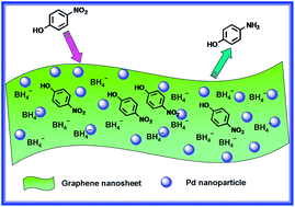
In this work, we described a simple and effective synthesis method for preparing well-dispersed Pd–graphene nanohybrids through simultaneous chemical reduction of functionalized graphene oxide and Pd2+ ions. The structure and physicochemical properties of the resulting nanohybrids were characterized in detail by Fourier transformation infrared spectroscopy, Raman, X-ray photoelectron spectroscopy, high-resolution transmission electron microscopy and scanning electron microscopy. The obtained results indicated that Pd nanoparticles distributed uniformly on the surface of the functionalized graphene when the loading amount of Pd is less than 1.0 wt%. Moreover, the so-formed Pd-FG hybrid material could be well dispersed in water, forming a homogeneous dispersion. This nanohybrid, combining the unique catalytic properties of Pd with the excellent adsorption and electron transfer ability of graphene, exhibited enhanced catalytic activity toward the reduction of 4-nitrophenol by NaBH4 even under lower Pd loading.
Ever since Novoselov et al. succeeded in extracting single atom-thick layer from bulk graphite in 2004, graphene has attracted unusual attention due to its unique structural and fascinating properties.8,9 Particularly, the large surface area (calculated value, 2630 m2 g−1) and tunable surface properties have made graphene highly desirable for use as a 2-D catalyst support in which the surface functional groups can act as favorable sites for the nucleation of the guest materials.10,11 It has been demonstrated that reduced GO as a conducting support can anchor semiconductor and metal nanoparticles and exhibit enhanced catalytic performance in electrocatalysis,12–14 redox catalysis,15 carbon–carbon bond formation16–18 and photocatalytic reactions.19–21 The enhanced catalytic performance can be ascribed to the increasing catalytic surface area, efficient capping and protection of catalyst materials, excellent adsorb ability and the conductivity of graphene which help to improve the separation and transportation of the charge carriers or electrons.22 Compared to pure graphene, chemically-oxidized GO exhibits a significant loss of conductivity. They need to be reduced to restore the sp2 hybrid network and thus reintroduce the conductive property. However, a crucial challenge in the synthesis of graphene by reduction is that the reduced GO itself is not soluble and tend to aggregate due to van der Waals force interactions.11 In view of most of their unique properties were only associated with well-dispersed nanosheets, keeping them well separated would be of importance in exploring their technological and engineering applications, in particular, use as a catalyst support. To this end, various strategies such as utilizing the surface oxygen-containing groups of GO,17 electrostatic interactions,23 hydrophobic interactions13 and surface functionalization of graphene24,25 have been proposed and proven to be useful in suppressing aggregation of reduced GO nanosheets and loading metal and semiconductor nanoparticles onto graphene. Though some methods mentioned above can be used to prepare graphene based composites of metal or semiconductor nanoparticles, to obtain solution-processable individual graphene sheet with well-dispersed nanoparticles decorated on its surface remains challenging particularly because of the poor solubility of graphene and the weak interactions between graphene and catalyst nanoparticles. Therefore, development of a general, synthetically versatile and cost effective complement is still highly desired.
In this paper, we report on a simple method for the synthesis of well dispersible Pd-functionalized graphene (Pd-FG) nanohybrids by using dispersible functionalized graphite oxide (FGO) and PdCl2 as starting agents. As shown in Scheme 1, graphite oxide (GO) was covalently modified with hydrophilic benzene sulfonic groups using a simple and effective method. The surface functional groups can on the one hand improve the water dispersibility upon reduction of GO,26 on the other hand they act as favorable sites for the nucleation and stabilization of Pd nanoparticles.27 Thus, well dispersed Pd nanoparticles stabilized by sulfonic groups on the surface of functionalized graphene could be obtained by simultaneous reduction of Pd2+ ions and FGO using NaBH4. Importantly, the resultant Pd-FG nanohybrids could be well dispersed in water, forming an almost homogeneous solution. It was demonstrated that this water dispersible Pd-FG nanohybrid could function as an effective catalyst to activate the reduction of 4-nitrophenol (4-NP) to 4-aminophenol (4-AP) in the presence of excess NaBH4 following pseudo-first-order kinetics, otherwise unfeasible if only the strong reducing agent NaBH4 was employed.
![]() | ||
Scheme 1 Schematic process of surface functionalization of GO and simultaneous reduction of Pd2+ and FGO for Pd-FG nanohybrid. |
After completion of the reaction the catalyst was recovered by simple filtration. The filtrate was washed repeatedly with dimethyl sulfoxide, deionized water, and acetone in consecutive washing cycles. Ultrasonic treatment was used in every cycle in order to re-disperse the FGO and remove adsorbed impurities. After washing, the sample was dried at 60 °C in vacuum for 24 h.
Fig. 1 shows the water dispersibility of GO, FGO and their Pd composites. As shown in Fig. 1a, the as-prepared GO is highly soluble in water forming a transparent brown solution. After functionalization by benzene sulfonic groups, the dispersibility improved obviously and concomitant color change from dark brown to black occurred, indicating the alteration of surface chemical composition (Fig. 1b). Although the surface oxygen-containing groups or structural defects of GO were often used to anchor metal nanoparticles, in this study we found just GO could not offer stable protection to the in situ formed Pd nanoparticles and precipitation occurred immediately even for 0.1 wt% of Pd loading (Fig. 1c). Compared to GO, FGO showed better carrying capacity for Pd nanoparticles upon reduction. The obtained Pd-FG catalyst even with higher Pd loading amount is still dispersible in water forming an almost homogeneous solution without precipitation for at least several weeks (Fig. 1d).
Fig. 2 demonstrates the SEM observation of the thin exfoliated sheet structure of FGO and the uniform decoration of Pd nanoparticles on the surface of graphene with 0.5 wt% of Pd loading amount. Compared to the thin and crimple nanosheet of exfoliated FGO (Fig. 2a), the single-sheet nature of the Pd-FG hybrid is still presented except for the uniform decoration of Pd nanoparticles (Fig. 2b). This result confirms that well-dispersed Pd-FG hybrids have been successfully synthesized. To examine the effect of loading amount on the dispersing state of Pd nanoparticles on the surface of graphene, a series of Pd-FG hybrids with different Pd loading amounts were prepared. The dispersing state of Pd nanoparticles on the surface of graphene was observed by SEM images for different samples. As shown in Fig. S1(a–c),† when the loading amount is less than 1.0 wt%, the spherical Pd nanodots with uniform distribution could be easily observed on the surface of graphene and with the increase of Pd loading, more well-dispersed Pd nanoparticles could be observed on the surface. When the loading amount is larger than 1.0 wt%, obvious aggregation occurred and with the continue increase of Pd loading, bigger aggregations appeared [Fig. S1(d and e)†]. The aggregation will result in the decrease of surface area which will further lead to the decrease of catalytic activity. This conclusion is testified by the catalytic results in the subsequent catalytic reduction of 4-NP. Based on these observations, we conclude that 0.5–1.0 wt% ratio of Pd nanoparticles to FGO should be appropriate for preparation of well-dispersed Pd-FG catalyst.
![]() | ||
Fig. 2 SEM observations of (a) FGO and (b) Pd-FG nanohybrid with 0.5 wt% of Pd loading amount. The scale bar in the images represents 100 nm. |
To further verify the uniform distribution of Pd nanoparticles, the Pd-FG nanohybrid with 0.5 wt% Pd loading was characterized by TEM. As shown in Fig. 3a, Pd nanoparticles are uniformly dispersed onto the clean graphene with a narrow size distribution. The average size of as-synthesized Pd nanoparticles is about 15–20 nm, which is consistent with the SEM observation. A representative HR-TEM image is shown in Fig. 3b where the lattice fringes are clearly visible with a space about 0.388 nm, corresponding to the lattice space of the (111) planes of Pd. Moreover, the presence of a large amount of Pd nanoparticles on graphene is also confirmed by EDX analysis (Fig. 3c). Although the existence of Pd is verified, it is worthy of note that the characteristic sulphur signal of sulfonic groups is absent from the EDX scan. This may be due to the screen of Pd nanoparticles. This deduction was confirmed when we re-scan the EDX on the blank region of the sample, where a well-marked signal corresponding to sulphur occurred (shown in Fig. S2†). This result also corroborates the coordination and stability role of sulfonic groups for Pd nanoparticles from another angle. The distributions of the constituting elements in the nanohybrid were also observed vividly by STEM image and the EDS elemental mapping (shown in Fig. S3†).
![]() | ||
Fig. 3 TEM image of Pd-FG dispersed in water (a), corresponding HRTEM image (b) and (c) EDX detection result for the Pd-FG hybrid (the Cu peaks come from the copper grid). |
The wide-angle X-ray diffraction was conducted to trace the crystalline structure and nanoscale size of the supported Pd nanoparticles. As shown in Fig. 4, four prominent peaks at 2θ values of about 40°, 46°, 68° and 81°, corresponding to the characteristic of face centered cubic crystalline Pd (JCPDS, Card no. 05-0681), i.e. (111), (200), (220) and (311) confirmed the success reduction of Pd2+ ions. The average size of the Pd nanoparticles, estimated from the half-widths of (111) peaks by using the Scherrer equation,32 is about 17 nm, which is approximate to the TEM and SEM observations.
![]() | ||
Fig. 4 XRD pattern of the Pd-FG nanohybrid showing intense peaks indexed to the cubic Pd structure (40° (111), 46° (200), 68° (220) and 82° (311)). |
The extent of surface covalent modification was further examined based on the percentage of weight loss by TGA. As shown in Fig. 6, a significant amount of weight loss from the FGO occurs primarily in the temperature range of 400–600 °C compared to the GO due to the decomposition of the surface grafted benzene sulfonic groups. According to the TGA traces, the weight ratio of grafted benzene sulfonic groups was approximately 25.3 wt%. Based on this data we can estimate that the molar ratio of benzene sulfonic groups to carbon atoms of graphene is approximately 2.5%.
Raman spectroscopy is a powerful tool to characterize the structural changes of carbon-based materials, including disorder and defect structures. As shown in Fig. 7, the Raman spectra of GO and FGO are characterized by two main peaks compared with that of graphite precursor. The G peak at approximately 1580 cm−1 originates from the in-plane vibration of sp2 carbon atoms and is a doubly degenerate phonon mode (E2g symmetry),37 the D peak around 1355 cm−1 is a breathing mode of A1g symmetry involving phonons.38 The presence of a D band in two spectra suggests that both samples hold considerable amounts of defects associated with oxygen-containing groups on the surface. However, the FGO has an increased D/G intensity ratio compared to that of GO. Since the D/G intensity ratio is inversely proportional to the average size of the sp2 domains, the increase of the D/G intensity ratio suggests that smaller in-plane sp2 domains are formed during the surface functionalization of GO, indicating effective surface functionalization by benzene sulfonic groups. A 2D band, which is the characteristic band of graphene, is usually used to determine the number of layers of graphene in the sample. This band originated from a two-phonon double-resonance Raman process associated with the band structure of graphene. A 2D band observed in GO and enhanced in FGO indicates that the nanosheets contain only a few layers of graphene.39
For further investigating the changes of surface element composition upon modification and reduction, X-ray photoelectron spectroscopy (XPS) measurements for GO, FGO and Pd-FG were conducted and shown in Fig. S4 (a–c).† Compared to GO, a well-marked peak at 168 eV, representing the characteristic signal of sulphur, occurred for the FGO and Pd-FG, which further corroborates the successful modification by sulfonic groups. Moreover, comparing the scans of sulphur region (S 2p) of the FGO with that of Pd-FG, we found that the intensity of the S 2p peak of the Pd-FG was stronger than that of the FGO (Fig. S4d†). This enhancement should ascribe to the reduction of surface oxide-containing groups during the process of co-reduction, which caused the increase of relative ratio of sulfonic groups to carbon. This conclusion is further verified by the records of carbon region (C 1s) scans. As shown in Fig. 8, after oxidation of the graphite into GO, two dominant peaks were observed at 284.7 and 286.8 eV (Fig. 8a). The peak at 284.7 eV is a characteristic peak for CC/C–C bonding of graphite. The other peak at 286.8 eV shows a broad tail to higher binding energy region, which is attributed to emission from the oxidized carbon atoms in the GO. The three fitted peaks at 284.7, 286.8 and 287.9 eV can be assigned to the binding energies of carbon in C
C/C–C, C–O (epoxy/hydroxyls), and C
O (carbonyl/ketone), respectively. For the FGO (Fig. 8b), the characteristic peak for C
C/C–C bonding of graphite was observed at the same binding energy position and the peak intensity increased obviously compared with the GO spectrum. However, the high binding energy peak for oxidized carbon atoms decreased dramatically in the normalized peak areas, indicating that surface functionalization resulted in the decrease of peak intensity of the surface oxygen functionalities.
After reduction of the Pd2+ ions and FGO to Pd-FG hybrid by NaBH4, a strong peak at 284.7 eV (CC/C–C) was dominant and the peak intensities of C–O and C
O decreased significantly compared to the spectrum of FGO (Fig. 9a), indicating that most oxy-functional groups have been removed after the reduction process. XPS data further indicate that Pd precursors and GO are reduced simultaneously. Besides, it was noteworthy that a new fitted C 1s peak at 290.0 eV appeared compared to that of GO and FGO, indicating that Pd nanoparticles may have an important influence on the surface carbon of graphene. Theory calculations by density functional theory (DFT) have also shown considerable affinity between the metals and pristine graphene or carbon nanotubes.34,35 Similar observation has also been reported by Yan and coworkers, where they for the first time confirmed covalent interaction between the Pd particles and graphene by IR spectroscopy.36 Based on these analyses, we believed that this new C 1s peak should also originate from the covalent interaction of Pd nanoparticles and the surface carbon atom of FG. Further studies to better understand the mechanism remain important. Furthermore, the XPS measurement for the Pd 3d regions of the Pd-FG hybrid was also performed to elucidate the oxidation state of the Pd nanoparticles as shown in Fig. 9b. The peaks at 335.0 and 340.2 eV in the Pd XPS spectrum are attributed to the binding energies of Pd 3d5/2 and Pd 3d3/2, respectively. These binding energies are comparable to those of Pd metal, confirming the zerovalent state of the Pd.6
In the reduction process, the overall concentration of NaBH4 was 10 mM and 4-NP was 0.1 mM. Considering the much higher concentration of NaBH4 compared to that of 4-NP, it is reasonable to assume that the concentration of BH4− remains constant during the reaction. In this context, pseudo-first-order kinetics could be used to evaluate the kinetic reaction rate of the current catalytic reaction, together with the UV-vis absorption data in Fig. 10a. The absorbance of 4-NP is proportional to its concentration in solution; the absorbance at time t (At) and time t = 0 (A0) are equivalent to the concentration at time t (Ct) and time t = 0 (C0). The rate constant (k) could be determined from the linear plot of ln(Ct/C0) versus reduction time in seconds. As expected, a good linear correlation of ln(Ct/C0) versus time was obtained as shown in Fig. 10b, whereby a kinetic reaction rate constant k is estimated to be 2.35 × 10⁻³ s−1. This value is comparable to that of other metal catalysts for the reduction of 4-NP in the presence of NaBH4.
For the supported heterogeneous metal catalysts, the surface area of the catalyst which related to the particle size and dispersing state of nanoparticles on the support is one of the most important influent factors on the catalytic activity. To further prove the catalytic performance of as-prepared Pd-FG and examine the effect of dispersing state on the catalyst activity, control experiments were performed under the same conditions using different Pd-FG catalysts within a given reaction time of 20 min (the UV-vis absorbance data were shown in Fig. S5†). For better showing the effect of Pd loading on the catalytic activities, the plots of Ct/C0 versus reduction time for different catalysts were displayed in Fig. 11. It was found when the Pd loading amount was less than 0.5 wt%, the reduction reaction could not accomplish within 20 min. When the Pd loading amount increased to 0.5 wt%, the reaction rate enhanced significantly with the complete transformation of 4-NP in as little as 12 min. However, for the catalysts with 1.0 and 1.5 wt% Pd loading amounts, the reduction time did not reduce but increased to 14 and 16 min, respectively. When the loading amount of Pd reached up to 2.0 wt%, the reduction reaction cannot accomplish during 20 min. These results revealed that 0.5 wt% of Pd-FG showed the best catalytic activity which is consistent with the dispersing state of Pd nanoparticles on the surface of FG as demonstrated by SEM images.
![]() | ||
Fig. 11 Plot of Ct/C0 versus reaction time for the reduction of 4-NP Pd-FG catalysts with different Pd loading (–■– 0.25 wt%; –▲– 0.5 wt%; –◀– 1.0 wt%; –▼– 1.5 wt%; –▶– 2.0 wt%; –●– 3.0 wt%). |
Furthermore, we have also monitored the cycle stability of the Pd-FG catalyst by monitoring the catalytic activity during successive cycles of the reduction reactions. As shown in Fig. 12, the Pd-FG catalyst (0.5 wt% Pd) exhibits a similar catalytic performance in the conversion even after 5 cycles. Even so, a slight decrease in the activity (<10%) of the catalyst was found in the course of the recycling experiments. To examine the main reason of the decrease of activity, the catalyst was recovered by simple filtration after the reaction. The filtrate was analyzed for leaching of palladium metal using atomic absorption spectroscopy. The result showed that there was almost no leaching of palladium metal during the course of the reaction. Thus, small amount of decrease in the activity of the catalyst may be attributed to the alteration of morphology or distribution state of Pd nanoparticles during the reaction. To this end, TEM observation was conducted for the recovered catalyst. As shown in Fig. S6,† slight aggregation of Pd nanoparticles was observed after five cycles of reaction, which caused the decrease of surface area of Pd nanoparticles and resulted in the slight decline of catalytic activity. We deduce initially that the amino groups of on-site formed 4-AP may be responsible for the slight aggregation of Pd nanoparticles. Further study to well understand it is undergoing.
Footnote |
† Electronic supplementary information (ESI) available: SEM images of distribution of Pd nanoparticles for different Pd loading amounts, EDX scan for the blank region of Pd-FG, EDS elemental mapping of the constituting elements, XPS surveys of GO, FGO and Pd-FG, sulphur region (S 2p) scans of FGO and Pd-FG, UV-vis spectra for catalytic properties in the presence of different Pd-FG catalysts and TEM observations of the used catalyst. See DOI: 10.1039/c3ra47721e |
This journal is © The Royal Society of Chemistry 2014 |