Inseong Youa,
Young Chang Seob and
Haeshin Lee*ab
aGraduate School of Nanoscience & Technology, Korea Advanced Institute of Science and Technology (KAIST), Daejeon, 305-701, Republic of Korea. E-mail: haeshin@kaist.ac.kr; Fax: +82-42-350-2810
bDepartment of Chemistry KAIST, Daejeon, 305-701, Republic of Korea. E-mail: haeshin@kaist.ac.kr; Fax: +82-42-350-2810
First published on 3rd February 2014
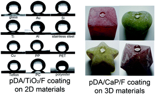
This study reports methods for general preparation of superhydrophobic surfaces on any type of material surface using mussel-inspired poly(dopamine) (pDA). The use of pDA presents several advantages over conventional superhydrophobic fabrication methods: development of superhydrophobicity in a material-independent manner, enhancement of mechanical stability, decreases in angle hysteresis, and applicability to 3D objects.
Inspired by the ability of marine mussels to adhere to nearly any material, polydopamine (pDA) coating has been identified as a useful substance for material-independent modification of surface chemistry.11–13 The in situ, spontaneous polymerization of dopamine hydrochloride effectively modifies any material surfaces, which have been widely applied in the inhibition of corrosion, in the controlled adhesion of cells, as a photothermal therapeutic agent in cancer therapy, as a mechanically stable lithium ion battery separator, and in the immobilization of biomolecules.14 Recently, Zhang et al. fabricated superhydrophobic nanoparticles, which were deposited onto material surface to develop superhydrophobic surfaces in a material-independent way by utilizing pDA coating. This study is meaningful in that it provides a general route for the preparation of superhydrophobic surfaces, but it is expected that the low-energy nanoparticles are difficult to form chemically stable, mechanically robust adhesion on surfaces in general.15 Thus, the material-independent fabrication of stable superhydrophobic surfaces by the pDA coating is fully addressed issue. The pDA coating provides abundant catechol groups on the surface, and the catechol moiety is known to interact with titanium oxide (TiO2).16 The catechol moiety can also bind calcium ions (Ca2+), as evidenced by the high concentration of Ca2+ found in the external coating layer of mussels' byssal threads.17 Utilizing this chemical principle, Ryu et al. developed a method to grow hydroxyapatite crystal in a material-independent manner.18 Because the surface-grown TiO2 and hydroxyapatite have nano-scale roughness, we hypothesized that we can fabricate superhydrophobic surfaces in a material-independent manner by depositing a low interfacial energy compound onto the grown oxide and ceramic layer. Here, we report the pDA-mediated, material-independent fabrication of superhydrophobic surfaces. Two approaches were developed. First, the in situ formation of TiO2 from titanium(IV) isopropoxide on a pDA layer followed by the deposition of perfluorophosphate surfactant (zonyl®FSE) resulted in a mechanically stable, superhydrophobic surface. When the titanium(IV) isopropoxide precursor solution was spin-coated onto the pDA-coated substrate (2 mg mL−1, Tris buffer, pH 8.5, 16 h coating), a rapid reaction with water vapor in the air triggered polymerization of the precursor, resulting in TiO2 formation. Modification with zonyl®FSE completed the superhydrophobic surface preparation (pDA/TiO2/F). The surfactant molecule has a phosphate functional group which forms a stable self-assembled monolayer on metal oxides such as TiO2 and bio-minerals such as calcium phosphate.19,20 Second, in situ growth of calcium phosphate (CaP) on the pDA layer followed by modification with zonyl®FSE yielded a superhydrophobic surface. The uniform growth of crystalline CaP on the pDA layer was achieved by immersion of the pDA-coated substrates in a supersaturated CaP solution at 38 °C for 24 h. Modification with zonyl®FSE completed the superhydrophobic surface preparation (pDA/CaP/F). Reasons for developing the two methods are that each of these methods has its own advantages and disadvantages. The first method (pDA/TiO2/F) is based on a spin-coating technique, making it quick to perform but largely inapplicable to 3D objects. The second method (pDA/CaP/F) requires longer times, but can be easily applied to 3D objects (please see Fig. S1† for the schematic explanation).
Static contact angle measurements of pDA/TiO2/F-functionalized materials demonstrated that almost all treated materials exhibited superhydrophobicity: 162 ± 1° (glass), 161 ± 1° (Au), 159 ± 1° (Si), 157 ± 1° (TiO2), 162 ± 2° (AlOx), 162 ± 1° (stainless steel), 168 ± 1° (Cu), 158 ± 2° (polypropylene, PP), 155 ± 1° (polyethylene terephthalate, PET), 160 ± 1° (Teflon), 155 ± 2° (polycarbonate, PC), and 157 ± 2° (polyester) (Fig. 1A). Successful superhydrophobic conversion was confirmed by X-ray photoelectron spectroscopy (XPS) (Fig. S2†). The characteristic peaks, such as the N1s peak from the pDA-coated substrate (Si), the Ti2p peak from pDA/TiO2, and the F1s peak from pDA/TiO2/F, were all observed, indicating that each step of modification was successful. The second strategy for fabricating superhydrophobic surfaces (pDA/CaP/F functionalization) can be applied to 3D objects because the key step, the growth of crystalline calcium phosphate, is performed in supersaturated CaP solution. While water droplets on the unmodified surfaces exhibited hydrophilicity (Fig. 1B, left), pDA coating followed by CaP formation and zonyl®FSE compound deposition resulted in superhydrophobic surfaces with contact angles over 150° (Fig. 1B, right). Characterization of the surface by XPS (Fig. S3†) clearly showed the characteristic N1s peak from pDA, the P2p peak from pDA/CaP, and the F1s peak from pDA/CaP/F.
![]() | ||
Fig. 1 (A) The static water contact angle images of the bare (left) and pDA/TiO2/F (right) substrates. (B) The contact images of the unmodified (left) and pDA/CaP/F (right) 3D objects (vol = 20 μL). |
The morphology of the pDA/TiO2/F surface was investigated by scanning electron microscopy (SEM). The TiO2 formation displayed hierarchical micro-/nano-structures (Fig. 2A) with a thickness of approximately 1 μm (Fig. S4A†), and the low-resolution SEM images show that micro-/nano-structured TiO2 is uniformly dispersed in a large scale (Fig. S5†). Random-yet-hierarchical structures are often found in nature, with examples including lotus leaves, legs of water striders, and integument of leafhoppers.1,21,22 Hierarchical structures have been shown to increase stability of superhydrophobicity. The stability of both the pDA/TiO2/F surfaces and the negative control surfaces (no-pDA/TiO2/F, treated with titanium(IV) isopropoxide spin-coating and zonyl®FSE modification without pDA coating) were tested. The surfaces were exposed to strong ultrasonication (50 kHz, 150 W, 60 min) to induce detachment of the coating layers and were subsequently observed by SEM. Defects on the functionalized surfaces were found. The TiO2 detachment percent (i.e. detached area/total area, %) was calculated by the following formula:
The total TiO2 area and the detached TiO2 area were determined with an image-processing program called Image J. The TiO2 portion of the pDA/TiO2/F Si surface remained stable after 1 h of ultrasonication. For Si, negligible detachment (∼0%) of TiO2 was observed for the samples functionalized with an underlying pDA layer (Fig. 2B). Other substrates, such as glass, PP, and PET functionalized by pDA/TiO2/F, revealed detachments of 2.6%, 3.0%, and 1.0%, respectively, after 1 h of ultrasonication. However, the surfaces functionalized with TiO2/F without the pDA coating exhibited significant increases in TiO2 detachment: 25.1% for Si, 46.5% for glass, 43.7% for PP, and 21.5% for PET (Fig. 2C). Furthermore, the ultrasonicated pDA/TiO2/F surfaces retained their initial superhydrophobicity. The contact angles after mechanical stress were 151.4 ± 1.6° for Si, 162.4 ± 0.4° for glass, 157.4 ± 1.0° for PP, and 155.5 ± 1.0° for PET (insets Fig. 2B). As expected, the ultrasonicated no-pDA/TiO2/F surfaces exhibited drastically decreased contact angles: 110.6 ± 0.8° for Si, 117.1 ± 2.5° for glass, 89.2 ± 1.5° for PP, and 110.7 ± 4.3° for PET (insets Fig. 2C). The enhancement of the adhesion stability of TiO2 by the underlying pDA layer is attributed to the strong coordination bonds between the catechol groups in pDA and TiO2.16 The dynamic contact angle difference (i.e., the hysteresis between advanced and receding angles) of the pDA/TiO2/F surfaces were generally found to be below 10° (glass: 6.9°, Au: 9.2°, Si: 4.9°, Al: 8.7°, Ti: 7.7°, Cu: 5.3°, PP: 6.7°, PET: 6.2°, and Teflon: 7.9°). The observed low hysteresis indicated a low adhesion force to water droplets, approaching the original concept of lotus effect. Interestingly, surfaces fabricated without a pDA layer showed large hysteresis: 48.1° for glass, 53.7° for Au, 38.1° for Si, 39.5° for Al, 33.6° for Ti, 48.2° for Cu, 39.1° for PP, 45.4° for PET, and 38.1° for Teflon (Fig. S6A†). The large hysteresis resulted in the pinning of water droplets on the surfaces. The underlying pDA layer is required for the generation of superhydrophobicity with low-hysteresis. Additionally, pDA/TiO2/F surfaces showed good water-repellence at extremely low temperatures (−60 °C). Surfaces were visually observed as super-cooled water (−60 °C) was poured onto pre-chilled pDA/TiO2/F Al, no-pDA/TiO2/F Al, and unmodified Al (all surface T was −60 °C) surfaces to determine whether the formation of solid ice occurred upon contact of the two phases (the surface tilting angle was 20°).23
The pDA/TiO2/F Al surface displayed nearly perfect anti-icing (Fig. S6B; video S1†), while the no-pDA/TiO2/F surface and unmodified substrate instantly exhibited ice formation upon liquid–solid contact as a result of the adhesion force between the super-cooled water and the material surfaces (Fig. S6C and D; video S1†). It is apparent that the pDA/TiO2/F surface is very effective in the prevention of ice formation.
The morphology of the pDA/CaP/F surface was investigated by SEM. The surface-grown CaP phase appears as broad, platelet-like structures. The high-density growth of crystalline CaP initiated by the pDA layer resulted in extremely rough surfaces with a steep angle relative to the underlying substrates. This method can also be applied to different categories of substrates (Au, glass, Ti, polyurethane (PU), PP, and teflon) (Fig. 3A). The thickness of a single CaP platelet was approximately 200–300 nm, while the thickness of the entire CaP layer was up to 10 μm (Fig. S4B†). The pDA layer plays an important role in CaP growth, as demonstrated by the fact that unmodified substrates did not induce the CaP formation even after one day of incubation (Fig. 3B). Fig. 3C shows the thin-film XRD (X-ray diffraction) results of the CaP. The reflection at 16° (2θ) is characteristic of octacalcium phosphate (OCP), and the reflections at 26° and 32° are characteristic of OCP and hydroxyapatite (HA).24 Thus, we concluded that the synthesized CaP is mixture of polycrystalline OCP and HA. The morphology of the CaP results in small solid–liquid contacts between water drops and the coating, resulting in superhydrophobicity. Contact angle analysis revealed angles higher than 150°: 160 ± 1° for Au, 160 ± 2° for glass, 156 ± 2° for Ti, 157 ± 1° for PU, 156 ± 2° for PP, and 159 ± 2° for teflon (Fig. S7†).
Footnote |
† Electronic supplementary information (ESI) available: Details of experimental procedures and additional data. See DOI: 10.1039/c3ra47626j |
This journal is © The Royal Society of Chemistry 2014 |