Colloidosomes from poly(N-vinyl-2-pyrrolidone)-coated poly(N-isopropylacrylamide-co-acrylic acid) microgels via UV crosslinking

Yi Gong, Ai Mei Zhu, Qiu Gen Zhang and Qing Lin Liu*
Department of Chemical and Biochemical Engineering, College of Chemistry and Chemical Engineering, Xiamen University, Xiamen 361005, P.R. China. E-mail: qlliu@xmu.edu.cn; Fax: +86-592-2184822; Tel: +86-592-2188072

Received 8th December 2013 , Accepted 20th January 2014

First published on 27th January 2014


Abstract

We first report the synthesis of colloidosomes via UV crosslinking of PVP-coated microgels. The photo-crosslinkable poly(N-vinyl-2-pyrrolidone) (PVP) was coated as a layer on the surface of poly(N-isopropylacrylamide-co-acrylic acid) (P(NIPAM-co-AAc)) microgels at pH = 3.5. A toluene-in-water Mickering emulsion was further fabricated based on the resulting PVP/P(NIPAM-co-AAc) microgels and then exposed to UV irradiation leading to the formation of colloidosomes. Since only the microgels on the surface of emulsion droplets could be crosslinked by UV irradiation, the yield and the mean size of the colloidosomes can be controlled by the amount of toluene used in the preparation. The proposed method presents a new platform to fabricate colloidosomes since photo-responsive comonomers or/and chemical crosslinkers are not essential in the system. The colloidosomes can be used as a vehicle for both hydrophilic and hydrophobic dyes. The release of the encapsulated dyes can be controlled by temperature.

Introduction

As a kind of microcapsules with a shell consisting of packed colloidal particles,1 colloidosomes have attracted considerable attention because of their promising applications in the field of medicine2,3 and biotechnology.4–6 They are usually made by ‘locking’ the colloidal particles or microgels7 which self-assemble on the oil/water interface (interfacial particles) in a Pickering or Mickering7 emulsion together by specific technologies. The locking of the interfacial particles in the emulsion to make a dense shell is crucial for colloidosomes preparation. Several techniques such as annealing,8 polyelectrolyte complexation,9,10 and covalent crosslinking11,12 have been proposed for the locking process. Colloidosomes or microcapsules prepared on the basis of double Pickering emulsion templates were also reported.13,14 With continued research, facile, fast and biocompatible approaches have been attracting researchers' attention. Nomura et al.15 reported the aggregation of the interfacial poly (methylmethacrylate-co-butyl acrylate) latex particles induced by the diffusion of ethanol in the W/O emulsions to form colloidosomes. Ferritin-(poly(N-isopropylacrylamide)/2-(dimethylmaleinimido)-N-ethyl-acrylamide) hybrid microgels were locked to prepare colloidosomes using UV irradiation.16 Due to its rapidness, ease of use and free of toxic substances, UV crosslinking is widely used in polymer chemistry and biotechnology.17–19 These advantages are also applicable to the fabrication of colloidosomes.

Since UV crosslinking is desirable to lock the interfacial particles in a Pickering or Mickering emulsion, the way to obtain photo-crosslinkable microgels is of paramount importance. It is accomplished by the introduction of a photo-responsive comonomer into the polymer network in the literature.16,19,20 However, this route to prepare photo-crosslinkable microgels has also some drawbacks. The introduction of a photo-responsive comonomer will change the original structure of the polymer chain which is usually specially designed for a particular application. This is inevitable and could possibly cause some side effects when scientists get photo-crosslinkable microgels by this method. What is more important, it is difficult to introduce a photo-responsive comonomer into the existing microgels. Therefore, to explore an alternative method without bringing any side effects is of relevance. To the best of our knowledge, there have rare reports on preparation of photo-crosslinkable microgels via photo-responsive polymer coating for colloidosomes preparation. PVP has been reported as one of the UV crosslinkable materials.21,22 In the present work, we have presented a new technique in which the photo-crosslinkable PVP contained microgels are crosslinked to form microcapsule with the aid of UV irradiation.

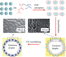
Stimuli responsive materials have been currently attracting intensive attention due to their interesting perspectives in biomedicine. As one of the temperature responsive materials, P(NIPAM-co-AAc) has been widely studied18,23–25 and the emulsion stabilized by the particles of PNIPAM or its derivative has been reported.26–29 So, as depicted in Fig. 1, P(NIPAM-co-AAc) was chosen as a template microgel in this work. As a cheap and available polymer with excellent biocompatibility,30 PVP is a desirable material to endow the photo-responsiveness to P(NIPAM-co-AAc) microgels. A PVP layer was formed on the microgel surface via electrostatic adsorption. The PVP/P(NIPAM-co-AAc) composite microgels dispersion was mixed with an oil phase to get a Mickering emulsion. The microgels dispersed at the oil–water interface owing to their stabilizing effect. After UV irradiation, these microgels were crosslinked together resulting in the formation of microcapsule. Photo-responsive comonomer or/and chemical crosslinkers are not essential when the photo-crosslinkable PVP is used in this system. Thus, the proposed method could avoid the possible side effects associated with the addition of photo-responsive comonomer. The synthesis will provide significant contributions to colloidosomes preparation.


image file: c3ra47405d-f1.tif
Fig. 1 Illustration of the synthesis of colloidosomes.

Experimental

Materials

N-Isopropylacrylamide (NIPAM) was purchased from Tokyo Chemical Industry Co. Ltd. Poly(N-vinyl-2-pyrrolidone) (PVP, K30) was purchased from Shanghai Chemical Reagent Store (China). N,N-Methylenebisacrylamide (MBA, analytical grade) was provided from the 5th Plant of Shenyang Reagent Co. Ltd. Acrylic acid (AAc), fluorescein isothiocyanate labeled dextran (FITC-Dex, 4.4 kDa), Nile red (NR) and rhodamine B were purchased from Sigma Aldrich and used as received. The water used in this work was purified by a Milli-Q reagent grade system. All the other chemicals were supplied from Shanghai Chemical Reagent Store (China) and used without further purification.

Synthesis of PVP/P(NIPAM-co-AAc) microgels

1.2500 g of NIPAM, 112 μl of AAc, 125 mg of MBA and 20 mg of ammonium persulfate (APS) were dissolved in 100 g of deionized water. The resulting solution was then poured into a flask equipped with a stirrer and a N2-inlet. Then the whole apparatus was put into a microwave reactor. The polymerization was triggered after bubbling with N2 for 15 min with the microwave power setting to 800 W and a constant temperature of 70 °C. After 5 min, the formed microgels were isolated via centrifugation (centrifugal force = 12[thin space (1/6-em)]787g) and then washed by deionized water for 3 times. The resultant products were dispersed in 20 ml of PVP solution (20 mg ml−1) with the pH adjusted to 3.5 using 0.1 M HCl and 0.1 M NaOH. After 12 h, the as-prepared products were further isolated via centrifugation (centrifugal force = 12[thin space (1/6-em)]787g) and then washed using deionized water for 3 times.

Synthesis of colloidosomes

3.0 g of P(NIPAM-co-AAc) microgels dispersion (20 mg ml−1) and toluene (3.0 g, oil phase) were used as the water phase and the oil phase respectively. The two phases were then mixed in a microtube (20 ml) and emulsified using a vortex mixer (QL-901, 400 W, Haimen City Qilin Medical Instrument Co., China) for 20 s. The resulting emulsion was exposed to a UV lamp with a radiant flux of 1000 W cm−2 (Blue Sky Special Lamps Development Co. Ltd., China) for a certain time. Then ethanol was added into the emulsion. After the mixture became clear, the mixture was centrifuged (centrifugal force = 1420g) to yield the final product.

Loading and release of dye

Loading of NR. 20 mg of NR was dissolved in 100 ml of toluene to make a solution used as an oil phase. The other procedure was the same as that described in the above section. The NR-containing colloidosomes can be made after UV crosslinking.
Loading of FITC-Dex. FITC-Dex was loaded (20 mg/100 ml) by the method reported in the literature.31 Considering the good solubility of ethanol in both water and toluene, the formed colloidosomes were washed by ethanol with the aid of rotation, isolated by centrifugation and then re-dispersed in fresh ethanol. This process was repeated once a day for a week to remove the toluene. Then 30 mg of the colloidosomes and 3 ml of the FITC-Dex solution (20 mg/100 ml) were mixed with rotation for 36 h. FITC-Dex-loaded colloidosomes were finally obtained via natural sedimentation and freeze drying.
Release of FITC-Dex. 30 mg of the FITC-Dex-loaded colloidosomes were dispersed into 3 ml of a phosphate buffer saline (PBS solution, pH = 7.4). The resulting dispersion was divided into two parts. One was then placed in a thermostatic shaker bath at 25 °C and the other at 50 °C. Fluorescence intensity of the supernatant was recorded at different time intervals during the release of the FITC-Dex from the colloidosomes.
Characterization. Transmission electron microscopy (TEM) images were recorded using a JEM-2100 (JEOL), and the samples for the TEM characterization were prepared by placing one drop of the sample on copper grids coated with carbon. Scanning electron microscopy (SEM) characterization was carried out using LEO1530 (Germany). For this, a drop of the sample was put onto a silicon wafer substrate and sputter coated with a homogeneous gold layer for charge dissipation during the SEM imaging. The hydrodynamic diameters of the P(NIPAM-co-AAc) microgels (3 ml, 20 mg ml−1) were measured by dynamic light scattering (DLS) (Malvern Autosizer 4700). The DLS measurements were done at 25 ± 0.1 °C with a fixed scattering angle of 90° and a refractive index of 1.3. Confocal laser scanning microscopy (CLSM) (Leica TCS SP5, Germany) was used to characterize the emulsion and the colloidosomes. Fluorescein isothiocyanate labeled dextran (FITC-Dex, 20 mg/100 ml) and Nile red (NR, 20 mg/100 ml) were dissolved in water and toluene, respectively. The microgels were stained by rhodamine B (20 mg/100 ml) for 24 h. Rhodamine B, Nile red, and FITC-Dex were excited using both a HeNe 561 nm laser and a 488 argon ion laser together. Emission was detected from 580 to 650 nm for both rhodamine B and Nile red, and from 500 to 545 nm for FITC-Dex. The average diameter and size distribution of the colloidosomes were acquired from the CLSM images. Fourier transform infrared (FTIR) spectra of the samples were recorded by Nicolet Avatar 330 for structure analysis. Fluorescence spectrophotometer (F-7000, Hitachi, Ltd., Japan) was used to record the fluorescence intensity of different samples (exciting wavelength: 490 nm; detection wavelength: 500–550 nm).

Results and discussion

Synthesis of PVP/P(NIPAM-co-AAc) microgels

The prerequisite for fabricating colloidosomes relies on synthesizing microgels with desirable size and narrow size distribution. Microwave reactor can provide a uniform heating which is beneficial to get microgels with a narrow size distribution. To this end, microwave assisted polymerization was employed in this work. As shown in Fig. 2a, the as-prepared microgels exhibited a spherical morphology and a narrow size distribution. For various applications, the mean size of microgels is an important factor leading to different performance of colloidosomes.11 In this work, the size of microgels can be easily controlled by the reaction time. Fig. 2b shows the relationship between the hydrodynamic diameter of the microgels and the reaction time. Cold water was added to the reaction solution after a specific time. Consequently, the temperature and the concentration of the monomer decreased sharply to cease the reaction. Then the microgels were centrifuged and washed. Therefore, the size of microgels can be controlled by reaction time in this way. Generally, it can be observed that the mean size of the microgels increased with the reaction time. But this phenomenon becomes weak when the reaction proceeding more than 25 min. The components of the microgels were verified by FTIR. Microgels prepared by different AAc monomer contents (mole ratio of AAc/(AAc + NIPAM)) were studied in this work (Fig. S1, ESI). The characteristic peaks of both PNIAPM and AAc can be found in the FTIR spectra of the microgels (10% and 15% AAc contents). This suggests the microgels contain both NIPAM and AAc component.
image file: c3ra47405d-f2.tif
Fig. 2 (a) SEM image of the P(NIPAM-co-AAc) microgels, (b) relationship between the hydrodynamic diameter of the microgels and the reaction time, TEM image of the P(NIPAM-co-AAc) microgels (c) before and (d) after PVP coating. The inserts are the same sample in a higher magnification.

At certain pH conditions, the carboxyl of P(NIPAM-co-AAc) can be H+ donator and exhibit a negative charge, PVP can accept H+ and exhibit a positive charge. When the above conditions are both satisfied, it is reasonable that PVP can be adsorbed onto the surface of P(NIPAM-co-AAc) microgels due to electrostatic attraction. The zeta potentials of the P(NIPAM-co-AAc) and PVP in different pHs were measured. The data (Fig. S2, ESI) show that pH range from 2.5 to 5.0 is suitable for PVP adsorption. A pH of 3.5 was thus adopted for the adsorption in this work. Fig. 2c shows TEM image of the P(NIPAM-co-AAc) microgels. The blurry nature of the border of the microgels is not caused by the focus misoperation but the structure of the microgels. The crosslinking density of the PNIPAM microgels prepared by precipitation polymerization decreased with increasing distance off the core.32 As such, the outmost layer of the microgels is too loose leading to a blur boundary. After PVP adsorption, a new layer was formed on the border of the microgels as depicted in the TEM image (Fig. 2d). The FTIR (Fig. S3, ESI) provides the evidence for PVP adsorption. As it can be observed, after PVP adsorption the microgels revealed a new characteristic peak at 1050 cm−1 associated with the stretching (C–C) of the ring of PVP. This result suggests the success of PVP coating process.

Synthesis of colloidosomes

It has been proved that the microgels could be dispersed on the oil/water interface to stablize a emulsion. The emulsion can be obtained when the oil phase and the water phase (microgels dispersion) are mixed together. In order to clearly distinguish between the water phase and the toluene phase in the emulsion, fluorescein isothiocyanate labeled dextran (FITC-Dex) and Nile red (NR) were dissolved in the water and the toluene, respectively. With red emission (NR) in the droplet and green emission (FITC-Dex) outside of the droplet, the toluene-in-water emulsion could be clearly identified by the merged CLSM image (Fig. 3a). This indicates that the type of the emulsion is oil in water. To verify the distribution of the microgels in the emulsion, the microgels were stained by rhodamine B. Thus, the emulsion droplets were observed as a spherical ring by CLSM (Fig. 3b). The toluene/water interface consists of the microgels which can be observed as red dots in the image. This indicates that the microgels tend to situate on the surface of the emulsion droplets.
image file: c3ra47405d-f3.tif
Fig. 3 The CLSM images of the Mickering emulsion in which the oil and the water phase was stained by (a) FITC-Dex and NR respectively and (b) the microgels were stained by rhodamine B. SEM images of the microgels under UV irradiation for different time: (c) 5, (d) 10 and (e) 15 min.

After being exposed under UV irradiation, ethanol was added into the emulsion. As a result, the emulsion droplets disappeared because both water and toluene are mutually ethanol-soluble. The resultant samples were isolated and then characterized by SEM to verify the crosslinking. The effect of irradiation time on this process was studied. As shown in Fig. 3d and e, the microgels are slightly linked via UV irradiation for 5 min. With irradiation for 10 min, some fragments can still be observed. However, the microgels are completely linked together to form a dense structure (colloidosomes) after irradiation for 15 min. Control experiments show that the microgels without PVP coating in the emulsion cannot be crosslinked by UV irradiation.

Formation mechanism of colloidosomes

During the preparation of colloidosomes, the PVP coated P(NIPAM-co-AAc) microgels in the emulsion are usually more than that are required to stabilize the emulsion. As a result, the excessive part is dispersed in the water. Consequently, some of the microgels are on the interface of emulsion droplets (interfacial microgels in Fig. 4a) and the others are dispersed in the continuous phase (dispersed microgels in Fig. 4a). To study the behavior of those two kinds of microgels under UV light, the microgels in the water dispersion was used as a control. Fig. 4b shows that the composite microgels can still keep monodisperse after UV irradiation for 15 min. So the microgels on the surface of emulsion droplets rather than those in the continuous phase can be crosslinked by UV light. This phenomenon can be explained as follows. The crosslinking reaction between the microgels can take place only when they contact each other. Because of the existence of the repulsion among the microgels in the water phase due to the same surface charge, they keep off in the solution to avoid crosslinking. However, on the surface of emulsion droplets, the microgels packed closely to stabilize the emulsion. It is reported that the microgels adopt a “fried-egg like” structure, with a protruding core and a flat shell made out of long ramified digitations that cover the oil–water interface in a Mickering emulsion.7,25 One can thus conclude that, to minimize the free energy of the oil–water interface, the deformed microgels should situate as tightly as possible on the surface of the emulsion droplets. Since the photo-crosslinkable PVP is located on the surface of microgels which contact each other tightly, it is reasonable that the crosslinking can also occur between these microgels under UV irradiation. The mechanism of PVP self-crosslinking under UV irradiation was studied in our previous work.33
image file: c3ra47405d-f4.tif
Fig. 4 (a) Illustration of Mickering emulsion, (b) SEM image of PVP/P(NIPAM-co-AAc) microgels in the dispersion after UV irradiation for 15 min, (c) Effect of the amount of toluene used in the preparation on the yield of colloidosomes.

Based on the above theory, microgels can be crosslinked to form microcapsules only when they locate on the surface of toluene droplets. Thus, the amount of toluene should play an important role in the preparation. To confirm this assumption, the effect of the amount of toluene in the emulsion on the colloidosomes was studied by keeping all the other parameters constant. As shown in Fig. 4c, the yield of colloidosomes increased with the amount of toluene (yields = mass of the used microgels/mass of the obtained colloidosomes). This is because the number of emulsion droplets increased with increasing the amount of toluene. As a result, more surface and crosslinkable microgels can be provided. These lead to an increase in the yield of colloidosomes after UV crosslinking. But the effect was almost negligible when the amount of toluene arrived at 2.5 g. This is because the amount of toluene (2.5 g) exceeds the emulsifying capacity of the microgels dispersion.

Characterization of colloidosomes

The contours of the shell were visualised using CLSM. In solution, the colloidosomes are spherical (Fig. 5a). The contours are clearly visible and in the center of the capsule, wrinkles can be seen which is typical for a soft matter capsule. The soft matter composition of the capsules was further investigated using SEM. The magnified SEM image (Fig. 5b) shows that the shell of colloidosome is comprised of nanospheres. In comparison with the SEM of the dried emulsion (Fig. 5c and d), the similar size suggests that those nanospheres in Fig. 5b are the microgels which stabilize the emulsion. The microgels in Fig. 5d pile together with clear contours. After UV irradiation, the microgels (Fig. 5b) were crosslinked together to form the shell of colloidosome. This suggests the success of the formation of collodosomes. The mean size of the colloisomes is ca. 10.6 μm and the size distribution can be found in Fig. 5e.
image file: c3ra47405d-f5.tif
Fig. 5 (a) CLSM image of the colloidosomes, (b) SEM images of the shell of the colloidosomes, (c and d) the dried Mickering emulsion, and (e) Histogram of the measured colloidosome sizes.

Loading and release of dye

Nile red and FITC-Dex were used as a hydrophobic dye and a hydrophilic dye respectively in this work to study the performance of colloidosomes in dye delivery. As a hydrophobic dye, Nile red is dissolved in toluene which is the dispersed phase of the emulsion. After UV irradiation, the Nile red was encapsulated in the formed colloidosomes. As shown in Fig. 6a, red fluorescent signal can be detected on the colloidosomes. This indicates that the Nile red was successfully encapsulated in the microcapsule. FITC-Dex was loaded into the colloidosomes via diffusion. The colloidosomes were washed by ethanol to remove the toluene and then transferred into FITC-Dex solution. The hydrophilic dye can diffuse across the shell leading to the formation of FITC-Dex-loaded colloidosomes (Fig. 6b). Since only UV irradiation is employed to trigger the crosslinking reaction and both of the hydrophobic and hydrophilic dyes can be loaded, the proposed method provides a facile, green and rapid encapsulation pathway for a wide range of dyes.
image file: c3ra47405d-f6.tif
Fig. 6 CLSM images of (a) the NR- and (b) FITC-Dex-loaded colloidosomes.

Temperature sensitivity of PNIPAM is caused by the different solubility above and below its lower critical solution temperature (LCST) in water. The LCST of the colloidosomes is approximately 45 °C confirmed by studying the relationship between temperature and the absorbance of colloidosomes dispersion (Fig. S4, ESI). Since the Nile red is poorly water soluble, the release of the loaded FITC-Dex from colloidosomes was examined under a simulated physiological condition in phosphate buffer saline (PBS, pH 7.4). Fig. 7 shows the fluorescence intensity observed at different time intervals during the release of the loaded FITC-Dex from colloidosomes at 25 and 50 °C. Temperature sensitivity of the colloidosomes was evidenced by studying their performances below (25 °C) and above (50 °C) the LCST (45 °C). It can be seen from Fig. 7 that the FITC-Dex releases much faster at 50 °C than at 25 °C. The reason is that the crosslinked microgels in the shells of the colloidosomes are thermal sensitive. They swelled at 25 °C and the dye released into the aqueous solution mainly by diffusion, but at 50 °C the microgel in the shells became hydrophobic and then shrunk. The water inside together with the dyes was squeezed out of the shells into the environmental solution, so the dyes release faster at 50 °C. It is known that, in the shell of the colloidosomes, the P(NIPAM-co-AAc) microgels dispersed in the crosslinked PVP matrix. The shrinkage of the P(NIPAM-co-AAc) microgels would stretch its surrounding which will become loose in the matrix. This structure is beneficial to the diffusion which can explain the faster release of the dye after the shrinkage of the microgel.


image file: c3ra47405d-f7.tif
Fig. 7 Fluorescence intensity at 515 nm observed at different time intervals during the release of the loaded FITC-Dex from colloidosomes at 25 and 50 °C.

Conclusion

In summary, a novel method for the synthesis of PVP/P(NIPAM-co-AAc) colloidosomes via UV crosslinking has been demonstrated. The photo-crosslinkable PVP was coated as a layer on the surface of the P(NIPAM-co-AAc) microgels. The Mickering emulsion stabilized by the resulting microgels was prepared and characterized. On the surface of emulsion droplets, the photo-responsive PVP coated P(NIPAM-co-AAc) microgels were crosslinked via UV irradiation to form colloidosomes. The yield and mean size of the colloidosomes can be controlled by the amount of toluene used in the preparation. Since photo-responsive comonomer or/and chemical crosslinkers are not required when the photo-crosslinkable and biocompatible PVP is used in our system, the methodology revealed in this study can avoid the possible side effects imposed by the addition of a photo-responsive comonomer. The colloidosomes can be used as a vehicle for both hydrophobic and hydrophilic dyes. The release of FITC-Dex can be controlled by temperature in the PBS solution.

Acknowledgements

Financial support from National Nature Science Foundation of China Grant Nos. 21376194, 21107089 and 21076170, Nature Science Foundation of Fujian Province of China Grant no. 21107089, and the research fund for the Priority Areas of Development in Doctoral Program of Higher Education (no. 20130121130006) in preparation of this article are gratefully acknowledged.

References

  1. D. Lee and D. A. Weitz, Adv. Mater., 2008, 20, 3498 CrossRef CAS.
  2. S. Shilpi, A. Jain, Y. Gupta and S. K. Jain, Crit. Rev. Ther. Drug Carrier Syst., 2007, 24, 361 CrossRef CAS.
  3. A. San Miguel and S. H. Behrens, Soft Matter, 2011, 7, 1948 RSC.
  4. N. A. Eze and V. T. Milam, Soft Matter, 2013, 9, 2403 RSC.
  5. P. H. Keen, N. K. Slater and A. F. Routh, Langmuir, 2011, 28, 1169 CrossRef PubMed.
  6. P. H. Keen, N. K. Slater and A. F. Routh, Langmuir, 2012, 28, 16007 CrossRef CAS PubMed.
  7. W. Richtering, Langmuir, 2012, 28, 17218 CrossRef CAS PubMed.
  8. A. Dinsmore, M. F. Hsu, M. Nikolaides, M. Marquez, A. Bausch and D. A. Weitz, Science, 2002, 298, 1006 CrossRef CAS PubMed.
  9. V. D. Gordon, X. Chen, J. W. Hutchinson, A. R. Bausch, M. Marquez and D. A. Weitz, J. Am. Chem. Soc., 2004, 126, 14117 CrossRef CAS PubMed.
  10. Z. Ao, Z. Yang, J. Wang, G. Zhang and T. Ngai, Langmuir, 2009, 25, 2572 CrossRef CAS PubMed.
  11. R. K. Shah, J. W. Kim and D. A. Weitz, Langmuir, 2010, 26, 1561 CrossRef CAS PubMed.
  12. Y. Yang, Z. J. Wei, C. Y. Wang and Z. Tong, ACS Appl. Mater. Interfaces, 2013, 5, 2495 CAS.
  13. D. Lee and D. A. Weitz, Adv. Mater., 2008, 20, 3498 CrossRef CAS.
  14. Y. Ning, Y. Yang, C. Y. Wang, T. Ngai and Z. Tong, Chem. Commun., 2013, 49, 8761 RSC.
  15. T. Nomura and A. F. Routh, Langmuir, 2010, 26, 18676 CrossRef CAS PubMed.
  16. P. van Rijn, N. C. Mougin, D. Franke, H. Park and A. Böker, Chem. Commun., 2011, 47, 8376 RSC.
  17. W. B. Tsai, Y. R. Chen, H. L. Liu and J. Y. Lai, Carbohydr. Polym., 2011, 85, 129 CrossRef CAS PubMed.
  18. T. Trongsatitkul and B. M. Budhlall, Langmuir, 2011, 27, 13468 CrossRef CAS PubMed.
  19. X. Liu, C. Yi, Y. Zhu, Y. Yang, J. Jiang, Z. Cui and M. Jiang, J. Colloid Interface Sci., 2010, 351, 315 CrossRef CAS PubMed.
  20. Z. Cheng, S. Liu, H. Gao, W. Tremel, N. Ding, R. Liu, P. W. Beines and W. Knoll, Macromol. Chem. Phys., 2008, 209, 1145 CrossRef CAS.
  21. A. Sionkowska, M. Wisniewski, J. Skopinska, S. Vicini and E. Marsano, Polym. Degrad. Stab., 2005, 88, 261 CrossRef CAS PubMed.
  22. L. C. Lopérgolo, A. B. Lugão and L. H. Catalani, Polymer, 2003, 44, 6217 CrossRef.
  23. Y. Dong, Y. Ma, T. Zhai, F. Shen, Y. Zeng and H. Fu, Macromol. Rapid Commun., 2007, 28, 2339 CrossRef CAS.
  24. F. Zhang and C. C. Wang, Langmuir, 2009, 25, 8255 CrossRef CAS PubMed.
  25. R. Mohsen, G. J. Vine, N. Majcen, B. D. Alexander and M. J. Snowden, Colloids Surf., A., 2013, 428, 53 CrossRef CAS PubMed.
  26. S. Tsuji and H. Kawaguchi, Langmuir, 2008, 24, 3300 CrossRef CAS PubMed.
  27. M. Destribats, V. Lapeyre, M. Wolfs, E. Sellier, F. Leal-Calderon, V. Ravaine and V. Schmitt, Soft Matter, 2011, 7, 7689 RSC.
  28. T. Ngai, H. Auweter and S. H. Behrens, Macromolecules, 2006, 39, 8171 CrossRef CAS.
  29. S. Wiese, A. C. Spiess and W. Richtering, Angew. Chem., Int. Ed., 2013, 125, 604 CrossRef.
  30. O. Higa, S. Rogero, L. Machado, M. Mathor and A. Lugao, Radiat. Phys. Chem., 1999, 55, 705 CrossRef CAS.
  31. O. J. Cayre, J. Hitchcock, M. S. Manga, S. Fincham, A. Simoes, R. A. Williams and S. Biggsa, Soft Matter, 2012, 8, 4717 RSC.
  32. M. Stieger, W. Richtering, J. S. Pedersen and P. Lindner, J. Chem. Phys., 2004, 120, 6197 CrossRef CAS PubMed.
  33. Q. G. Zhang, W. W. Hu, A. M. Zhu and Q. L. Liu, RSC Adv., 2013, 3, 1855 RSC.

Footnote

Electronic supplementary information (ESI) available: Fig. S1 FTIR of the samples prepared at different AAc contents, Fig. S2 zeta potentials of the P(NIPAM-co-AAc) microgels and PVP in different pH solutions, Fig. S3 FTIR of different samples: (a) P(NIPAM-co-AAc) microgels before PVP coating, P(NIPAM-co-AAc) microgels after PVP coating with AAc content of (b) 10% and (c) 15%, Fig. S4 relationship between the temperature and absorbance of the colloidosomes (1 mg ml−1) in PBS (pH = 7.4) solution.

This journal is © The Royal Society of Chemistry 2014
Click here to see how this site uses Cookies. View our privacy policy here.