DOI:
10.1039/C3RA47359G
(Paper)
RSC Adv., 2014,
4, 12958-12963
A novel photosensitized Fenton reaction catalyzed by sandwiched iron in synthetic nontronite†
Received
6th December 2013
, Accepted 21st February 2014
First published on 26th February 2014
Abstract
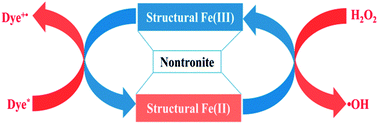
The conventional photo-Fenton reaction often suffers from the constraints of operation pH, low iron loading, ultraviolet availability in solar light and instability of iron-based catalysts. Here we report a novel heterogeneous Fenton reaction which works with a dye-photosensitized structural Fe(III)/Fe(II) redox cycling mechanism. The synthesized nontronite catalyst (NAU) was characterized by powder X-ray diffraction (XRD), transmission electron microscopy (TEM), Fourier transform infrared spectra (FTIR), X-ray photoelectron spectroscopy (XPS) analysis, and thermal gravimetric analysis (TG). NAU exhibited excellent catalytic activity over a wide pH range (3.0–8.0) for highly efficient degradation of Rhodamine B by hydrogen peroxide (H2O2) under visible light irradiation (λ > 420 nm). The excited dye molecule donates electrons to structural iron sandwiched in NAU which further catalyzes H2O2 to generate highly reactive ˙OH radicals. This iron-rich clay mineral (total Fe, 24.4 wt%) is chemically and mechanically stable. There are no measurable iron leaching, nor any noticeable loss of activity and damage to the clay structure observed after 6 recycles. Therefore, NAU clay has outstanding merits for the practical treatment of organic dye pollutants at large scale.
1. Introduction
The heterogeneous photo-Fenton reaction has been widely applied in wastewater treatment, with the important advantages of working in a broad pH range without formation of Fe sludges.1–3 A wide range of solid materials, including bulk iron-containing catalysts (e.g. hematite,4 goethite5,6) and loaded catalysts impregnated with iron or iron oxides (e.g. bentonite clay-based Fe nanocomposite film,3 Fe-exchanged zeolite,7 Fe-activated carbon fibers8 and nano-composite Fe-catalyst9), have been proposed as heterogeneous catalysts for the oxidative degradation of recalcitrant organic compounds. These iron-based catalysts can directly react with H2O2 to generate ˙OH, which is a powerful, nonselective oxidant responsible for removal of organic contaminants. However, in most cases, UV illumination is used to excite Fe oxides or Fe(III) complexes and accelerate photoreduction of Fe(III) to Fe(II) in a heterogeneous photo-Fenton process.5–7 The UV light only occupies 3–5% of solar light energy that reaches the earth, whereas artificial UV apparatus typically consumes large quantities of electrical power which limits further large scale of industrial application. Therefore, it is highly desirable to develop a new heterogeneous photo-Fenton system in which sunlight or visible light is used to drive reactions instead of UV.1,2
Most of iron oxides do not effectively utilize visible light,10 while some industrial contaminants such as dyes can absorb visible light. Dyestuff pollutants in textile effluents raise a significant environmental concern due to their massive discharge, toxicities and non-biodegradable nature. Zhao and his coworkers proposed an alternative approach to the treatment of dye-polluted waters using visible light or sunlight.11–14 Fe3+ ions can be readily reduced by excited dyes via intermolecular electron transfer process. The rates of ˙OH generation were significantly enhanced in this dye-sensitised Fenton system due to the accelerated Fe3+/Fe2+ redox cycles. To overcome the pH constraints of homogeneous photo-Fenton process, they developed a variety of solid supports such as resin11 and clays.2 Smectite clays have been widely used in a range of processes, including contaminants adsorption,15 catalytic degradation of organic compounds,16 selective oxidation,17 and catalytic reduction of compounds.18,19 The recent investigations indicated that the intrinsic structural iron in layered clay such as montmorillonite acts as an electron shuttle by which excited dyes can donate electron to H2O2 and Cr(VI), with simultaneous removal of dyes pollutants.20,21 The proposed mechanisms are illustrated by eqn (1)–(7). However, the low iron loading (ca. 2%) in natural montmorillonite restricts the rates of dye degradation in such dye–clay–H2O2 Fenton system.2,20
|
dye + visible light → dye˙+ + e−
| (1) |
|
Fe(III) + e− → Fe(II)
| (2) |
|
Fe(II) + H2O2 → Fe(III) + ˙OH + OH−
| (3) |
|
Fe(III) + H2O2 → Fe(II) + O2˙−/HO2˙
| (4) |
|
Fe(III) + O2˙−/HO2˙ → Fe(II) + O2
| (5) |
|
Fe(II) + O2˙−/HO2˙ → Fe(III) + H2O2
| (6) |
|
˙OH + dye˙+ → degraded products
| (7) |
Nontronite is a kind of iron-rich smectite in nature, but all natural nontronites contain amounts of unsubstituted Al3+ either in their tetrahedral and/or octahedral sheets.22 Iron content in nontronite is assumed to increase further if Fe3+ occupies all tetrahedral/octahedral sites of Al3+ by substitution via a chemical synthetic pathway. Therefore, synthetic iron-rich nontronite is expected to be a highly efficient Fenton catalyst, although its feasibility, to the best of our knowledge, has not been tested in dye wastewater treatment.
The aim of this study is to synthesize pure nontronite (NAU) by hydrothermal method and investigate its applicability for decoloration of dyes solutions under visible light. The effects of initial pH, H2O2 dosage and O2 on decoloration kinetics of dyes were also examined. This work is valuable for the practical application of iron-bearing clays in the removal of organic dye pollutants.
2. Experimental section
2.1. Chemicals
Rhodamine B (RhB, pure), hydrogen peroxide (H2O2, 30% aqueous solution), sodium metasilicate (SiO2·Na2O·5H2O) and ferrous chloride (FeCl2) are of analytical reagent grade and purchased from Sinopharm Chemical Reagent Co., Ltd. N2 gas (99.999% purity) was used for deaerating suspensions. Barnstead Ultra Pure water (18.25 MΩ cm) was used throughout the study. The diluted solutions of NaOH or HClO4 were used to adjust the pH of solutions.
2.2. Synthesis of nontronite (NAU)
The starting material is a gel prepared using dissolved SiO2·Na2O·5H2O and FeCl2·4H2O (theoretical composition Si/Al/Fe = 4/0/2) according to the reaction:22–24 |
 | (8) |
After precipitation for one week, the solid phase (gel) was recovered by filtration. The gel was dehydrated at 60 °C for 48 hours and then grinded into powder. The initially dark-blue gel became brown-yellow after dehydration owing to the iron oxidation. One gram of powered gel and 60 mL of ultrapure water were placed in Teflon coated autoclave. The pH was adjusted to 12.3 by adding 1 M NaOH. Syntheses were conducted at 150 °C (±2 °C), under equilibrium water pressure for 4 weeks. At the end of synthesis, the solid phase was washed and filtrated for several times by centrifugation to remove NaCl, then dried overnight at 40 °C.
2.3. Clay characterization
The Fe concentrations were determined by atomic absorption spectrometry AAS (Hitachi Z-2000). Before measurement, the sample was digested completely with a solution of the 0.125 g NAU and 10 mL strong acid (HCl
:
HNO3 = 3
:
1 (v/v)). X-ray diffraction (XRD) patterns (2θ, 5–90°) of air-dried powders were obtained by D/max-2550 PC, equipped with Cu Kα radiation (λ = 1.54056 Å), operating at 40 kV and 200 mA. Experimental measurement parameters were 0.06 s counting time per 0.02° 2θ step. Nicolet 6700 FTIR spectrometer was used to record Fourier transform infrared (FTIR) spectra at lower than 0.09 cm−1 resolution in transmission mode in the 4000–400 cm−1 range. The KBr pellets (1 cm in diameter) were prepared by mixing 1 mg of sample with 100 mg of KBr. Morphological observations of dispersed samples were performed by transmission electron microscopy (TEM) using a JEM-2100. Thermal analyses were carried out with a TG 209 F1. Thermal gravimetric analysis (TGA) data were collected in air using a heating rate of 20 °C min−1 over the 20–1000 °C temperature range. To confirm the oxidation state of Fe(III), dried powder samples were also analyzed on a X-ray photoelectron spectroscopy (XPS) (Escalab 250Xi) with Al Kα radiation as the exciting source. Brunauer–Emmett–Teller (BET) surface area was measured by a nitrogen adsorption/desorption method in a TriStar II 3020 BET surface analyzer system at 77 K. The mesopore size distribution was determined by using the Barret–Joyner–Halenda (BJH) method to the adsorption branch of the isotherm.
2.4. Reaction procedures
All batch photochemical experiments were performed in a photochemical reactor (Nanjing Xujiang Electromechanical Plant, China). The irradiation source was a 500 W metal halide lamp positioned inside a cylindrical Pyrex jacket and cooled by circulating water to maintain the temperature at 56 ± 2 °C. A cut-off filter was placed outside the water jacket to cut off the light of wavelengths below 420 nm, ensuring the irradiation only by visible light. The catalytic activity of NAU was studied using Rhodamine B (RhB) as a model pollutant. All photocatalytic experiments were conducted in a cylindrical Pyrex vessel (60 mL). The reaction suspensions contained 0.2 g L−1 clay sample, 20 μM RhB and 10 mM H2O2. The pH of the suspensions was adjusted to 3.0 ± 0.1 by dilute HClO4 and NaOH solution. By a stirring in dark for ca. 1 h, the degradation of RhB was initialized after the adsorption/desorption equilibrium of RhB on the clay, and were stirred magnetically throughout the photolysis. Deaerated suspensions were prepared by purging nitrogen gas continuously before and during the illumination.
The iron content in the clay and solutions were determined by atomic absorption spectrometry on a Hitachi Z-2000 instrument. At a given time interval, about a 1 mL aliquot from the reaction vessel was sampled and centrifuged. Then the precipitate were suspended in 1 mL acetone solution and centrifuged. The solvent extraction procedure of residual dye was repeated for three times to ensure the sufficient desorption of dye prior to the absorbance measurements. The extracted dye was analyzed immediately on a Hitachi Model U-2910 spectrophotometer at λ = 554 nm. To test the stability and recyclability of NAU, the catalyst was collected by centrifugation after the added dye was almost completely degraded. The obtained catalyst was resuspended into a fresh solution of RhB and H2O2. Dye degradation was continued as the second run. This process was repeated 6 times.
3. Results and discussion
3.1. Characterization of catalyst
X-ray diffraction patterns reveal that nontronite mineral is the only crystalline phase existing in NAU sample (Fig. 1), similar to that of a dioctahedral ferric smectite.24 This phase was clearly identified by a narrow (00l) reflections at 12.645 Å and dissymmetric (hk) bands at 4.5487 Å, 3.0829 Å, 2.6139 Å, 1.7415 Å and 1.5340 Å. A d001 distance of 12.645 Å is due to a single water layer, in accordance with a sodic smectite.22
 |
| Fig. 1 Powder XRD pattern of synthesized nontronite. | |
TEM observations indicate that synthesized nontronite appears as crumpled flakes with rolled edges, associated in micron-sized aggregates (Fig. 2).22,24 The average particle size was estimated as 46.5 nm by BET method. The BET area of the synthetic nontronite was measured as 128.9 m2 g−1 by BET surface area plot (ESI-Fig. 1†), much higher than the reported values for natural nontronite minerals25,26 (33.4 m2 g−1 or 52.8 m2 g−1 for NAu-1, 11.7 m2 g−1 for NAu-2). The internal lamellar structure and porosity (ESI-Fig. 2,† pore total volume, 0.12 m3 g−1) of clay comprises the majority of the specific surface area, which favors the adsorption of dyes and subsequent electron transfer processes. XPS Fe 2p3/2 spectra for NAU catalyst (Fig. 3a) presents a major contribution occurring at near 711.7 eV, which corresponds to the binding energy of Fe(III)–O. The absence of peaks with lower binding energy in Fig. 3a indicated Fe(II) content in this sample was negligible.27 The structural formulae of obtained NAU sample was estimated as NaFeSi3O7(OH)2·nH2O according to the atomic ratios of Na, Si, Fe and O in XPS measurement.
 |
| Fig. 2 TEM images of synthesized nontronite. | |
 |
| Fig. 3 X-ray photoelectron spectra (a) and FTIR spectra (b) of synthesized nontronite. | |
A FTIR spectrum (400–4000 cm−1) of the NAU sample is presented in Fig. 3b. In the 3000–4000 cm−1 region, the broad band at 3433 cm−1 is attributable to OH-stretching of the remaining interlayer water. An OH-stretching adsorption band was present at 3560 cm−1 in the frequency range of Fe3+–OH–Fe3+ stretching vibrations in nontronite.28 A peak at 997 cm−1 should be assigned to the strong Si–O stretching band which often shifts from 1004 to 991 cm−1 when the octahedral Fe3+ content,23 and especially the tetrahedral Fe3+ content increases.24 The 814 cm−1 band is clearly attributed to the Fe23+–OH-bending mode. The occurrence of a band near 850 cm−1 is related to a second Fe23+–OH bending mode.21,23 Bands near 428, 452, 488, 598, and 673 cm−1 are often observed in IR spectra of nontronites.28 A new band at 712 cm−1 (Fig. 3b) is probably related to a high level of Fe-for-Si tetrahedral substitution of NAU, because this band is absent in IR spectra of ferric smectites without Fe for-Si tetrahedral substitution.21
The TGA curve of NAU exhibits two endothermic events (Fig. 4): the first event below 100 °C corresponds to the loss of adsorbed water. The second event in the range 373–393 °C should be attributed to the dehydroxylation of the smectite. For nontronites, Brigatti observed a good negative correlation between the dehydroxylation temperature and the Fe content.29 The low dehydroxylation temperatures (<400 °C) measured here would be correlated with the high Fe contents of this pure nontronite (total Fe, 24.4 wt%).
 |
| Fig. 4 The DTA-TG curves of the Na-saturated nontronite sample. | |
3.2. Photocatalytic activity
3.2.1. Photodegradation of RhB. The photodegradation of RhB was carried out after the adsorption/desorption equilibrium had been reached between RhB and the catalyst NAU. The degradation of RhB was found to be negligible in the ternary or binary systems in the dark (Fig. 5, curve b, d and g), or in the absence of the catalyst (Fig. 5, curve e) and only exist RhB (Fig. 5, curve f) under visible light irradiation. As the suspension of RhB, NAU and H2O2 was irradiated with visible light, RhB was promptly degraded (Fig. 5, curve a), demonstrating the necessity of nontronite, H2O2 and visible irradiation for the decoloration of RhB. It is worthy to note that dioxygen was not necessarily required for dye decoloration, as RhB could be completely photo-degraded with the catalyst in the deaerated solution (ESI-Fig. 3†).
 |
| Fig. 5 The decoloration of RhB in the different systems under visible irradiation and in the dark. [RhB] = 20 μM, [H2O2] = 10 mM, NAU = 0.2 g L−1, pH = 3. | |
To differentiate the contribution of structural iron and iron oxide impurity in synthetic NAU to dye degradation, NAU was treated by CBD procedure (sodium citrate (0.3 M), bicarbonate (0.1 M), and dithionite (0.1 M)) to remove iron oxides.30 The iron oxide content in the treated NAU (denoted as C-NAU) was found to be less than 1.3 wt%. C-NAU catalyst exhibited the nearly same catalytic activity as NAU (Fig. 6). This demonstrates that the structural iron as a major Fe species in NAU mineral was responsible for activating H2O2 and subsequent dye degradation. The unique layer structure of NAU facilitates the intact adsorption of dye with iron-bearing tetrahedral and/or octahedral sheets. Then the sandwiched structural iron in NAU serves as an efficient redox mediator for decomposing H2O2 to ˙OH radical.
 |
| Fig. 6 Decay of RhB in the NAU and C-NAU system under visible irradiation. [RhB] = 20 μM, [H2O2] = 10 mM, NAU = 0.2 g L−1, C-NAU = 0.2 g L−1, pH = 3. | |
3.2.2. Effect of initial pH. The effect of initial solution pH on rates of RhB degradation was also investigated (Fig. 7). NAU exhibited good catalytic activity over a wide pH range (pH 3.0–8.0), which was distinct from the homogeneous Fenton system.31 About 95% of RhB was removed within 30 min at pH 3.0–5.0. The reduced rates of decoloration of RhB at higher pH should be due to the less oxidizing ability of H2O2 at a higher pH. Therefore, pH = 3 was chosen as an optimum pH in this heterogeneous photo-Fenton process for further experiments.
 |
| Fig. 7 Effect of initial pH on the decolorization of RhB by heterogeneous photo-Fenton process. [RhB] = 20 μM, [H2O2] = 10 mM, NAU = 0.2 g L−1. | |
3.2.3. Effect of H2O2 concentration. In a photo-Fenton system, H2O2 concentration is a key factor that can significantly affect the degradation of organic pollutants, since it is directly related to the amounts of hydroxyl radicals generated, and thus to the performance achieved.8,31 The effect of the hydrogen peroxide concentration was tested by varying its initial concentration between 0 and 20 mM. As shown in Fig. 8, increasing H2O2 concentration from 0 to 20 mM led to a significant increase in the degradation efficiency. The inhibitory effect of higher H2O2 concentration due to its ˙OH scavenging ability, which have been found in most photo-Fenton catalytic degradation,31,32 were not observed in the present study possibly because the low dosages of H2O2 and dye.
 |
| Fig. 8 Effect of initial H2O2 concentration on the degradation of RhB in NAU–H2O2 system under visible irradiation. [RhB] = 20 μM, NAU = 0.2 g L−1, pH = 3. | |
3.3. FT-IR spectra
The FT-IR spectroscopy is an effective means for qualitative characterizing dye degradation by determining the adsorbed dye and its intermediates on clay. The FT-IR spectra of dye, NAU, NAU with adsorbed RhB and NAU after used for a photoreaction are shown in Fig. 9. The peak at 997 cm−1 in curve a (the spectrum of NAU) is assigned to the uniquely characteristic vibrations of Si–O and Si–O–Si in the clay lattice. Peaks at 3560 and 3433 cm−1 are due to the clay lattice –OH stretching vibrations and adsorbed H2O deformation, respectively. Comparing curve b (the spectrum of pure RhB) with curve c (the spectrum of RhB loaded on NAU), some characteristic peaks of RhB,33 such as 1590 cm−1 (benzene C
C stretching vibration), 1468 cm−1 (aromatic ring vibrations), 1341 cm−1 (stretching vibration of C–N linked benzene rings), 1249 cm−1 (stretching vibration of C–N in a –N(C2H5) group), 1181 cm−1 (asymmetry stretching vibration of C–O–C) and 1132 cm−1 (–C–O–H stretch vibration), were observed in curve c. After photoreaction, the characteristic peaks at 1590, 1341, 1181 and 1132 cm−1 shown in curve c disappeared completely in curve d, indicating the degradation of RhB. The TOC removal rate of RhB was 52.7% after 2 h reaction. Based on the FTIR results, a heterogeneous photosensitization mechanism of dye degradation was proposed. The excited RhB34 (E0(RhB+˙/RhB*) = −1.09 V vs. NHE) can reduce the structural Fe(III) to structural Fe(II)35 (E0(structural FeIII/FeII) = 0.44 V vs. NHE) in clay, which further reacts with H2O2 to produce ˙OH radicals. The redox cycling of Fe(III)/Fe(II) driven by excited dye and H2O2 leads to continuous generation of ˙OH radicals and consequent dye degradation. The similar reaction mechanism has been proposed by Zhao and his coworkers,2,20 with measurement of DMPO–OH ESR signal and fluorescence quenching of dye. In consistent with their previous work, the present study provides a much efficient heterogeneous Fenton catalyst working in a photosensitization mode.
 |
| Fig. 9 The FTIR spectra of (a) NAU, (b) pure RhB, (c) NAU with adsorbed RhB, and (d) NAU–RhB hybrid after photoreaction. | |
3.4. Catalyst stability
The stability and recyclability of NAU catalyst was evaluated by the additional degradation process of fresh RhB–H2O2 solution with the used catalyst from the previous runs. It was found that NAU was able to be reutilized for at least 6 runs and the reused catalyst almost retained the catalytic activity as efficient as the fresh one (Fig. 10).
 |
| Fig. 10 Stability of the NAU catalyst for the degradation of RhB. The experiments were conducted under conditions: [Rhb] = 20 μM, [H2O2] = 10 mM, NAU = 0.2 g L−1, pH = 3. | |
The tested recyclability of as-prepared NAU is superior to those of the conventional Fenton systems and TiO2, because it is difficult to separate and recover their catalysts in solutions. As demonstrated in FTIR spectrum (Fig. 9), NAU catalyst remained intact after reaction with dye. In contrast, the ruining of iron oxides in photo-Fenton systems is mostly inevitable due to the photodissolution mechanism.10 For example, transformation of goethite to crystalline phase and leaching of iron ions after reaction may occur.36 In the present study, no measurable leaching of Fe from NAU mineral was detected by AAS during the RhB degradation process at pH 3.0, indicating that the synthesized nontronite is really stable and an excellent catalyst for the removal of RhB.
4. Conclusions
An efficient heterogeneous photo-Fenton catalyst nontronite (NAU) was successfully synthesized by hydrothermal method. The NAU catalyst was characterized by XRD, TEM, XPS and BET analyses. The NAU catalyst showed good catalytic activity for the degradation of RhB in the presence of H2O2 under visible light irradiation (λ > 420 nm). The rapid degradation of the organic dye is attributed to the generation of highly oxidative ˙OH radicals during the photosensitized Fenton reaction. The degradation can occur efficiently over a wide pH range of 3.0–8.0. The as-prepared NAU exhibited good stability and recyclability for the degradation of RhB after six recycles. Therefore, NAU catalyst shows great promise for the treatment of colored organic pollutants owing to its low cost and eases for separation.
Acknowledgements
The authors gratefully acknowledge the financial support from the Fundamental Research funds for Central Universities Central (12D11317, 13D111312). This work was partially supported by National Science Foundation of China (no. 21007009, 41273108 and 21377023) and “Chen Guang” project (10CG34).
Notes and references
- A. N. Soon and B. H. Hameed, Appl. Catal., A, 2013, 450, 96 CrossRef CAS PubMed.
- M. M. Cheng, W. J. Song, W. H. Ma, C. C. Chen, J. C. Zhao, J. Lin and H. Y. Zhu, Appl. Catal., B, 2008, 77, 355 CrossRef CAS PubMed.
- J. Y. Feng, X. J. Hu and P. L. Yue, Water Res., 2005, 39, 89 CrossRef CAS PubMed.
- X. M. Zhou, H. C. Yang, C. X. Wang, X. B. Mao, Y. S. Wang, Y. L. Yang and G. Liu, J. Phys. Chem. C, 2010, 114, 17051 CAS.
- G. B. Ortiz de la Plata, O. M. Alfano and A. E. Cassano, Chem. Eng. J., 2008, 137, 396 CrossRef CAS PubMed.
- M. C. Lu, Chemosphere, 2000, 40, 125 CrossRef CAS.
- H. Kuŝić, N. Koprivanac and I. Selanec, Chemosphere, 2006, 65, 65 CrossRef PubMed.
- Y. Y. Yao, L. Wang, L. J. Sun, S. Zhu, Z. F. Huang, W. Y. Lu and W. X. Chen, Chem. Eng. Sci., 2013, 101, 424 CrossRef CAS PubMed.
- J. Y. Feng, X. J. Hu, P. L. Yue, H. Y. Zhu and G. Q. Lu, Ind. Eng. Chem. Res., 2003, 42, 2058 CrossRef CAS.
- C. Chen, Z. Wang, W. Ma and J. Zhao, in Biophysico-Chemical Processes of Anthropogenic Organic Compounds in Environmental Systems, ed. B. Xing, N. Senesi and P. M. Huang, John Wiley & Sons, Inc., Hoboken, 2011, p. 91 Search PubMed.
- M. M. Cheng, W. H. Ma, J. Li, Y. P. Huang and J. C. Zhao, Environ. Sci. Technol., 2004, 38, 1569 CrossRef CAS.
- Y. D. Xie, F. Chen, J. J. He, J. C. Zhao and H. Wang, J. Photochem. Photobiol., A, 2000, 136, 235 CrossRef CAS.
- K. Q. Wu, Y. D. Xie, J. C. Zhao and H. Hidaka, J. Mol. Catal. A: Chem., 1999, 144, 77 CrossRef CAS.
- J. C. Zhao, C. C. Chen and W. H. Ma, Top. Catal., 2005, 35, 269 CrossRef CAS PubMed.
- D. Nguyen-Thanh, K. Block and T. J. Bandosz, Chemosphere, 2005, 59, 343 CrossRef CAS PubMed.
- J. M. Tatibouët, E. Guélou, J. Barrault and J. Fournier, Top. Catal., 2005, 33, 1 CrossRef PubMed.
- K. Ebitani, M. Ide, T. Mitsudome, T. Mizugaki and K. Kaneda, Chem. Commun., 2002, 690 RSC.
- M. Elsner, R. P. Schwarzenbach and S. B. Haderlein, Environ. Sci. Technol., 2004, 38, 799 CrossRef CAS.
- J.-M. Zen, S.-H. Jeng and H.-J. Chen, J. Electroanal. Chem., 1996, 408, 157 CrossRef.
- W. J. Song, M. M. Cheng, J. H. Ma, W. H. Ma, C. C. Chen and J. C. Zhao, Environ. Sci. Technol., 2006, 40, 4782 CrossRef CAS.
- R. L. Liu, Y. G. Guo, Z. H. Wang and J. S. Liu, Chemosphere, 2014, 95, 643 CrossRef CAS PubMed.
- A. Decarreau, S. Petit, F. Martin, F. Farges, P. Vieillard and E. Joussein, Clays Clay Miner., 2008, 56, 322 CrossRef CAS.
- P. Andrieux and S. Petit, Appl. Clay Sci., 2010, 48, 5 CrossRef CAS PubMed.
- A. Decarreau and D. Bonnin, Clay Miner., 1986, 21, 861 CAS.
- D. P. Jaisi, H. Dong and C. Liu, Geochim. Cosmochim. Acta, 2007, 71, 1145 CrossRef CAS PubMed.
- M. Dogan, A. U. Dogan, F. I. Yesilyurt, D. Alaygut, I. Buckner and D. E. Wurster, Clays Clay Miner., 2007, 55, 534 CrossRef CAS.
- T. Fujii, F. M. F. de Groot, G. A. Sawatzky, F. C. Voogt, T. Hibma and K. Okada, Phys. Rev. B: Condens. Matter Mater. Phys., 1999, 59, 3195 CrossRef CAS.
- B. A. Goodman, J. Phys., Colloq., 1976, 37, 19 Search PubMed.
- M. F. Brigatti, Clay Miner., 1983, 18, 177 CAS.
- I. H. M. van Oorschot and M. J. Dekkers, Earth Planet. Sci. Lett., 1999, 167, 283 CrossRef CAS.
- E. G. Garrido-Ramírez, B. K. G. Theng and M. L. Mora, Appl. Clay Sci., 2010, 47, 182 CrossRef PubMed.
- Z. Wang, W. Ma, C. Chen and J. Zhao, J. Hazard. Mater., 2009, 168, 1246 CrossRef CAS PubMed.
- J. Li, W. Ma, P. Lei and J. Zhao, J. Environ. Sci., 2007, 19, 892 CrossRef CAS.
- J. C. Zhao, T. X. Wu, K. Q. Wu, K. Oikawa, H. Hidaka and N. Serpone, Environ. Sci. Technol., 1998, 32, 2394 CrossRef CAS.
- C. Swearingen, J. Wu, J. Stucki and A. Fitch, Environ. Sci. Technol., 2004, 38, 5598 CrossRef CAS.
- S. S. Lin and M. D. Gurol, Environ. Sci. Technol., 1998, 32, 1417 CrossRef CAS.
Footnote |
† Electronic supplementary information (ESI) available: BET measurement results (ESI-Fig. 1 and 2) and N2-purged experiment (ESI-Fig. 3). See DOI: 10.1039/c3ra47359g |
|
This journal is © The Royal Society of Chemistry 2014 |
Click here to see how this site uses Cookies. View our privacy policy here.