Rapid and selective detection of Cys in living neuronal cells utilizing a novel fluorescein with chloropropionate–ester functionalities

Dhiraj P. Muralea, Hwajin Kimb, Wan Sung Choib and David G. Churchill*a
aMolecular Logic Gate Laboratory, Department of Chemistry, Korea Advanced Institute of Science and Technology (KAIST), 373-1 Guseong-dong, Yuseong-gu, Daejeon, 305-701, Republic of Korea. E-mail: dchurchill@kaist.ac.kr; Fax: +82-42-350-2810; Tel: +82-42-350-2845
bDepartment of Anatomy and Neurobiology, Medical Research, Center for Neural Dysfunction, Institute of Health Science, School of Medicine, Gyeongsang National University, 92 Chilam-dong, Jinju, Gyeongnam 660-751, Republic of Korea

Received 4th December 2013 , Accepted 11th December 2013

First published on 20th December 2013


Abstract

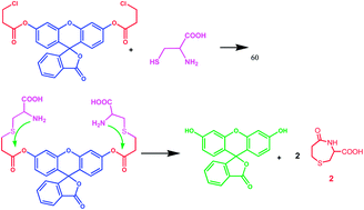
A chloropropionate-caged fluorescein probe allows for prompt detection of cysteine over other biothiols, e.g., homocysteine with a limit of detection of 12.8 μM.


Cysteine (Cys), homocysteine (Hcy) and glutathione (GSH) are bio-thiols that are essential in processes in living biological systems. These low molecular weight species are involved in many physiological processes. Some of these vital processes include protein synthesis, metabolism, detoxification, signal transduction and gene regulation. Also, thiols (and selenols) are demonstrably key players in regulating the redox state of proteins and in protein structure.1 These species are involved in many crucial biological functions; excess concentrations of thiols can be causal, or otherwise signify a variety of different disorders. Excess concentrations of Cys may lead to, or signal, neurotoxicity;2 deficiency may also lead to adverse effects such as skin lesions, edema, lethargy, muscle weakness, retarded growth, and promotion of liver damage.3 A heightened [Hcy] in human plasma has been correlated with incidence of Alzheimer's disease and cardiovascular disease; cellular GSH insufficiency leads to oxidative stress which is linked to neurodegenerative disorders. Cancers and AIDS are also related to biothiol concentration.4,5 Thus, detection of these small sulfur containing species is important and timely. To fully achieve understanding of cellular compartmentalization of free sulfur amino acid chemistry would be a significant achievement e.g., that seen in neurodegenerative disease progression. This requires further investigations into the engineering of selective small molecular fluorescent probes. One huge stumbling block in the selective detection of small biothiols is the structural similarity and chemical nature of Cys and Hcy. How fluorescent sensors can efficiently distinguish between these thiols comes down to gearing selective organic chemistry reactivity and fluorophore optical patterns that coordinate the scavenging of one over the other, kinetically or thermodynamically, with a concomitant dramatic and clear photophysical modulation. Ideally, fluorescence chemosensing can allow for excellent determinations and imaging of analytes such as cysteine.

The number of probes that detect Cys and Hcy in a relatively equal basis is quite high;6 but the achievement of excellent selective detection, e.g., between Cys and Hcy is rare.7 Chemodosimeters are currently heralded by synthetic chemists and biological practitioners alike as they offer inherent selectivity and sensitivity in biothiol detection over general types of chemosensors.8a,b Recently, efforts from our research group resulted in the development of a novel rechargeable meso-aryl BODIPY-based chemodosimeter for selective detection of Cys over other biothiols (e.g., Hcy and GSH).8a As an extension to this interest we subsequently reported novel probes to detect biothiols via ester hydrolysis.8b

Probe was synthesized (Scheme 1) and characterized by ruby NMR and ESI-Mass (Fig. S1–S3). Here, the probe design was based on aliphatic chloride substitution. This allows for intramolecular cyclization (substitution/cyclization reactions) of chloropropionate with the sulfhydryl group of cysteine, followed by cyclization which forms cyclized product 2 and free fluorescein (Fig. 1). Here, Cys and Hcy are able to be discerned based on size of ring formation. The side product formed independently after the reaction of Cys is a heterocycle involving a seven-membered ring, whose formation is kinetically favored over the formation of an eight-membered ring allowed by reaction with Hcy. Reaction time is also an important consideration for chemodosimeters seeing as kinetic products are involved; time also allows for discrimination between Cys and Hcy. The formation of the nine-membered ring will take longer. Fast detection of Cys is most likely because the intramolecular cyclization reaction to form a seven-membered ring is significantly faster compared to eight-membered ring formation. Thus, the chloropropionate group serves as a good detection moiety for Cys over Hcy. In this same perspective, Zhang et al.9a recently showed a chloroacetate-based probe for recognition of Cys and Hcy; Kim et al.,9b also showcased Cys sensing based on a bromoacetate version of fluorescein. When compared to previous reports, our probe is more selective and the detection time is very fast in living neuronal cells and human blood plasma. The chloroacetate version can sense both Cys and Hcy; whereas, this chloropropionate version detects only Cys but not Hcy. The bromoacetate version has a detection limit of 35 μM, whereas that for our probe was found to be 12.8 μM.


image file: c3ra47280a-s1.tif
Scheme 1 Synthesis of disubstituted probe 1.

image file: c3ra47280a-f1.tif
Fig. 1 Proposed chemical and photophysical mechanism of probe 1 with analyte being cysteine.

Photophysical properties of probe 1 were investigated under physiological conditions (buffered H2O[thin space (1/6-em)]:[thin space (1/6-em)]DMSO 80[thin space (1/6-em)]:[thin space (1/6-em)]20; pH 7.4 PBS). The probe first was dissolved in DMSO and then subsequently diluted in 80% PBS buffer, pH 7.4. The detection properties of probe can be accessed via UV-vis absorption and emission spectroscopy, since the fluorescein probe in its lactone form has no fluorescence.

The probe was first screened with dissolved forms of sulfur-containing amino acids (L-Cys, Hcy, N-acetyl-L-Cys, Met, GSH) and non-sulfur-containing amino acids (Ala, Arg, Asn, Asp, Gln, Glu, Gly, His, Ile, Leu, Lys, Phe, Pro, Ser, Thr, Trp, Tyr, Val). These analytes were prepared as aqueous solutions on the order of 66 μM. Here, 3.0 mL of 4.0 × 10⁻⁶ M, PBS buffer, pH 7.4 probe solution was used and incubated with ∼10 equiv. of amino acid. After ≤10 min, significant changes in the color of the probe solution was found with Cys; a slight change only was observed with Hcy. A dramatic fluorescence intensity increase was found when determined quantitatively with analyte via emission studies (Fig. 2). A clear 250-fold and enhancement in fluorescence intensity was quantified for Cys, compared to the starting probe fluorescence and 4-fold compared to Hcy (Fig. 2). When the concentration of Cys increased, a steady increase in fluorescence intensity was observed (Fig. 3a). On this same basis, the detection limit10 (3.3 × standard deviation (SD) of y/slope) was estimated to be 12.8 μM, indicating that the utility of the probe for detection of Cys in biological systems. Also, when the effect of intensity by probe was determined over the course of 1.0 h, an extremely smooth increase in fluorescence intensity was recorded (Fig. 3b) with a plateau shaping after ∼25 min (under said conditions). Importantly, a competition study was undertaken to validate the selectivity and practical utility of the probe specifically, we performed of 1 + Cys solution in the presence of other thiol-containing species: Hcy, N-acetyl-L-Cys, and GSH within 10 min. As a result, we found a ∼25% decrease in fluorescence intensity with GSH and N-acetyl-L-Cys, whereas with Hcy there was no interference (Fig. S5).


image file: c3ra47280a-f2.tif
Fig. 2 Emission spectra of probe 1 (4.0 × 10⁻⁶ M, buffered H2O[thin space (1/6-em)]:[thin space (1/6-em)]DMSO 80[thin space (1/6-em)]:[thin space (1/6-em)]20; pH 7.4 PBS) with amino acids (L-Cys, Hcy, N-acetyl-L-Cys, Met, GSH, Ala, Arg, Asn, Asp, Gln, Glu, Gly, His, Ile, Leu, Lys, Phe, Pro, Ser, Thr, Trp, Tyr, Val) (∼10 equiv. in water) incubated for 10 min at RT.

image file: c3ra47280a-f3.tif
Fig. 3 (A) Emission spectra of probe 1 (4.0 × 10⁻⁶ M, buffered H2O[thin space (1/6-em)]:[thin space (1/6-em)]DMSO 80[thin space (1/6-em)]:[thin space (1/6-em)]20; pH 7.4 PBS) with increasing concentration of L-Cys (16.6–166.5 μM in water) incubated for 10 min at RT. (B) Time-dependent emission spectra of compound 1 (2.0 × 10⁻⁶ M, buffered H2O[thin space (1/6-em)]:[thin space (1/6-em)]DMSO 80[thin space (1/6-em)]:[thin space (1/6-em)]20; pH 7.4 PBS) with Cys (∼10 equiv. in water), λexci = 490 nm.

A separate reaction was carried out with probe 1 (1.0 equiv.) and Cys (2.5 equiv.) in 30 mL of a MeOH[thin space (1/6-em)]:[thin space (1/6-em)]H2O (90[thin space (1/6-em)]:[thin space (1/6-em)]10, v/v) solution was performed to support the presumed mechanism. The reaction mixture was stirred at RT for 1 h. Then, Et3N (100 μL) was added and the solution stirred for 1 h. The solvent was then removed and the crude product was checked directly with ESI-MS revealing a value corresponding to fluorescein as a major product (Fig. S4). The main product was the highly fluorescent fluorescein (518 nm).

Experiments with human blood plasma (Sigma-Aldrich) were carried out to check additional utility of the probe to detect cysteine in relevant biological matrices. Firstly, standard solutions of probe with Cys were prepared at different concentrations (5 μM, 10 μM, 15 μM and 20 μM). Further, to determine actual concentration of Cys plasma, probe 1 was incubated 10 μL and 20 μL of human blood plasma for 10 min. Finally, through the use of a standard deviation plot using standard solutions of probe with Cys, the actual concentration of Cys in blood plasma was calculated to be 11.7 μM (Fig. 4). Here, the detection limit for human blood plasma is lower as compared to the detection limit mentioned above. Here, we believe the medium present in the blood plasma assists the substitution reaction compared to the cuvette experiment milieu. The estimation of the detection limit was made using using a greater portion of plasma. The amount of Cys added is quite high (a concentration of 10 μL). 20 μL may be a cause for a lowered detection limit. The main aim behind this experiment was to check further potential bioapplicability of probe Cys detection.


image file: c3ra47280a-f4.tif
Fig. 4 (A) Relative fluorescence intensity of Cys (5 μM, 10 μM, 15 μM and 20 μM) with probe 1 (4.0 × 10⁻⁶ M, buffered H2O[thin space (1/6-em)]:[thin space (1/6-em)]DMSO 80[thin space (1/6-em)]:[thin space (1/6-em)]20; pH 7.4 PBS). (B) Relative fluorescence of human blood plasma (10 μL, 20 μL) with probe 1 (4.0 × 10⁻⁶ M, buffered H2O[thin space (1/6-em)]:[thin space (1/6-em)]DMSO 80[thin space (1/6-em)]:[thin space (1/6-em)]20; pH 7.4 PBS). (C) Standard deviation plot of Cys conc. (5.0 μM, 10 μM, 15 μM and 20 μM) with the emission intensity.

Finally, the probe was subjected to Cys concentration in live neuronal cells. As a result there was a quick response to intracellular cysteine with 1 μM of probe within 10 min. These results were further confirmed by NEM experimental conditions (Fig. 5). From these results it is clear that the probe can be used to detect cysteine in living neuronal cells within 10 minutes.


image file: c3ra47280a-f5.tif
Fig. 5 Fluorescence microscopic images of living SH-SY5Y cells. (A) SH-SY5Y cells were incubated with vehicle (DMSO). (B) Probe (1 μM) for 10 min. (C) SH-SY5Y cells were pre-incubated with NEM (1 mM) for 30 min. (D) Probe (1 μM) for 10 min. (λexc = 490 nm) Scale bar = 50 μm.

In conclusion, a novel chloropropionate ester modality of Fluorescein was synthesized which acts as a highly selective ratiometric, fluorescent and sensitive probe for the detection of Cys over other thiol-containing naturally occurring amino acids and related molecules. A detection limit of 12.8 μM and 250-fold increase in fluorescence intensity within 10 minutes were determined. Sensing was also demonstrated in living neuronal cells and in human blood plasma milieu.

Notes and references

  1. (a) J. B. Schulz, J. Lindenau, J. Seyfried and J. Dichgans, Eur. J. Biochem., 2000, 267, 4904 CrossRef CAS; (b) S. Seshadri, A. Beiser, J. Selhub, P. F. Jacques, I. H. Rosenberg, R. B. D'Agostino, P. W. F. Wilson and P. A. Wolf, N. Engl. J. Med., 2002, 346, 476 CrossRef CAS PubMed; (c) R. O. Ball, G. Courtney-Martin and P. B. Pencharz, J. Nutr., 2006, 136, 1682S CAS; (d) R. Hong, G. Han, J. M. Fernandez, B. J. Kim, N. S. Forbes and V. M. Rotello, J. Am. Chem. Soc., 2006, 128, 1078 CrossRef CAS PubMed; (e) Z. A. Wood, E. Schroder, J. R. Harris and L. B. Poole, Trends Biochem. Sci., 2003, 28, 32 CrossRef CAS.
  2. X. F. Wang and M. S. Cynader, J. Neurosci., 2001, 21, 3322 CAS.
  3. S. Shahrokhian, Anal. Chem., 2001, 73, 5972 CrossRef CAS.
  4. H. Refsum, P. M. Ueland, O. Nygard and S. E. Vollset, Annu. Rev. Med., 1998, 49, 31 CrossRef CAS PubMed.
  5. (a) G. Y. Wu, Y. Z. Fang, S. Yang, J. R. Lupton and N. D. Turner, J. Nutr., 2004, 134, 489 CAS; (b) D. M. Townsend, K. D. Tew and H. Tapiero, Biomed. Pharmacother., 2003, 57, 145 CrossRef CAS; (c) L. A. Herzenberg, S. C. DeRosa, J. G. Dubs, M. Roederer, M. T. Anderson, S. W. Ela, S. C. Deresinski and L. A. Herzenberg, Proc. Natl. Acad. Sci. U. S. A., 1997, 94, 1967 CrossRef CAS; (d) W. W. Huber and W. Parzefall, Curr. Opin. Pharmacol., 2007, 7, 404 CrossRef CAS PubMed.
  6. (a) Y. M. Yang, Q. Zhao, W. Feng and F. Y. Li, Chem. Rev., 2013, 113, 192–270 CrossRef CAS PubMed; (b) X. Chen, Y. Zhou, X. J. Peng and J. Yoon, Chem. Soc. Rev., 2010, 39, 2120–2135 RSC; (c) H. S. Jung, X. Chen, J. S. Kim and J. Yoon, Chem. Soc. Rev., 2013, 42, 6019–6031 RSC; (d) C. Yin, F. Huo, J. Zhang, R. Martinez-Manez, Y. Yang, H. Lv and S. Li, Chem. Soc. Rev., 2013, 42, 6032–6059 RSC.
  7. (a) W. H. Wang, J. O. Escobedo, C. M. Lawrence and R. M. Strongin, J. Am. Chem. Soc., 2004, 126, 3400–3401 CrossRef CAS PubMed; (b) X. F. Yang, Y. X. Guo and R. M. Strongin, Angew. Chem., Int. Ed., 2011, 50, 10690–10693 CrossRef CAS PubMed; (c) H. L. Li, J. L. Fan, J. Y. Wang, M. Z. Tian, J. J. Du, S. G. Sun, P. P. Sun and X. J. Peng, Chem. Commun., 2009, 5904–5906 RSC; (d) H. L. Chen, Q. Zhao, Y. B. Wu, F. Y. Li, H. Yang, T. Yi and C. H. Huang, Inorg. Chem., 2007, 46, 11075–11081 CrossRef CAS PubMed; (e) H. P. Wu, C. C. Huang, T. L. Cheng and W. L. Tseng, Talanta, 2008, 76, 347–352 CrossRef CAS PubMed; (f) J. Mei, Y. J. Wang, J. Q. Tong, J. Wang, A. J. Qin, J. Z. Sun and B. Z. Tang, Chem. – Eur. J., 2013, 19, 612–619 CrossRef PubMed; (g) H. L. Wang, G. D. Zhou, H. W. Gai and X. Q. Chen, Chem. Commun., 2012, 48, 8341–8343 RSC; (h) L. G. Wang, Q. Zhou, B. C. Zhu, L. G. Yan, Z. M. Ma, B. Du and X. L. Zhang, Dyes Pigm., 2012, 95, 275–279 CrossRef CAS PubMed; (i) Z. Guo, S. W. Nam, S. Park and J. Yoon, Chem. Sci., 2012, 3, 2760–2765 RSC; (j) X. Yang, Y. Guo and R. M. Strongin, Org. Biomol. Chem., 2012, 10, 2739–2741 RSC.
  8. (a) O. G. Tsay, K. M. Lee and D. G. Churchill, New J. Chem., 2012, 36, 1949 RSC; (b) D. P. Murale, H. Kim, W. S. Choi and D. G. Churchill, Org. Lett., 2013, 15, 3630 CrossRef CAS PubMed.
  9. (a) B. Zhu, Y. Zhao, Q. Zhou, B. Zhang, L. Liu, B. Du and X. Zhang, Eur. J. Org. Chem., 2013, 888 CrossRef CAS; (b) K. H. Hong, S. Y. Lim, M. Y. Yun, J. W. Lim, J. H. Woo, H. Kwon and H. J. Kim, Tetrahedron Lett., 2013, 54, 3003 CrossRef CAS PubMed.
  10. See the website for the U.S. Food and Drug Administration (FDA), http://www.fda.gov/downloads/Drugs/GuidanceComplianceRegulatoryInformation/Guidances/ucm073384.pdf.

Footnote

Electronic supplementary information (ESI) available: Experimental details, in-line spectral data, MS spectra, See DOI: 10.1039/c3ra47280a

This journal is © The Royal Society of Chemistry 2014
Click here to see how this site uses Cookies. View our privacy policy here.