A highly sensitive SERS method for the determination of nitrogen oxide in air based on the signal amplification effect of nitrite catalyzing the bromate oxidization of a rhodamine 6G probe

Qingye Liu a, Jinchao Donga, Yanghe Luoab, Guiqing Wena, Lin Weib, Aihui Liang*a and Zhiliang Jiang*a
aKey Laboratory of Ecology of Rare and Endangered Species and Environmental Protection of Ministry Education, Guangxi Normal University, Guilin 541004, China. E-mail: zljiang@mailbox.gxnu.edu.cn; ahliang2008@163.com
bHezhou University, Hezhou 542899, China. Fax: +086-0773-5846201; Tel: +086-0773-5846141

Received 4th December 2013 , Accepted 6th January 2014

First published on 7th January 2014


Abstract

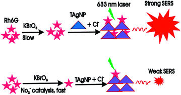
Rhodamine 6G adsorbed onto a triangular plate-type nanosilver aggregate exhibits a strong SERS peak at 1508 cm−1. Based on NO2 catalysis of the bromate oxidization of Rh6G and SERS peak quenching, trace nitrogen oxides in air samples were analyzed by a catalytic amplification SERS method.


1. Introduction

Nitrogen oxides (NOx), mainly NO2 and NO, are a type of atmospheric pollutant involved in acid rain formation, nitrogen oxides fog accidents, ozonosphere destruction and the greenhouse effect, which have aroused worldwide attention.1–3 Within cities, NOx mainly comes from car emissions and combustion gases. When the NOx content exceeds a certain value, its fatal effects are greatly enhanced. It is therefore necessary to strictly monitor the NOx draining volume and concentration in cities. At present, several methods have been reported to assay nitrogen oxide including spectrophotometric,4 chromatographic,5 electrochemical,6 and chemiluminescence methods.7 The sensitivity of spectrophotometry is low, with detection of 0.08 mg L−1 NO2. Chromatography was used to determine concentrations as low as 0.04 mg L−1 NO2. Electrochemical and chemiluminescence methods have the advantage of low cost, but have some restrictions for ultratrace detection. SERS is a very efficient spectral detection technique which is rapid, sensitive, information-rich and highly selective.8–10 However, a routine SERS quantitative method is not commonly used due to few active substrates with good reproducibility and high sensitivity.11–15 An optofluidic SERS device for on chip detection of vasopressin was developed by Williams et al., based on an L-DNA aptamer.16 Vasopressin was fluorescein isothiocyanate (FITC)-labeled, and the aptamer was incorporated in a metallic multilayer nanotube SERS active substrate. SERS signals from the FITC-labeled vasopressin, which was bound to the incorporated aptamers, were measured with a detection limit of 5.2 μU ml−1 vasopressin in PBS buffer solution. Wen et al.17 combined ssDNA with AgNPs to obtain a stable SERS probe for the quantitative analysis of 6.3–403.6 μg L−1 melamine. Rh6G as a SERS molecular probe exhibited strong SERS activity and has been utilized in inorganic and organic analysis.18–21 Furthermore, it has been utilized to improve sensitivity by combining with catalysis reactions.22–24 Nanoparticles have novel properties that have been utilized in spectral and electrochemical analysis.25–29 For example, nanogold and nanosilver sols have been commonly used as SERS substrates, but the stable triangular plate-type nanosilver aggregate (TAgNPA) is rarely reported as a SERS substrate.30 As far as we know, there are no reports on a SERS quantitative method for detecting trace NO2 and NOx in air, based on the catalytic reaction of Rh6G as the SERS probe and TAgNP sol as the SERS substrate. In this article, using stable TAgNP aggregates as the most sensitive SERS substrate, Rh6G as the SERS molecular probe, and a catalytic reaction to amplify the signal, a new catalytic SERS method which is accurate, selective and sensitive, has been developed for the determination of trace NOX in air samples.

2. Experimental

The following equipment was used: a DXR smart Raman spectrophotometer (Thermo Fisher, USA) with a laser of 633 nm, collect time of 5 s and a power of 3.5 mW, a Cary Eclipse fluorescence spectrophotometer (Varian Company, USA), a TU-1901 double beam UV-Vis spectrophotometer (Beijing Purkinje General Instrument Co., China), a JEM-2100F field emission transmission electron microscope (Japan), a 360 FT-IR spectrometer (Nicolet, USA), and a 3 K–15 high-speed refrigerated centrifuge (Sigma Co., Germany). 1.0% HAuCl4·4H2O, 1.0 mol L−1 H3PO4, 1.0 mol L−1 HCl, 1.0% trisodium citrate, 0.010 mol L−1 Rh6G, 1 mol L−1 NaCl, 10 mmol L−1 KBrO3, PEG 10000, 2.4 × 10⁻² mol L−1 AgNO3, 0.25 mg mL−1 NaBH4 and 1.45 mmol L−1 NaNO2 were used. All reagents were of analytical grade and the water was doubly distilled.

The preparation of TAgNPs was considered and polyvinylpyrrolidone was not added as it strongly inhibits the SERS effect. The recommended procedure is as follows; add 500 μL 10 mmol L−1 AgNO3, 1.5 mL 60 mmol L−1 trisodium citrate and 120 μL 30% H2O2, to a 100 mL round-bottom flask containing 47 mL water on a magnetic stirrer, at this point the mixture is colorless. After the addition of 200 μL 0.1 mol L−1 NaBH4 solution, the color changed quickly from pale yellow to blue with the appearance of lots of bubbles. After standing for 12 h, the mixture was stored in a refrigerator at 4 °C, the concentration of TAgNPs was 5.4 mg L−1 with an average side length of 40 nm and an absorption peak at 600 nm which remained constant for 20 days (see, Fig. 1S).

The general detection procedure is as follows; 100 μL 1.0 × 10⁻⁵ mol L−1 Rh6G solution, 200 μL 10 mmol L−1 KBrO3 solution, 100 μL 1 mol L−1 H3PO4 and a certain amount of NO2 solution were added into a 5 mL marked test tube, diluted to 0.8 mL and mixed well. After 15 min at 50 °C, the reaction was stopped by cooling with tap water. 120 μL 1.0 mol L−1 NaCl solution and 1.1 mL 5.4 mg L−1 TAgNPs (or 300 μL 57 μg mL−1 AuNPs or 300 μL 19.9 μg mL−1 AgNPs) were then added, diluted to 2.0 mL and mixed well. The mixture was transferred into a 1 cm quartz cell. The SERS spectrum and the SERS intensity at 1507 cm−1 (I1507 cm−1) were recorded. Meanwhile, a blank reagent solution (I1507 cm−1)0 without NO2 was recorded, and a value of ΔI = (I1507 cm−1)0I1507 cm−1 was calculated.

3. Results and discussion

In 50 mmol L−1 H3PO4 solution, TAgNPs were stable and dispersed in the solution, and there was no SERS single after adding Rh6G. Upon the addition of 60 mmol L−1 NaCl, TAgNPs aggregated to form a stable nanosilver-aggregate (TAgNPA) that could conjugate with Rh6G to obtain a TAgNPA–Rh6G SERS probe with 13 peaks at the Raman shift of 225 cm−1, 301 cm−1, 346 cm−1, 561 cm−1, 603 cm−1, 761 cm−1, 1122 cm−1, 1174 cm−1, 1306 cm−1, 1358 cm−1, 1506 cm−1, 1565 cm−1 and 1647 cm−1 (see, Fig. 2S). According to ref. 31 these SERS peak enhanced factors (Ef) were in the range of 2.7 × 10⁶–3.5 × 10⁷. The SERS of the oxidization of Rh6G by KBrO3 (ORh6G) was also examined, and weak SERS effects were observed (Fig. 3S) at the Raman shift of 162 cm−1, 341 cm−1, 530 cm−1, 620 cm−1, 768 cm−1, 1195 cm−1, 1340 cm−1, 1481 cm−1, 1497 cm−1, 1547 cm−1 and 1630 cm−1, and the Ef was in the range of 5.0 × 10²–4.6 × 10⁴.

Under the chosen conditions, the redox reaction of Rh6G–KBrO3 is slow. Trace NO2 has strong catalytic enhanced effects on the redox reaction (Fig. 1). When the NO2 concentration increased, the SERS peak at 1508 cm−1 decreased due to the decrease in Rh6G, and the decreased SERS intensity was directly proportional to the nitrite concentration (Fig. 2). As the NOx content in air is directly proportional to the nitrite concentration, a new catalytic SERS method was established to detect trace NOx in the air.


image file: c3ra47279e-f1.tif
Fig. 1 The principle of catalytic SERS detection of NO2 using Rh6G as a probe.

image file: c3ra47279e-f2.tif
Fig. 2 SERS spectra of the Rh6G–NO2–KBrO3–TAgNP system: (a) 0.5 μmol L−1 Rh6G + 1 mmol L−1 KBrO3 + 50 mmol L−1 H3PO4 + 60 mmol L−1 NaCl + 2.97 μg mL−1 TAgNPs; (b) a + 7.2 nmol L−1 NO2; (c): a + 18 nmol L−1 NO2; (d): a + 43.2 nmol L−1 NO2-; (e) a + 72 nmol L−1 NO2.

In 50 mmol L−1 H3PO4 solution, TAgNPs were stable, the color was blue and there were three surface plasmon resonance (SPR) absorption peaks at 330 nm, 395 nm and 700 nm. When NaCl was added, the TAgNPs aggregated into big particles, the color changed from blue to gray and the SPR absorption peak decreased greatly. Rh6G has a strong molecular absorption peak at 525 nm. The oxidation reaction between KBrO3 and Rh6G takes place when the NO2 catalyst was added, the absorption peak intensity at 525 nm decreased and shifted to 540 nm simultaneously (Fig. 4S). In 50 mmol L−1 HCl solution, Rh6G has a strong fluorescence peak at 564 nm when λex is at 498 nm. When the concentration of NO2 increased, the catalytic effect was enhanced and the Rh6G concentration decreased by KBrO3 oxidization resulted in a fluorescence peak at 564 nm reducing the intensity significantly (Fig. 5S).

As shown in the IR spectra (Fig. 6Sa), the characteristic peak at 3226 cm−1 is due to the stretching vibration absorption of the N–H groups, 1716 cm−1 is the C[double bond, length as m-dash]O group, 1646 cm−1 is the C[double bond, length as m-dash]N group, 1607 cm−1, 1529 cm−1, 1501 cm−1 and 1443 cm−1 are the benzene groups, 1312 cm−1 is the C–N group and 1179 cm−1 is the C–O group (in contact with the aromatic rings). Fig. 6Sb and 6Sc are the FTIR spectra of ORh6G gained through different methods. The product of Fig. 6Sb was achieved using NO2 as a catalyst and heating at 50 °C for 4 h, while the product of Fig. 6Sc was achieved by not adding NO2 solution as a catalyst and heating at 50 °C for 7 h. By comparing with Fig. 6Sa, we found that the characteristic peak shifts of N–H, C[double bond, length as m-dash]O, C[double bond, length as m-dash]N, benzene and C–O basically remain stable in the product of Fig. 6Sb after the oxidation reaction, which indicates that these groups remained in ORh6G. Meanwhile, the characteristic absorption peak of the Ar–N(H)–R group at 1312 cm−1 disappeared and the characteristic absorption peak of the C–N[double bond, length as m-dash]O group at 1287 cm−1 and C–NO2 group at 1339 cm−1 were present instead. In addition, although the oxidation product described in Fig. 6Sc was prepared without a NO2 catalyst, it still had the characteristic absorption peaks from C–N[double bond, length as m-dash]O groups and C–NO2 groups, which illustrate that the final product was a mixture of NRh6G and ORh6G.

Solid and nanosol substrates are used commonly in SERS detection. However, the reproducibility of solid substrates is not good, and they are difficult to utilize in routine quantitative analysis. Nanosols have good reproducibility and low-cost using modern nanosynthesis, and their shape can also be controlled accurately. In this article, stable TAgNPs with an average side length of 40 nm, AgNPsd with an average size of 10 nm (Fig. 3) and AuNPs with 10 nm sols were prepared and considered as SERS substrates. Results showed that the TAgNP substrate is the most sensitive, the preparation is simple and rapid, and the stability is very good. Thus, it was chosen for use.


image file: c3ra47279e-f3.tif
Fig. 3 TEM of the TAgNPs (a) and AgNPs (b).

H3PO4, H2SO4 and HCl were considered as the acidic medium. The results showed that H3PO4 gave the highest sensitivity, and it was therefore selected for use. We studied the effect of H3PO4 concentration on ΔI. The results indicated that the catalytic rate became fast when the concentration of H3PO4 was 50 mmol L−1 and the value of ΔI reached its maximum. When the concentration of H3PO4 exceeded 50 mmol L−1, the acidity was too high resulting in a decreased catalytic rate, and the value of ΔI tended to decline. We therefore chose 50 mmol L−1 as the final concentration of H3PO4. We have studied the effect of Rh6G concentration on ΔI. The results showed that when the concentration of Rh6G was 0.5 μmol L−1, the value of ΔI reached its maximum. When the concentration of Rh6G exceeded 0.5 μmol L−1, the concentration was too high resulting in an increase in the blank values, and the value of ΔI tended to decline (Fig. 7S). We therefore chose to use 0.5 μmol L−1 Rh6G. The effect of KBrO3 concentration on ΔI was also studied. The results indicated that when the concentration of KBrO3 was 1.0 mmol L−1, ΔI reached maximum (Fig. 8S). 1.0 mmol L−1 KBrO3 was therefore chosen for the assay. Reaction temperature had an important effect on the catalytic reaction. When the bath temperature was 50 °C, the ΔI values reached the maximum. When the bath temperature exceeded 50 °C, the non-catalyzed rate increased and the values of ΔI tended to decline. A bath temperature of 50 °C was therefore used for the reaction. In order to completely investigate the catalytic reaction, the effect of bath time on ΔI was also studied. The results showed that when the bath time was in the range of 15–30 min, the ΔI values reached the maximum and remained basically stable. 15 min was therefore chosen as the bath time.

The concentration of NaCl had an important effect on the aggregation extent of the TAgNPs, so the effect of NaCl concentration on ΔI was studied. The results indicate that when the concentration of NaCl was 60 mmol L−1, the value of ΔI reached its maximum. We therefore chose 60 mmol L−1 as the optimal concentration of NaCl. The effect of TAgNP concentration on ΔI was also studied. When the concentration of TAgNPs increased within a certain range, the SERS signal of the system increased. When the concentration of TAgNPs was 2.97 μg mL−1, the value of ΔI reached its maximum. However, when the TAgNP concentration exceeded 8.55 μg mL−1, the blank values of the system increased, and led to a decrease in ΔI (Fig. 9S). We therefore chose to use 2.97 μg mL−1 TAgNPs.

According to the procedure, the influence of coexistent substances (CS) on the determination of 36.0 nmol L−1 NO2 was tested, with a relative error of ±5%. The results indicated that 500 times more SO32−, Br, ClO4, PO43−, SO42−, CO32−, NO3 and F, 300 times more Co2+, Zn2+, Cd2+, Mg2+ and Ca2+, and 100 times more Fe3+, Ba2+ and Cu2+ do not interfere with the determination (Fig. 4). It was shown that this new SERS method had good selectivity.


image file: c3ra47279e-f4.tif
Fig. 4 Influence of coexistent substances (CS) on the determination of nitrite (NO2).

Under the selected conditions, the TAgNP substrate system is most sensitive, and the AgNP is more sensitive than the AuNP (Table 1). Thus, the Rh6G–KBrO3–TAgNP system was chosen to determine NOx in air, with a linear range (LR) of 3.6–72 nmol L−1 NO2 and a detection limit (DL) of 1.2 nmol L−1 NO2 (Fig. 5). The accuracy was examined and found to be 8.6% and 6.0% for 7.2 nmol L−1 NO2 and 43.2 nmol L−1 NO2 respectively. Compared with the fluorescence32 and the standard method of N-(1-naphthyl)ethylene diamine dihydrochloride spectrophotometry, this catalytic SERS method is more sensitive, and a smaller air sampling volume was used.

Table 1 Analysis features for the three nanosol substrates
Nanosol Regression equation LR (nmol L−1 NO2) Coefficient DL (nmol L−1 NO2)
AuNPs ΔI = 0.46 C + 4.1 14.4–144 0.9917 7
AgNPs ΔI = 3.60 C + 26.9 7.2–72 0.9782 3
TAgNPs ΔI = 3.36 C + 5.4 3.6–72 0.9932 1.2


image file: c3ra47279e-f5.tif
Fig. 5 Working curve.

Air samples were collected using the air sampling device (Fig. 10S) and analyzed for their NOx content. The results (Table 1S) show that the SERS results were in agreement with those obtained by the fluorescence method and the standard method of N-(1-naphthyl)ethylene diamine dihydrochloride. The relative standard deviation was in the range of 5.5–8.4% and the recovery was in the range of 96.3–103.8%.

4. Conclusion

Trace NO2 has a strong catalytic effect on the slow redox reaction of KBrO3 and Rh6G that causes SERS quenching at 1605 cm−1 in the TAgNPs nanosol substrate. Using Rh6G as the SERS probe, we established a new catalytic reaction SERS method to detect NOx in air samples, which has high sensitivity, good selectivity, simplicity and rapidity.

Acknowledgements

This work was supported by the National Natural Science Foundation of China (no. 21267004, 21367005, 21307017, 21365011), the Research Funds of Guangxi Key Laboratory of Environmental Pollution Control Theory and Technology, the Natural Science Foundation of Guangxi (no. 2013GXNSFFA019003, 2013GXNSFAA019046), and the Research Funds of Guangxi Education Department (no. 2013YB234, 2013YB035).

References

  1. R. M. Heck and R. J. Farrauto, Comm. Technol., 1995, 6, 161 Search PubMed.
  2. L. J. Muzio and G. C. Quartucy, Prog. Energy Combust. Sci., 1997, 23, 233 CrossRef CAS.
  3. V. I. Parvulescu and P. Grange, Cat. Today, 1998, 46, 233–316 CrossRef CAS.
  4. Y. N. Shan and S. Q. Gao, Environ. Monit. China, 1988, 4(3), 2–4 Search PubMed.
  5. H. M. Peng, M. J. Wu, F. L. Ren, Z. H. He and W. L. Lian, Chin. J. Anal. Chem., 2007, 35, 289–292 CAS.
  6. C. Y. Ren, H. Y. Jiang and S. G. Yu, Chin. J. Anal. Chem., 2006, 34, 514–516 CAS.
  7. R. A. Jenkins and B. E. Gill, Anal. Chem., 1980, 52, 925 CrossRef CAS.
  8. Z. Zhou, G. G. Huang, T. Kato and Y. Ozaki, J. Raman Spectrosc., 2011, 42, 1713 CrossRef CAS.
  9. D. V. Lierop, I. A. Larmour, K. Faulds and D. Graham, Anal. Chem., 2013, 85, 1408 CrossRef PubMed.
  10. J. A. Dougan and K. Faulds, Analyst, 2012, 137, 545 RSC.
  11. A. M. Michaels, M. Nirmal and L. E. Brus, J. Am. Chem. Soc., 1999, 121, 9932 CrossRef CAS.
  12. R. A. Halvorson and P. J. Vikesland, Environ. Sci. Technol., 2010, 44, 7749 CrossRef CAS PubMed.
  13. P. L. Stiles, J. A. Dieringer, N. C. Shah and R. P. Van Duyne, Annu. Rev. Anal. Chem., 2008, 1, 601–626 CrossRef CAS PubMed.
  14. D. Cialla, A. März, R. Böhme, F. Theil, K. Weber, M. Schmitt and J. Popp, Anal. Bioanal. Chem., 2012, 403, 27 CrossRef CAS PubMed.
  15. W. Xie, P. H. Qiu and C. B. Mao, J. Mater. Chem., 2011, 21, 5190–5202 RSC.
  16. K. P. Williams, X. H. Liu, T. N. Schumacher, H. Y. Lin, D. A. Ausiello and P. S. Kim, Proc. Natl. Acad. Sci. U. S. A., 1997, 94, 11285 CrossRef CAS.
  17. G. Q. Wen, L. P. Zhou, T. S. Li, A. H. Liang and Z. L. Jiang, Chin. J. Chem., 2012, 30, 869 CrossRef CAS.
  18. Z. L. Jiang, D. M. Yao, G. Q. Wen, T. S. Li, B. Chen and A. H. Liang, Plasmonics, 2013, 8, 1 CrossRef.
  19. J. A. Dieringer, K. L. Wustholz, D. J. Masiello, J. P. Camden, S. L. Kleinman, G. C. Schatz and R. P. Van Duyne, J. Am. Chem. Soc., 2008, 131, 849 CrossRef PubMed.
  20. J. Chen, T. Martensson, K. A. Dick, K. Deppert, H. Q. Xu, L. Samuelson and H. X. Xu, Nanotechnology, 2008, 19, 275712 CrossRef PubMed.
  21. Y. H. Luo, K. Li, G. Q. Wen, Q. Y. Liu, A. H. Liang and Z. L. Jiang, Plasmonics, 2012, 7, 461 CrossRef CAS.
  22. Z. L. Jiang, H. M. Qin and A. H. Liang, Chin. J. Chem., 2012, 30, 59 CrossRef.
  23. C. N. Jiang, S. M. Ling, P. F. Wang, A. H. Liang, B. Chen, G. Q. Wen and Z. L. Jiang, Luminescence, 2011, 26, 500 CrossRef CAS PubMed.
  24. A. H. Liang, Y. Zhang, Y. Y. Fan, C. Q. Chen, G. Q. Wen, Q. Y. Liu, C. Y. Kang and Z. L. Jiang, Nanoscale, 2011, 3, 3178 RSC.
  25. Y. Liu and C. Z. Huang, Nanoscale, 2013, 5, 7458 RSC.
  26. S. J. Xiao, P. P. Hu, L. Q. Chen, S. J. Zhen, L. Peng, Y. F. Li and C. Z. Huang, PLoS One, 2013, 8, e53935 CAS.
  27. Y. Wu, S. Zhan, H. Xing, L. He, L. Xu and P. Zhou, Nanoscale, 2012, 4, 6841 RSC.
  28. J. J. Peng, S. P. Liu, L. Wang and Y. Q. He, Spectrochim. Acta, Part A, 2010, 75, 1571 CrossRef PubMed.
  29. J. Su, W. Zhou, Y. Xiang, R. Yuan and Y. Chai, Chem. Commun., 2013, 49, 7659 RSC.
  30. B. Guo, Y. J. Tang, J. S. Luo and J. P. Cheng, Precious Met., 2008, 29(2), 5 CAS.
  31. E. C. Le Ru, E. Blackie, M. Meyer and P. G. Etchegoin, J. Phys. Chem. C, 2007, 111, 13794 CAS.
  32. C. L. Lu, H. Y. Song, Z. H. Cai, H. O. Qiu and Z. Y. Tang, Metall. Anal., 2009, 29(7), 49 CAS.

Footnotes

Electronic supplementary information (ESI) available. See DOI: 10.1039/c3ra47279e
Author contributions, these authors (JC Dong and QY Liu) contributed equally to this work.

This journal is © The Royal Society of Chemistry 2014
Click here to see how this site uses Cookies. View our privacy policy here.