DOI:
10.1039/C3RA47074A
(Paper)
RSC Adv., 2014,
4, 5382-5388
An environmentally friendly approach to functionalizing carbon nanotubes for fabricating a strong biocomposite Film†
Received
27th November 2013
, Accepted 13th December 2013
First published on 16th December 2013
Abstract
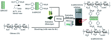
Water-soluble, highly functionalized multiwalled carbon nanotubes (MWNTs) were prepared via a facile, environmentally benign method. The effectiveness of the highly functionalized MWNTs as a reinforcing filler in a water-soluble mechanically weak chitosan biopolymer was evaluated. We observed a substantial improvement in the mechanical properties of the film; the stretchability of the film was maintained when 5 wt% MWNTs was added. In addition, the biocomposite film exhibited long-term antibacterial activity. The reinforcing effect can be explained by the homogeneous dispersion of the nanotubes in the polymer matrix through the strong hydrogen bonding between the sulfonic acid groups (–SO3H) on the sidewalls of the MWNTs and the amine (–NH2) and hydroxyl (–OH) groups on the chitosan backbone. The improvement in the mechanical properties of the MWNT–chitosan biocomposite may make it suitable for many environmental and clinical applications.
1. Introduction
The outstanding electronic, optical, thermal, and mechanical properties of carbon nanotubes (CNTs) have prompted their extensive investigation for use in novel materials such as composites, electronics, optical devices, and biological applications.1–5 However, their potential use in clinical applications has been hampered by their low biocompatibility.6–8 Some additional problems are difficulties in dispersing CNTs in the polymer matrix of biocomposites and sustaining strong interactions between the nanotubes and the matrix.9 The dispersion of CNTs is inhibited by the strong van der Waals interactions between adjacent tubes, thus making the tubes to aggregate and prevent the transfer of the CNT physical properties to the polymer matrix.10,11
CNTs can be dispersed in a polymer matrix by covalently functionalizing their sidewalls. Previous studies have shown that the functionalization of CNTs has a direct effect on their antimicrobial activity and their ability to penetrate the cells of microorganisms.12–14 CNTs may be rendered more compatible with a polymer matrix by strengthening their interfacial adhesion via covalent bonds, dipolar interactions, π–π interactions, or hydrogen bonds. These effects may be achieved through covalent functionalization via acid treatment, oxidation, esterification, amidation, radical coupling, and anionic coupling.15 These methods typically involve hazardous solvents that necessitate lengthy additional purification procedures that require expensive equipment. In addition, the possibility that toxic solvents may remain trapped in the carbon nanotube sample means that these methods are not suitable for biomedical applications.16 Thus, a simple, clean protocol for functionalizing the sidewalls of carbon nanotubes is required.
We have developed a facile, environment-friendly route for functionalizing the sidewalls of multiwalled carbon nanotubes (MWNTs) with sulfonic acid groups, via a diazonium reaction using water as the solvent. Chitosan was used as a model biopolymer because of its excellent biocompatibility, biodegradability, and antimicrobial properties.17,18 The water-soluble MWNTs were added to the mechanically weak chitosan matrix to examine their reinforcing effect. The mechanical and antibacterial properties of the biocomposite film were superior to that of pure chitosan because of the strong hydrogen bonding between the sulfonic acid groups on the sidewalls of the MWNTs and the amine and hydroxyl groups on the backbones of the chitosan molecules. We expect that this mechanically tough, antimicrobial biocomposite film will be suitable for many environmental and clinical applications.
2. Experimental sections
2.1. Starting materials
Low-molecular-weight chitosan (85% deacetylated, Sigma-Aldrich) was purified according to a literature procedure.19,20 The MWNTs were purchased from Iljin Nanotech Co., Ltd. Their diameter was 10–20 nm and the average length was about 20 μm. Sulfanilic acid, acetic acid, and (3-methylbutyl) nitrite (Aldrich) were used without further purification.
2.2. Functionalization of MWCNTs
The MWNTs were chemically functionalized using a diazonium reaction in water. Sulfanilic acid (5.77 g) was dissolved in deionized water (90 mL) at 85 °C in a 100 mL round-bottomed flask. The MWNTs (100 mg) were dispersed in deionized water (10 mL) in a 20 mL vial by sonication for 30 min. The MWNT dispersion was added to the round-bottomed flask. After 30 min, (3-methylbutyl) nitrite (2.93 g) was added dropwise and the mixture was stirred vigorously at 80 °C for 24 h. The nanotube dispersion was filtered (pore size: 0.45 μm), and the filter cake was washed several times with copious water and acetone. The product was re-dispersed in DMF to remove any impurities and filtered through a 0.45 μm membrane filter. The final sulfonated multiwalled carbon nanotube (S-MWNT) product was dried under vacuum at room temperature.
2.3. Fabrication of biocomposite film
The biocomposite films were fabricated by adding 1.0, 3.0, or 5.0 wt% water-soluble MWNTs to pure chitosan. Chitosan (200 mg) was dissolved in 10 mL of aqueous acetic acid solution (8.5 × 10⁻³ N) aqueous acetic acid and then mixed with an aqueous MWNT solution (10 mL). The mixture was sonicated for 30 min to achieve homogeneous nanotube dispersion. The mixture was placed in a Petri dish and the solvent was allowed to evaporate at 60 °C for 4 days under vacuum to remove the trapped water from the biocomposite film.
2.4. Characterization and measurements
The functionalized MWNTs were characterized by Fourier transform infrared (FT-IR) spectroscopy (FT-IR 300E, Jasco). X-ray photoelectron spectroscopy (XPS; ESCA 2000, Kratos Analytical) was used to investigate the surface composition of the carbon nanotubes. Raman spectroscopy (HR 514 nm Ar-ion laser, LabRam, Jobin-Yvon) was used to confirm the change in crystallinity of the MWNTs before and after chemical functionalization. Ultraviolet visible (UV-vis) spectra of the samples were recorded on a UV-vis spectrometer (U2001, Hitachi). The surface morphologies and dispersion states of the S-MWNTs in chitosan were observed by transmission electron microscopy (TEM; JEM 2100F, JEOL). The mechanical properties of the composite films were measured at an elongation rate of 5 mm min−1 at room temperature using a tensile testing machine (4468, Instron) for a dumbbell-type specimen. The dimensions of the specimens were 60 (length) × 10 (width) × 10 (narrow portion length) × 3 (narrow portion width) × 0.50 (thickness) mm. The following measurement conditions were used: gauge length of 25 mm; crosshead speed of 10 mm min−1; and loading of 2.5 kN. At least three samples were tested and an average measurement was used. Thermogravimetric analysis was carried out using a thermal analyzer (Q50, TA Instruments) with a heating rate of 10 °C min−1 under a nitrogen gas flow (30 mL min−1).
2.5. Determination of antimicrobial activity
Escherichia coli (E. coli) was used in this study to determine the antimicrobial properties of the biocomposite films. The bacterial strain was cultured in Luria–Bertani (LB) broth for 24 h at 37 °C. A fresh colony of E. coli grown overnight on an agar plate was scraped and grown in LB medium. The mid-logarithmic phase cell suspension (1 × 10⁸ CFUs mL−1) was diluted ten-fold with fresh sterile medium. The sample solution (500 μg mL−1) was prepared in the medium, inoculated in 15 mL tubes, and incubated at 37 °C for 18 h. After incubation, the solution was mixed using a Vortex–Genie mixer to distribute the inoculums evenly in the test tube. The OD600 was measured every 4 h to determine the bacterial growth. All the experiments were performed in triplicate and the averages were calculated. Bacteria were grown in tryptic soy broth (TSB) supplemented with the samples (500 μg mL−1) for 18 h. The cell suspensions were serially diluted from 10−1 to 10−6. Brain–heart infusion agar plates were inoculated with the diluted cell suspension (100 μL) and incubated for 18 h.
3. Result and discussion
3.1. Functionalization of MWCNT
Scheme 1 shows the protocol for functionalizing MWNTs using a mild diazonium reaction in aqueous media at 80 °C for 24 h. This approach was simpler, more effective, and more environmentally benign than previously reported methods, which typically used expensive organic solvents and strong acids under harsh reaction conditions. A characteristic S
O stretching peak was observed at 1111 cm−1 in the FT-IR spectrum (Fig. 1(a)), indicating the incorporation of SO3– groups on the side wall of the MWNTs.21 The structural changes caused by chemical functionalization were evaluated with Raman spectroscopy. The R value, ID/IG, which is the integrated intensity of the D band divided by that of the G band, increased after chemical modification (Fig. 1(b)), indicating the covalent bonding of functional groups on the sidewalls of the MWNTs.22 The surface properties of the MWNTs before and after chemical functionalization (Fig. 1(c)) were determined by XPS. Pristine MWNTs showed two strong distinctive peaks at 285.0 (C 1s) and 531.6 eV (O 1s).
 |
| Scheme 1 Environment-friendly method for functionalizing MWNTs and fabricating antimicrobial biocomposite films. | |
 |
| Fig. 1 Characterization of S-MWNTs. (a) FT-IR spectra, (b) Raman spectra, (c) wide-scan XPS spectra, and (d) thermogravimetric curves of pristine and functionalized S-MWNTs. (e) Zeta potentials and (f) UV-absorption spectra of pristine and functionalized S-MWNT suspension in distilled water. (g) FE-SEM image, and corresponding sulfur and oxygen EDS maps and energy-dispersive X-ray spectra of functionalized MWNTs. | |
However, two additional peaks at 170.1 (S 2p) and 233.0 eV (S 2s) in the chemically modified MWNTs indicated that sulfur groups had been introduced via covalent functionalization.23–25 In addition, thermogravimetric analysis was used to quantitatively evaluate the degree of functionalization (Fig. 1(d)). The weight loss in pristine MWNTs was less than 1.0 wt% at 800 °C. Interestingly, the mass loss from the chemically functionalized sample at 800 °C was ∼13.8 wt%, which may arise from the thermal decomposition of the aryl sulfonic acid (–C6H5–SO3H) groups on the sidewalls of the MWNTs.26 In addition, the stability of the MWNT suspension was evaluated using the zeta potential (Fig. 1(e)). The sulfonic acid covalently attached to the sidewalls of MWNTs was ionized, and thus, the zeta potential of the S-MWNTs was higher than that of the pristine MWNTs over a wide pH range (2 to 11).
These results demonstrate that the functionalized S-MWNTs were highly stable in distilled water up to a concentration of 0.05 mg mL−1.27 Furthermore, UV absorption was used to quantitatively evaluate the dispersing ability of the MWNTs in distilled water before and after chemical functionalization (Fig. 1(f)). Both nanotube suspensions showed absorption profiles similar those of conventional carbon materials.
According to the Lambert–Beer law, the real intensity of the absorbance is directly proportional to the amount of dispersed nanotubes. Thus, the S-MWNTs showed higher dispersibility than MWNTs in distilled water because of the sulfonic acid groups.28,29 Energy-dispersive X-ray spectroscopy (EDS) elemental mapping of the bulk S-MWNTs was used to identify the spatial distribution of the elemental composition (Fig. 1(g)). The EDS maps of oxygen (dark green) and sulfur (maroon) and the EDS spectra showed the presence of sulfur (1.7 at%) and oxygen (8.85 atom%). The morphological change in the MWNTs caused by the chemical modification was observed in detail using high-resolution TEM. The pristine MWNTs consisted of layered concentric tubules and had relatively smooth outer surfaces (Fig. 2(a)). However, the S-MWNTs (Fig. 2(b) and (c)) had rough external surfaces which also confirmed the chemical modification of the surface.30 The sulfanilic acid preferentially reacted with the near-surface region of the tubes and partially perturbed the surface structure.28,31 However, mild conditions meant that no substantial structural changes occurred.
 |
| Fig. 2 TEM images of (a) pristine MWNTs and (b and c) functionalized S-MWNTs. (d–f) TEM images and (g–i) SEM images of biocomposite films. S-MWNTs were individually dispersed in chitosan matrix. | |
TEM was used to examine the effect of the sulfonic acid groups on the dispersion of the S-MWNTs in the chitosan biocomposite films at different magnifications. Homogeneous, individually dispersed S-MWNTs were observed (Fig. 2(d)–(f)), even with 5 wt% S-MWNTs. A fractured composite film was prepared by dipping the film in liquid nitrogen and breaking it. Cross-sectional SEM images of the fractured surface of the film are shown in Fig. 2(g)–(i). There were few aggregated tubes, indicating that the S-MWNTs were well dispersed in the chitosan matrix.
The bright dots and lines in the SEM images (Fig. 2(g)–(i)) are the broken ends of S-MWNTs, suggesting that the S-MWNTs were broken rather than pulled out, because of the strong interfacial hydrogen bonding interactions between the sulfonic acid groups on the MWNT sidewalls and the hydroxyl and amine groups on the chitosan backbone.32 However in the case of pristine MWCNT chitosan composite aggregation took place which inhibits the load transfer from filler to matrix (Fig. S1†). To confirm the chemical interaction between the S-MWNTs and chitosan, FT-IR and XRD spectra of biocomposite films were obtained (Fig. 3(a) and (b)). Pure chitosan has two characteristic bands at 1650 and 1562 cm−1 which are assigned to the C–O stretching of –NHCO and the N–H bending of –NH2 groups, respectively. However, for the biocomposite films, the C–O stretching and the –NH bending bands shifted to lower frequencies because of the formation of strong hydrogen bonds with the –SO3H groups on the S-MWNT sidewalls.33 XRD was used to evaluate the effect of the S-MWNTs on the crystallinity of the chitosan in the biocomposite films (Fig. 3(b)). The chitosan showed characteristic peaks at around 11.6° and 18.3° corresponding to the hydrated crystalline structure, and at 23.8° for the amorphous structure. Therefore, the incorporation of carbon nanotubes did not alter the crystalline structure of chitosan.
 |
| Fig. 3 (a) FT-IR spectra and (b) XRD spectra of pure chitosan and S-MWNT-filled chitosan composite films. | |
3.2. Mechanical and thermal properties
Black carbon nanotube-filled chitosan composite films were prepared using a simple casting method. A photograph of the films is shown at the bottom right of Fig. 4(a). The tensile properties of the biocomposite films were compared with those of pure chitosan. The stress–strain curves are shown in Fig. 4(a). The S-MWNTs increased the modulus and breaking stress of the composite films; thus, they successfully reinforced the chitosan matrix (Fig. 4(b)). The strengthening effect was linearly proportional to the amount of S-MWNTs added. The improvement in the mechanical properties was attributed to the homogeneous dispersion of the S-MWNTs in the chitosan polymer and the strong hydrogen bonding between the two components, which efficiently transferred the load between the filler and the matrix.33–35 Moreover, our biocomposite films retained their stretchability, as demonstrated by the elongation-at-break measurements (Fig. 4(c)), even when the amount of S-MWNTs added was 5 wt%. As compared to pristine MWCNT chitosan composites, the S-MWCNT composite exhibits a large difference in its mechanical properties as the filler content increases (Fig. S2 and Table S1†). The thermal stabilities of the S-MWNT biocomposite films were also superior to those of chitosan (Fig. 4(d)), because the S-MWNTs acted as thermal barriers in the chitosan, and thus, prevented or delayed the permeation of oxygen and the escape of volatile degradation products. However in the case of pristine MWCNT chitosan composite such effect is not obvious due to the aggregation of pristine MWCNT in the matrix (Fig. S3†).
 |
| Fig. 4 (a) Stress–strain curves, (b) breaking stress and modulus, (c) elongation-at-break, and (d) thermal properties of pure chitosan and S-MWNT-filled chitosan composite films. | |
Thus, we propose that the aryl sulfonic acid groups on the sidewalls of the MWNTs allowed the tubes to be individually dispersed in the chitosan matrix, thereby maximizing the surface area of the reinforcing S-MWNTs, which significantly improved the mechanical and thermal properties of the chitosan matrix.
3.3. Antimicrobial properties
The antimicrobial activity of our biocomposite films against E. coli was measured. Planktonic cell growth experiments showed that the biocomposite films inhibited cell growth. The decrease in optical density demonstrated that the biocomposite film showed substantial antimicrobial activity against E. coli (Fig. 5(a) and (b)), which was greater than that of pure chitosan.
 |
| Fig. 5 (a) Optical density growth curves of bacteria after 6, 10, 14, and 18 h for pure chitosan, and MWNT-filled chitosan biocomposite films. (b) Images show the evaluation of the antibacterial activity of pure chitosan and S-MWNT-filled chitosan films. | |
A progressive inhibition effect of the biocomposite films on E. coli growth was observed as the amount of S-MWNTs increased. For the film with 5 wt% S-MWNTs, E. coli growth was reduced to less than 25% after 18 h. Cell growth on the biocomposite film strongly depended on the amount of S-MWNTs added. The inhibition of cell growth is strongly dependent on the surface characteristics and morphology of solid chitosan.36–38 The NH2 and OH groups on the chitosan backbone probably reacted with the SO3H groups on the S-MWNT sidewalls, which increased the hydrophobicity of the composite film surface.
Conclusions
We fabricated high-performance biopolymer composite films consisting of functionalized carbon nanotubes and chitosan with a simple solution casting method. The carbon nanotubes were chemically functionalized through application of our environment-friendly protocol; the reaction took place in water in the presence of sulfanilic acid. The sulfonic acid groups introduced onto the sidewalls of the MWNTs formed strong hydrogen bonds with the amine and hydroxyl groups on the backbone of chitosan. The homogeneous dispersion of nanotubes in the chitosan matrix caused by the strong hydrogen bonding between the S-MWNTs and the chitosan dramatically enhanced the mechanical properties of the biocomposite film. In addition, the biocomposite film exhibited better antimicrobial activity against E. coli than pure chitosan. The development of an environmentally benign method of functionalizing carbon nanotubes may contribute to the development of high-performance composites. We expect that this mechanically tough, electrically conductive, antimicrobial biocomposite film will be suitable for many environmental and clinical applications.
Acknowledgements
We acknowledge the supports from a National Research Foundation of Korea (NRF) grant funded by the Korean government (MEST) (no. 2012R1A2A2A01015155), Global Research Laboratory (K2090300202412E010004010) through the National Research Foundation of Korea (NRF) funded by the Ministry of Science, ICT (Information and Communication Technologies) and Future Planning, Korea. Y.C.J and C.-M.Y acknowledge the support from the KIST Institutional Program (2Z03870 and 2Z03910).
Notes and references
- M. S. Dresselhaus, G. Dresselhaus and P. C. Eklund, Science of Fullerenes and Carbon Nanotubes, Academic Press, Tokyo, 1995 Search PubMed.
- P. G. Collins, A. Zettl, H. Bando, A. Thess and R. E. Smalley, Science, 1997, 278, 100–102 CrossRef CAS.
- P. M. Ajayan, L. S. Schadler, C. Giannaris and A. Rubio, Adv. Mater., 2000, 12, 750–753 CrossRef CAS.
- R. H. Baughman, A. A. Zakhidov and W. A. de Heer, Science, 2002, 297, 787–792 CrossRef CAS PubMed.
- M. F. L. De Volder, S. H. Twafick, R. H. Baughman and A. J. Hart, Science, 2013, 339, 535–539 CrossRef CAS PubMed.
- S. K. Smart, A. I. Cassady, G. Q. Lu and D. J. Martin, Carbon, 2006, 44, 1034–1047 CrossRef CAS PubMed.
- C. Grabinski, S. Hussain, K. Lafdi, L. Braydich-Stolle and J. Schlager, Carbon, 2007, 45, 2828–2835 CrossRef CAS PubMed.
- Y. Liu, Y. Zhao, B. Sun and C. Chen, Acc. Chem. Res., 2013, 46, 702–713 CrossRef CAS PubMed.
- Z. Spitalsky, D. Tasis, K. Papagelis and C. Galiotis, Prog. Polym. Sci., 2010, 35, 357–401 CrossRef CAS PubMed.
- S. F. Wang, L. Shen, W. D. Zhang and Y. J. Tong, Biomacromolecules, 2005, 6, 3067–3072 CrossRef CAS PubMed.
- S. S. Mahapatra, S. K. Yadav, H. J. Yoo and J. W. Cho, Mater. Chem., 2011, 21, 7686–7691 RSC.
- S. Kang, M. S. Mauter and M. Elimelech, Environ. Sci. Technol., 2008, 42, 7528–7534 CrossRef CAS.
- A. S. Brady-Estévez, S. Kang and M. Elimelech, Small, 2008, 4, 481–484 CrossRef PubMed.
- J. D. Schiffman and M. Elimelech, ACS Appl. Mater. Interfaces, 2011, 3, 462–468 CAS.
- N. Karousis, N. Tagmatarchis and D. Tasis, Chem. Rev., 2010, 110, 5366–5397 CrossRef CAS PubMed.
- B. K. Price and J. M. Tour, J. Am. Chem. Soc., 2006, 128, 12899–12904 CrossRef CAS PubMed.
- B. Krajewska, P. Wydro and A. Jaczyk, Biomacromolecules, 2011, 12, 4144–4152 CrossRef CAS PubMed.
- J. Fan, Z. Shi, Y. Ge, Y. Wang, J. Wang and J. Yin, Polymer, 2012, 53, 657–664 CrossRef CAS PubMed.
- H. Bao, L. Li and H. Zhang, J. Colloid Interface Sci., 2008, 328, 270–277 CrossRef CAS PubMed.
- Q. Gan, T. Wang, C. Cochrane and P. McCarron, Colloids Surf., B, 2005, 44, 65–73 CrossRef CAS PubMed.
- F. Liu, J. Sun, L. Zhu, X. Meng, C. Qi and F. Xiao, J. Mater. Chem., 2012, 22, 5495–5502 RSC.
- L. G. Cancado, K. Takai, T. Enoki, M. Endo, Y. A. Kim, H. Mizusaki, L. N. Coelho, R. M. Paniago, A. Jorio and M. A. Pimenta, Appl. Phys. Lett., 2006, 88, 163106 CrossRef PubMed.
- Y. Lu, Q. Gong, F. Lu, J. Liang, L. Ji, Q. Nie and X. Zhang, J. Mater. Sci.: Mater. Med., 2011, 22, 1855–1862 CrossRef CAS PubMed.
- L. Zhang, T. Shi, S. Wu and H. Zhou, Colloid Polym. Sci., 2013, 291, 2061–2068 CAS.
- G. Chena, S. Zhaib, Y. Zhaia, K. Zhanga, Q. Yuea, L. Wanga, J. Zhaoa, H. Wanga, J. Liua and J. Jia, Biosens. Bioelectron., 2011, 26, 3136–3141 CrossRef PubMed.
- C. Li and R. Mezzenga, Langmuir, 2012, 28, 10142–10146 CrossRef CAS PubMed.
- B. Zhao, H. Hu, A. Yu, D. Perea and R. C. Haddon, J. Am. Chem. Soc., 2005, 127, 8197–8203 CrossRef CAS PubMed.
- Y. C. Jung, H. H. Kim, Y. A. Kim, J. H. Kim, J. W. Cho, M. Endo and M. S. Dresselhaus, Macromolecules, 2010, 43, 6106–6112 CrossRef CAS.
- J. H. Kim, M. Kataoka, K. Fujisawa, T. Tojo, H. Muramatsu, S. M. Vega-Daz, F. Tristnlpez, T. Hayashi, Y. A. Kim, M. Endo, M. Terrones and M. S. Dresselhaus, J. Phys. Chem. B, 2013, 115, 14295–14300 CrossRef PubMed.
- Y. C. Jung, H. J. Yoo, Y. A. Kim, J. W. Cho and M. Endo, Carbon, 2010, 48, 1598–1603 CrossRef CAS PubMed.
- Y. C. Jung, H. Muramatsu, K. Fujisawa, J. H. Kim, T. Hayashi, Y. A. Kim, M. Endo, M. Terrones and M. S. Dresselhaus, Small, 2011, 7, 3292–3297 CrossRef CAS PubMed.
- S. Rana, N. Karak, J. W. Cho and Y. H. Kim, Nanotechnology, 2008, 19, 495707 CrossRef PubMed.
- R. K. Layek, S. Samanta and A. K. Nandi, Polymer, 2012, 53, 2265–2273 CrossRef CAS PubMed.
- Y. L. Liu, W. H. Chen and Y. H. Chang, Carbohydr. Polym., 2009, 76, 232–238 CrossRef CAS PubMed.
- S. K. Yadav, I. J. Kim, H. J. Kim, J. Kim, S. M. Hong and C. M. Koo, J. Mater. Chem. C, 2013, 1, 5463–5470 RSC.
- M. Kong, X. G. Chen, K. Xing and H. J. Park, Int. J. Food Microbiol., 2010, 144, 51–63 CrossRef CAS PubMed.
- S. K. Yadav, S. S. Mahapatra, M. K. Yadav and P. K. Dutta, RSC Adv., 2013, 3, 23631–23637 RSC.
- S. K. Yadav, Y. C. Jung, J. H. Kim, Y. Ko, H. J. Ryu, M. K. Yadav, Y. A. Kim and J. W. Cho, Part. Part. Syst. Charact., 2013, 30, 721–727 CrossRef CAS.
Footnote |
† Electronic supplementary information (ESI) available: Fig. S1–S3 and Table S1. See DOI: 10.1039/c3ra47074a |
|
This journal is © The Royal Society of Chemistry 2014 |
Click here to see how this site uses Cookies. View our privacy policy here.