Characterization and applications of cyclic β-(1,2)-glucan produced from R. meliloti

Geetha Venkatachalam, Venkatesan Nandakumar, Ganesan Suresh and Mukesh Doble*
Bioengineering and Drug design Lab, Department of Biotechnology, IIT-Madras, Chennai-600036, India. E-mail: mukeshd@iitm.ac.in; Tel: +91-44-2257-4107

Received 27th November 2013 , Accepted 7th January 2014

First published on 13th January 2014


Abstract

Cyclic β-(1,2)-glucan, with a degree of polymerization ranging from 17–28, without any substitution and a molar mass of 3101.5 Da, was produced from Rhizobium Meliloti MTCC-3402 in glutamic acid and a mannitol medium. The size of this glucan was less than those reported from oats and yeast. The main fraction was with 19 glucose residues (cavity size of 0.92 nm) and a melting temperature of 134.1 °C. Glucan encapsulates drugs (85–99%) such as curcumin, dexamethasone, reserpine, 6-methylcoumarin, 4-hydroxycoumarin and 4 methyl umbelliferone very efficiently. The encapsulation efficiency was better for hydrophobic than hydrophilic drugs (correlation coefficient of 0.92). Glucan was not cytotoxic towards L6 myoblast and 3T3 fibroblast cells and could be produced on the nanometer scale (average particle size 50–200 nm). Glucan exhibited dose dependent radical scavenging antioxidant activity. It was able to bind to dyes including methyl violet, trypan blue and bromocresol green indicating that the latter could be used in vivo at very low concentrations. Glucan decolorized coomassie brilliant blue R, bromophenol blue and bromocresol purple opening up applications in effluent treatment industries.
Introduction

The family of Rhizobiaceae (Mesorhizobium, Rhizobium, Sinorhizobium and Bradyrhizobium japonicum) form symbiotic associations with host plants including alfalfa, clover, and soy bean, and produce cyclic β-(1,2)-glucans as extracellular polysaccharides which help the organism during osmoregulation and plant infection.1–3 In general, these oligosaccharides are D-glucose residues connected by β-(1,2) linkages with a degree of polymerization (DP) ranging from 17–25 and in some species it reaches up to 40 glucose units. Cyclic β-(1,2)-glucans produced from Rhizobium and Agrobacterium species have flexible backbone structures and narrow cavity sizes with varying molecular weights.4,5 These glucans are biocompatible and more water soluble (250 g L−1) than cyclodextrin (solubility 18 g L−1) and have negligible cytotoxicity.6

Linear glucans are biopolymers (β-1,3) produced by species of Agrobacterium, Rhizobium and Alcaligenes faecalis. They are well characterized and hence used extensively as gelling and bio-thickening agents in food for the improvement of texture, for example in tofu (bean curd), bean jelly and fish pastes.7 In Japan, curdlan is widely used in the food industry for its water-retention capacity. Curdlan possesses properties like anti-tumorigenicity, anti-inflammatory, immunomodulating, anti-infective activities against fungus, bacteria, protozoa and viruses, wound repair, protection against radiation and anti-coagulant activity. In contrast, reports on the applications of cyclic β-(1,2)-glucan are very limited since its physico-chemical and biological properties have not been fully elucidated. A detailed characterization of cyclic β-(1,2)-glucan produced from R. meliloti is reported here. This may help the biopolymer find applications in medical, pharmaceutical, food and flavouring industries.

Drugs for anticancer, antipsychotic, blood pressure, inflammatory and autoimmune conditions are hydrophobic and have poor water solubility. Polymeric nanoparticles including lactic acid and glycolic acid (PLGA), cyclodextrin, dextrin, chitosan, zein nanoparticles, albumin, micelles, pullulan and peptides have been modified by ionic crosslinking, covalent crosslinking, and a layer-by-layer method for encapsulation of such drugs. Deoxycholic acid, cholesterol, carboxylic acids, and hydrophobic polymers are used as hydrophobic segments in polysaccharide nanoparticles for hydrophobic drug release.8,9 Use of synthetic polymers for drug encapsulation is limited due to their poor water solubility and may require a prolonged stay in the body before they undergo degradation. In contrast, the cyclic glucan reported here is isolated from a natural source, not modified or cross linked and it is biocompatible. In this paper the encapsulation efficiency of glucan towards several drugs is reported.

The interaction and binding of glucan with several dyes including methyl violet (MV), tryphan blue (TB) and bromocresol green (BCG) are investigated here. These dyes are used in medical imaging and are reported to be cytotoxic. Paper mills, color photography and textile industries produce synthetic dyes as a major pollutant in their waste water. These dyes are carcinogenic, hazardous and affect the gas solubility in water, aesthetics and aquatic life.10 The decolorization of dyes using biological methods is an environmentally acceptable technique. Here the decolorizing ability of glucan is investigated against dyes such as coomassie brilliant blue R (CBBR), bromophenol blue (BPB) and bromocresol purple (BCP).

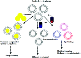
The present study explores three possible applications of cyclic β-(1,2)-glucan namely as a drug carrier, as a dye binder and as a dye decolorizer. The cytotoxicity of this polymer is also investigated, since this knowledge is necessary if it is to be tested for pharmaceutical applications.

Results and discussion

Purification of the cyclic β-(1,2)-glucan

A chromatogram of the ethanolic extract (after fermentation) purified by size exclusion chromatography is shown in the ESI (Fig. S1) and it is visualized by TLC as a single black spot (Fig. S2).

Physicochemical characteristics

The 1H NMR spectrum indicates the presence of methylene (δ 3.79), hydroxyl (δ 3.72, 3.70, and 3.40) and methine protons (δ 3.57, 3.68, 3.60, and 3.44) (Fig. S3). The chemical shift at 4.78 ppm (H-1) is the characteristic peak of the β-configuration of all the glucose residues. The shift at δ 3.44 ppm (H-2) corresponds to the H-2 protons involved in the β-(1,2) glycosidic linkage in glucan (Table S1). The methylene protons from the succinyl substitution which appear as signals at 2.44 and 2.66 ppm are absent here. From the 1H NMR spectrum it could be concluded that the cyclic β-(1,2)-glucan isolated from R. meliloti is similar to the one produced by other Rhizobial strains and is neutral without any substitution.5 The 2D NMR spectra also confirm the proton–proton and proton–carbon coupling of glucan at 3.44 ppm which includes 1H-1H COSY (H6 ↔ H5, H4 ↔ H5, H4 ↔ H3), HSQC (H5 ↔ 63.19, H6 ↔ 59.31, H4 & H3 ↔ 70.79, 69.23) and HMBC (H4 ↔ 70.79, H2 ↔ 59.31, H5 ↔ 68.23, H3 ↔ 70.79, H6 ↔ 63.19) (S4–S6).

Table S2 lists the bands present in the FT-IR spectrum of the cyclic β-(1,2)-glucan (hydrogen bonded hydroxyl groups and glycosidic bonds) which match well with data reported in the literature for similar glucans from Rhizobium meliloti.11 Molecular ions at a m/z of 2777.17, 2939.85, 3101.46, 3263.82, 3425.0, 3587.92, 3749.22, 3911.03, 4073.23, 4234.61, 4397.82, 4560.53 Da in the MALDI-TOF MS spectra indicate the presence of glucan corresponding to a degree of polymerization (DP) ranging between 17 and 28 (Fig. 1). These molecular ions are identical to the values reported for glucan produced by Mesorhizobium loti which are reported to have DPs ranging from 17–28.5 There is no substitution in the glucan structure which is confirmed from TLC and MALDI-TOF MS analysis. The present product is different from the ones produced by other bacterial species of Rhizobiaceae which have substitutions including acetyl, succinyl, phosphoglycerol, methylmalonyl or phosphocholine.12 The ion at a m/z of 3101.46 is the most intense peak, which represents the main component with a ring size of 19 glucose residues. It is interesting to note that the major glucan species containing 19 glucose residues reported in this study, is close to the range typical for the cyclic β-(1,2)-glucan predominantly produced by Rhizobium (DP of 17–28 glucose residues per ring).1,13 Our data also suggest that the β-glucan produced by Rhizobium meliloti shares a similar structure with those isolated from Mesorhizobium loti,5 Mesorhizobium huakuii13 and Brucella abortus.14


image file: c3ra47073c-f1.tif
Fig. 1 MALDI-TOF MS positive-ion mass spectrum of cyclic β-(1,2)-glucan. Values indicated on the peaks are the masses of sodium-cationized glucan.

DSC (differential scanning calorimetry) shows an endothermic transition peak at 134.08 °C (Fig. S7). This is an indication of its thermal stability, which depends on the degree of polymerization. Transitions may be due to the changes in its size.15 Two conformational transitions in the triple helix and triple helix–single coil of scleroglucan are observed as two endothermic peaks, which are due to the changes in the structure.16 This could be attributed to the differences in the DP between the two.17 To date there is no report on the thermal stability of cyclic β-glucan. Thermogravimetric analysis shows the degradation pattern of the glucan, which occurs in four temperature ranges (Fig. S8). An initial weight loss (5.36%) is observed between 42 and 200 °C. A sharp and major weight reduction is observed between 200 and 400 °C with a maximum occurring at 300 °C. The percentage weight loss from 200 to 400 °C is 50%, and from 400 to 600 °C it is 15%. The final weight reduction from 600 to 850 °C is 30.5%. No residue is observed from the TGA, which indicates complete degradation of the polymer.

The percentage of carbon and hydrogen (from CHN analysis) is 33.2 and 4.6 respectively, which indicates an average degree of polymerization of 19. The ratios of C and H in β-glucan from oats (100–200 kDa) and yeast (105 kDa) are reported to be 37.9 and 5.8%, and 43.8 and 6.2% respectively. These glucans are larger in size than the bacterial glucan reported here (DP = 17–28). The percentage of carbon and hydrogen identified through elemental analysis reported here is lower than the levels reported for (1,3)-β-glucan from brewer's yeast and oats.18,19 This may be due to the major differences in their molecular weight and shape. Glucan from oats is reported to form aggregates with sizes ranging from 5–100 μm (20 μm on average).20 The morphology of cyclic β-(1,2)-glucan in pure form appears as rod like structures, but upon simultaneous sonication and acetone precipitation it appears (in TEM) as spherical particles of 50 to 200 nm in diameter.

Encapsulation of hydrophobic drugs by cyclic β-(1,2)-glucan

The FTIR spectra of the cyclic β-(1,2)-glucan (Fig. 2a), and drug encapsulated (Fig. 2b curcumin, c. dexamethasone, d. reserpine, e. 6-methylcoumarin, f. 4-hydroxycoumarin, g. 4-methylumbelliferone) show changes in the 1000–1200 cm−1 region as well as the appearance of new peaks corresponding to the encapsulated drugs (Table S2). The 1H NMR spectra of the encapsulated product show resonances corresponding to glucan along with the drugs. In some regions the drug peaks merge with the glucan peaks (Fig. S9).
image file: c3ra47073c-f2.tif
Fig. 2 FTIR spectra of drug encapsulated cyclic β-(1,2)-glucan. (a) glucan alone, (b) curcumin, (c) dexamethasone, (d) reserpine, (e) 6-methylcoumarin, (f) 4-hydroxycoumarin, (g) 4-methylumbelliferone.

Glucan encapsulated curcumin appears as spheres (Fig. 3) with an average particle size of 58 nm (Fig. S10). The encapsulation efficiency is 98.7% which matches well with the literature reports for chitosan–poly(butyl cyanoacrylate) encapsulated curcumin nanoparticles.21 In the case of dexamethasone, the encapsulation efficiency here is 92.0%, whereas the literature reports only a 44.5–76.0% encapsulation efficiency with chitosan.22 97.8 and 87.5% of reserpine and 6-methylcoumarin respectively are encapsulated by cyclic β-(1,2)-glucan. Polylactide-co-glycolide acid (PLGA) nanoparticles are reported to encapsulate 88% of 4-methyl-7-hydroxy coumarin.23 85.2% and 92.2% of 4-hydroxycoumarin and 4-methylumbelliferone respectively are encapsulated by cyclic β-(1,2)-glucan. In our previous study we reported a 92.6% umbelliferone encapsulation by this polymer.2 The current study shows that cyclic β-(1,2)-glucan has a higher loading efficiency than other polymers reported in the literature. An excellent positive correlation (r = 0.92) exists between log[thin space (1/6-em)]P (hydrophilic–hydrophobic balance) of the drugs and the encapsulation efficiency (Fig. 4), indicating that this process is better with hydrophobic than with hydrophilic drugs. This is expected since the pocket of the cyclic glucan is hydrophobic in nature.


image file: c3ra47073c-f3.tif
Fig. 3 Transmission electron microscopy image of (A) cyclic β-(1,2)-glucan and (B) after curcumin encapsulation.

image file: c3ra47073c-f4.tif
Fig. 4 log[thin space (1/6-em)]P versus % of drug encapsulation (curcumin, dexamethasone, reserpine, 6-methylcoumarin, 4-hydroxycoumarin, 4-methylumbelliferone).

Cyclic β-(1,2)-glucan as a dye binder

Glucan does not absorb in the UV-visible range, but when it is used with the dyes, methyl violet (MV), trypan blue (TB) and bromocresol green (BCG), it increases the absorption maxima in a concentration dependant manner as well as shifts the λmax (Fig. 5A–C). The first two dyes are used to stain lens capsules for capsulorhexis creation (removal of the lens capsule during cataract surgery). When compared to MV, TB is reported to exhibit cytotoxicity at a concentration of 2.5 mg ml−1.24 So these dyes with glucan can be used in biomedical applications at a lower concentration than using the dye alone as a binder for staining the capsule. BCG is used as an inhibitor of the prostaglandin E2 transport protein.25 At a concentration of 30 μM in Krebs bicarbonate solution it is used for staining the tracheal segments in mice. Along with glucan its toxicity could be reduced and possibly be used in humans.
image file: c3ra47073c-f5.tif
Fig. 5 Cyclic β-(1,2)-glucan absorption of different dyes. (A) Methyl violet, (B) tryphan blue, (C) bromocresol green, (D) brilliant blue R, (E) bromophenol blue, and (F) bromocresol purple.

Cyclic β-(1,2)-glucan as a dye decolorizer

200 μg ml−1 of glucan when incubated with brilliant blue R (BBR), bromophenol blue (BPB) and bromocresol purple dyes, decreases the absorbance of the dyes (by 36.5, 22 and 10.9% respectively) which indicates that the polymer decolorizes them (Fig. 5D–F). Some natural polysaccharides such as galactomannans, locust bean gum, guar gum and cassia gum are reported to decolorize dyes from effluents and it is reported that the interaction is through van der Waals forces, hydrogen bonding and electrostatic attraction.26 These observations indicate that glucan can be used as a decolorizing agent in effluent treatment.

Cytotoxicity of glucan

L6 and 3T3 cells are viable even up to a concentration of 200 μg ml−1 of glucan, which proves that it is biocompatible (Fig. S11). NucBlue® stained fluorescence images of untreated and glucan treated L6 cells have intact and round nuclei (Fig. 6A and B respectively) which further confirms that the polymer is not toxic. It is reported that the viability of Human Embryonic Kidney 293 cell lines slightly decreased in the presence of cyclic β-(1,2)-glucan.27 However, no such inhibition is observed here with L6 cells. It is reported that glucan in general acts as an immune enhancer through the activation of macrophages and it accelerates the production of cytokines including TNF-α, IL-6 and IL-12.28
image file: c3ra47073c-f6.tif
Fig. 6 Fluorescence microscopy images of L6 cells stained with NucBlue®. (A) Untreated and (B) treated with cyclic β-(1,2)-glucan (magnification 10×, scale bar 85 mm).

Antioxidant properties

Cyclic glucan shows dose dependant antioxidant activity at increasing concentrations (from 100 to 500 μg ml−1) in DPPH (16.0 ± 1.8 to 40.8 ± 0.8%) and hydroxyl radical (24.0 ± 0.9 to 60.8 ± 1.9%) scavenging assays (Fig. S12a and b). The percentage of antioxidant activity of ascorbic acid (standard) at 100 μg is 82% ± 2.3 and 76% ± 3.1 in DPPH and hydroxyl radical scavenging assays respectively.

Experimental procedures

Chemicals

Curcumin, reserpine, dexamethasone, 6-methylcoumarin, 4-hydroxycoumarin and 4-methylumbelliferone were purchased from TCI chemicals, India, and dyes are from Himedia, Merck and Sigma Aldrich. Solvents used for encapsulation are of analytical grade (Merck, India). NMR solvents namely D2O, acetone D, ethanol D, methanol D and chloroform D were from Merck, Germany. All other chemicals used here were purchased from Merck, Himedia and Sigma, India and L6 cells were from the National Centre for Cell Sciences (NCCS), Pune, India. R. meliloti from the Microbial Type culture collection (MTCC 3402) was obtained from the Institute of Microbial Technology (IMTECH), Chandigarh, India.

Microorganism and cultivation

R. meliloti MTCC 3402 was used for the production of cyclic β-(1,2)-glucan. The media contained (in grams); glutamic acid – 2.0, mannitol – 10, K2HPO4 – 0.1, MgSO4·7H2O – 0.4, CaCO3 – 0.5, FeCl3·6H2O – 0.0025, MnCl2·4H2O – 0.001, Na2MoO4·2H2O – 0.00001, ZnSO4·7H2O – 0.00001, CuSO4·5H2O – 0.00001, H3BO3 – 0.00001, CoCl2·6H2O – 0.00001, biotin – 2 μg and thiamine – 5 mg in one litre (at a pH of 7.0).2 The cultivation was carried out on a 1 L scale for 8 days in a 3 L Erlenmeyer flask at 33 °C on a rotary shaker at 180 rpm. Cells and culture supernatants were separated by centrifugation at 35[thin space (1/6-em)]000 g for 30 min and the amount of cyclic β-(1,2)-glucan was quantified by the anthrone–sulfuric acid method.1

Characterization of the glucan

The aqueous ethanol phase was concentrated using a rotary evaporator and purified by size exclusion chromatography with Biogel P6 (Bio rad, USA) (24–96 cm) as the stationary phase and 5% acetic acid flowing at a rate of 1 ml min−1 as the mobile phase. The purity of the samples was assessed by thin layer chromatography (TLC) using a silica gel coated glass plate as the stationary phase and a butanol–ethanol–water (5[thin space (1/6-em)]:[thin space (1/6-em)]5[thin space (1/6-em)]:[thin space (1/6-em)]4 vol/vol) solvent mixture as the mobile phase. The active compounds were visualized by first spraying 5% H2SO4 in methanol and then heating it to 110 °C for 15 min.29

1H and 2D NMR (nuclear magnetic resonance) spectroscopy (Bruker 500 MHz spectrometer) was carried out with 10 mg of sample dissolved in D2O (2 ml). 5 mg of the sample was mixed with potassium bromide (KBr) at a ratio of 1[thin space (1/6-em)]:[thin space (1/6-em)]20 and pulverized, dried under vacuum for 1 h and compressed into a thin pellet and analyzed with a Fourier transform infrared spectrophotometer (FT-IR, Perkin Elmer ATR/FTIR) in the range of 400–4000 cm−1. The mass spectrum of the glucan was recorded with a matrix assisted laser desorption ionization time-of-flight mass spectrometer (MALDI-TOF MS Voyager-DE™ PRO Biospectrometry™ workstation, Applied Biosystems) in the positive ion mode using 2,5-dihydroxybenzoic acid as the matrix.5

The thermal stability of the degassed glucan was determined in the temperature range of 30 to 300 °C with a differential scanning calorimeter (DSC7 from Perkin Elmer, Q200 MDSC from TA instruments). The scans were performed at a rate of 10 °C min−1, and thermogravimetric analysis of the glucan was carried out with the TGA7, Perkin Elmer, Q500 Hi-Res TGA7 from TA instruments. Here, 1.06 mg of the sample was placed in an aluminum oxide crucible and was heated from 50 to 850 °C at a rate of 20 °C min−1.17 Measurements were carried out under a nitrogen atmosphere at a flow rate of 80 ml min−1. The amount of carbon and hydrogen in the lyophilized glucan was estimated with the help of a Perkin Elmer 2400 Series II CHN analyzer.30 The particle size and the zeta potential of the glucan were determined using a Zetatrac-Zeta potential analyzer (Microtrac Inc, USA) at a wavelength of 780 nm and a back scattering angle of 180°.

Encapsulation of poorly water soluble drugs by cyclic β-(1,2)-glucan

10 mg of curcumin, reserpine, dexamethasone, 6-methylcoumarin, 4-hydroxycoumarin and 4-methylumbelliferone (ESI Fig. S9) was solubilized in a suitable solvent (acetone or methanol) and added slowly to the glucan (100 mg) and kept in the dark for 24 h to attain equilibrium. Later the mixture was centrifuged at 14[thin space (1/6-em)]000 rpm for 30 min and the pellet was lyophilized. The loading efficiency of the encapsulated polymer was quantified by a method reported earlier.2,31 FTIR and NMR spectroscopy were used to characterize the polymer encapsulated drug. The particle size and changes in the morphology of the glucan were determined with a particle size analyzer and transmission electron microscope (Philips/Fei CM-20), respectively.

Cyclic β-(1,2)-glucan as a dye binder

The interaction and binding efficiency of glucan towards dyes such as methyl violet, trypan blue, Bromocresol green, brilliant blue R, bromophenol blue and bromocresol purple (ESI Fig. S13) were tested by a method reported earlier.2

Cytotoxicity of glucan

The biocompatibility of the glucan was tested against L6 myoblast and 3T3 cell lines. The cells were grown to confluence, trypsinized with 1X trypsin and the number of cells was counted with the help of a haemocytometer (Marienfeld, Germany). They were then diluted (104 cells per well) with Dulbecco's modified eagle's medium (DMEM) and seeded in 96 well plates and cultured for 24 h. The glucan was solubilized in DMEM, diluted to different concentrations (200, 150, 100, 75 and 50 μg ml−1) and added to each culture well and incubated for 24 hours.32 20 μl of 5 mg ml−1 (3-(4,5-dimethylthiazol-2-yl)-2,5-diphenyltetrazolium bromide (MTT) was added to each well and incubated for 4 hours. The precipitates were solubilized in DMSO (dimethyl sulfoxide) and the extent of the reduction of the MTT was quantified by measuring the absorbance at 570 nm using a multimode plate reader (Enspire, Perkin Elmer, Singapore) and the percentage of viable cells was calculated.

Simultaneously L6 cells were also cultured for 24 h in DMEM supplemented with 10% fetal bovine serum and 1% antibiotic at 37 °C in a 5% CO2 atmosphere on polystryrene petriplates with cover-slips. The cells were incubated with (200 μg ml−1) and without glucan for 24 h. Then the cells were stained with Nucblue® (NucBlue® Live ReadyProbes) and fixed in a glass slide and visualized under a fluorescence microscope (Leica Microsystems, Germany) to determine the changes in their morphology. DPPH (2,2-diphenyl-1-picrylhydrazyl)33 and hydroxyl radical scavenging assays34 was performed to determine the antioxidant potential of glucan, with ascorbic acid as the standard.

Structural features

The log[thin space (1/6-em)]P (hydrophilic to lipophilic ratio) of these drugs and dyes was calculated using ALOGPS 2.1 software (On-line Lipophilicity/Aqueous Solubility Calculation Software).

Conclusions

Cyclic β-(1,2)-glucan from Rhizobium Meliloti MTCC-3402 has a DP of 17–28 (cavity size of ∼0.83 to 1.36 nm) and it could be used as a drug carrier for poorly water soluble drugs. The encapsulation efficiency is 85 to 99% for hydrophobic drugs such as curcumin, dexamethasone, reserpine, 6-methylcoumarin, 4-hydroxycoumarin and 4-methylumbelliferone. The binding ability of glucan with dyes such as MV, TB and BCG demonstrates its possible application in medical imaging to decrease the cytotoxicity of the latter. The decolorizing ability of glucan with BBR, BPB and BCP indicates its possible application in effluent treatment industries. Glucan could be an ideal delivery system for hydrophobic drugs but not for hydrophilic ones. This property is very useful in cancer treatment where most of the drugs used are hydrophobic. High thermal stability and good biocompatibility of this polymer facilitates its use in in vivo applications including drug delivery and tissue engineering. Moreover, there is no substitution in the cyclic structure which could be exploited for preparing polymer blends to further improve its physico-chemical properties.

Acknowledgements

Geetha Venkatachalam gratefully acknowledges the financial support from the Department of Science and Technology, India, under the women scientist scheme (DST-SR/WOS-A/LS-15/2010). A special thanks to Mr Rakesh Nankar and Mrs Anju V Nair for helpful discussions. The authors also thank the Sophisticated Analytical Instrument Facility, IIT-Madras for analytical help.

References

  1. G. Venkatachalam, S. Gummadi and M. Doble, Cyclic β-Glucans from Microorganisms, Production, Properties and Applications, series: Springer briefs in Microbiology, Springer, Berlin Heidelberg, Berlin, 2012, p. 90 Search PubMed.
  2. G. Venkatachalam, D. Srinivasan and M. Doble, Process Biochem., 2013, 48, 1848–1854 CrossRef CAS PubMed.
  3. M. W. Breedveld and K. J. Miller, Microbiol. Rev., 1994, 58, 145–161 CAS.
  4. M. Hisamatsu, Carbohydr. Res., 1992, 231, 137–146 CrossRef CAS.
  5. Y. Kawaharada, H. Kiyota, S. Eda Minamisawa and H. Mitsui, Carbohydr. Res., 2008, 343, 2422–2427 CrossRef CAS PubMed.
  6. V. J. Stella and R. A. Rajewski, Pharm. Res., 1997, 14, 556–567 CrossRef CAS.
  7. C. Laroche and P. Michaud, Recent Pat. Biotechnol., 2007, 1, 59–73 CrossRef CAS.
  8. S. K. Nitta and K. Numata, Int. J. Mol. Sci., 2013, 14, 1629–1654 CrossRef CAS PubMed.
  9. R. Luo, S. S. Venkatraman and B. r. Neu, Biomacromolecules, 2013, 14, 2262–2271 CrossRef CAS PubMed.
  10. S. B. Jadhav and R. S. Singhal, Int. Biodeterior. Biodegrad., 2013, 85, 271–277 CrossRef CAS PubMed.
  11. S. Lee, D. Lee, H. Park, Y. Choi and S. Jong, Antonie van Leeuwenhoek, 2003, 84, 201–207 CrossRef CAS.
  12. S. Lee, E. Cho and S. Jung, BMB Rep., 2009, 42, 769–775 CrossRef CAS.
  13. A. Choma and I. Komaniecka, Acta Biochim. Pol., 2003, 50, 1273–1281 CAS.
  14. M. S. Roset, A. E. Ciocchini, R. A. Ugalde and N. Iannino Inon de, J. Bacteriol., 2006, 188, 5003–5013 CrossRef CAS PubMed.
  15. M. Sletmoen, S. N. Naess and B. T. Stokke, Carbohydr. Polym., 2009, 76, 389–399 CrossRef CAS PubMed.
  16. S. Kitamura and T. Kuge, Biopolymers, 1989, 28, 639–654 CrossRef CAS PubMed.
  17. O. M. Gazit and A. Katz, Langmuir, 2012, 28, 431–437 CrossRef CAS PubMed.
  18. M. S. Shin, S. Lee, K. Y. Lee and H. G. Lee, J. Agric. Food Chem., 2005, 53, 5554–5558 CrossRef CAS PubMed.
  19. S. Freimund, S. Janett, E. Arrigoni and R. Amado, Eur. Food Res. Technol., 2005, 220, 101–105 CrossRef CAS PubMed.
  20. K. Hunter Jr, R. Gault and M. Berner, Lett. Appl. Microbiol., 2002, 35, 267–271 CrossRef CAS.
  21. J. Duan, H. M. Mansour, Y. Zhang, X. Deng, Y. Chen, J. Wang, Y. Pan and J. Zhao, Int. J. Pharm., 2012, 426, 193–201 CrossRef CAS PubMed.
  22. A. Dustgani, E. V. Farahani and M. Imani, Iran. J. Pharm. Sci., 2008, 4, 111–114 Search PubMed.
  23. S. S. Bhattacharyya, S. Paul, A. De, D. Das, A. Samadder, N. Boujedaini and A. R. Khuda-Bukhsh, Toxicol. Appl. Pharmacol., 2011, 253, 270–281 CrossRef CAS PubMed.
  24. S. Thaler, J. Hofmann, K.-U. Bartz-Schmidt, F. Schuettauf, C. Haritoglou and E. Yoeruek, J. Cataract Refractive Surg., 2011, 37, 1147–1153 CrossRef PubMed.
  25. P. J. Henry, A. D'Aprile, G. Self, T. Hong and T. S. Mann, J. Pharmacol. Exp. Ther., 2005, 314, 995–1001 CrossRef CAS PubMed.
  26. R. S. Blackburn, Environ. Sci. Technol., 2004, 38, 4905–4909 CrossRef CAS.
  27. C. Kwon, Y. Choi, D. Jeong, J. G. Kim, J. M. Choi, S. Chun, S. Park and S. Jung, J. Inclusion Phenom. Macrocyclic Chem., 2012, 1–9 Search PubMed.
  28. K. Kofuji, Y. Huang, K. Tsubaki, F. Kokido, K. Nishikawa, T. Isobe and Y. Murata, React. Funct. Polym., 2010, 70, 784–789 CrossRef CAS PubMed.
  29. M. W. Breedveld, A. J. Benesi, M. L. Marco and K. J. Miller, Appl. Environ. Microbiol., 1995, 61, 1045–1053 CAS.
  30. V. Zechner-Krpan, V. Petravic-Tominac, P. Galovic, V. Galovic, J. Filipovic-Grcic and S. Srecec, Agric. Conspectus Sci., 2010, 75, 45–50 Search PubMed.
  31. V. Nandakumar, V. Geetha, S. Chittaranjan and M. Doble, Biomed. Pharmacother., 2013, 67, 431–436 CrossRef CAS PubMed.
  32. P. K. Prabhakar, S. Raj, S. N. Sawant and M. Doble, Colloids Surf., B, 2011, 86, 146–153 CrossRef CAS PubMed.
  33. Q. Yang, H. Chen, X. Zhou and J. Zhang, Carbohydr. Polym., 2013, 97, 736–742 CrossRef CAS PubMed.
  34. H. Chen, X. Xu and Y. Zhu, J. Microbiol. Biotechnol., 2010, 20, 835–843 CAS.

Footnote

Electronic supplementary information (ESI) available. See DOI: 10.1039/c3ra47073c

This journal is © The Royal Society of Chemistry 2014