Mohammad Hasanzadeha,
Mohammad Hossein Pournaghi-Azarb,
Nasrin Shadjouc and
Abolghasem Jouyban*a
aDrug Applied Research Center and Faculty of Pharmacy, Tabriz University of Medical Sciences, Tabriz 51664, Iran. E-mail: ajouyban@hotmail.com; Fax: +98 411 3363231; Tel: +98 411 3379323
bElectroanalytical Chemistry Laboratory, Faculty of Chemistry, University of Tabriz, Tabriz, Iran
cBiochemistry Lab, Pasteur Institute of Iran, Tehran, Iran
First published on 2nd January 2014
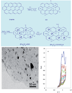
The electrochemical behavior of furosemide was investigated at the magnetic graphene oxide functionalized by chlorosulfonic acid-modified glassy carbon electrode (Fe3O4-GO-SO3H-GCE) in phosphate buffer solution (PBS), pH 6.8. Cyclic voltammetric study indicated that the oxidation process is irreversible and diffusion controlled. The number of exchanged electrons in the electro-oxidation process was obtained, and the data indicated that furosemide is oxidized via two one-electron steps. The results revealed that Fe3O4-GO-SO3H promotes the rate of oxidation by increasing the peak current. The diffusion coefficient and electron-transfer coefficient of furosemide were found to be 2.18 × 10⁻⁶ cm2 s−1 and 0.46, respectively. A sensitive, simple and time-saving differential-pulse voltammetric procedure was developed for the analysis of furosemide. The results show that by using the proposed method, furosemide can be determined with a detection limit of 0.11 μM. Finally, the applicability of the method to direct assays of spiked human serum and urine fluids is described.
In hypercalcemia treatment of, rapid reduction of serum calcium is required. The procedure includes rehydration with saline and diuresis with furosemide.5,6 Most patients presenting with severe hypercalcemia have a substantial component of prerenal azotemia owing to dehydration, which prevents the kidney from compensating for the rise in serum calcium by excreting more calcium in the urine. Therefore, the initial infusion of 500–1000 mL h−1 of saline to reverse the dehydration and restore urine flow can by itself substantially lower serum calcium. The addition of a loop diuretic such as furosemide not only enhances urine flow but also inhibits calcium re-absorption in the ascending limb of the loop of Henle.5,6 Therefore, careful medical supervision is necessary during treatment.
Monitoring of furosemide was requested in drug quality control and therapeutically drug monitoring investigations. Several methods have been used for the determination of furosemide in pharmaceutical formulations and biological fluids.7 Chromatography (especially LC/MS/MS) is now widely and routinely used for the analysis of furosemide.8 Chromatographic analyses are generally performed using expensive instruments. It requires extensive labor and analytical resources, and often results in a lengthy turn-around time. Given the low cost, ease of use, and sensitivity, electrochemical techniques are alternative methods for determining furosemide. But, surveying the literature revealed that electrochemical methods have been rarely applied to determination of pharmaceutical furosemide in serum and urine samples.9–14 Therefore, development of simple, sensitive, rapid and reliable electrochemical methods/sensors for the determination of furosemide is of great importance.
One of the best materials for construction electrochemical sensors is graphene oxide (GO). GO, as a basic material for the preparation of individual graphene sheets in bulk-quantity, has attracted great attention in recent years.15–17 In addition, the incredibly large specific surface area (two accessible sides), the abundant oxygen containing surface functionalities, such as epoxide, hydroxyl, and carboxylic groups, and the high water solubility afford GO sheets great promise for many more applications.15–17 The intrinsic oxygen-containing functional groups were used as initial sites for deposition of metal nanoparticles, such as Fe3O4, on the GO sheets, which opened up a novel route to multifunctional nanometer scaled catalytic, magnetic, and electronic materials.18–21 However, few studies about the drug monitoring by GO have been reported to date.
In the present work, we used Fe3O4-GO-SO3H as modifier material and studied the electrochemical oxidation of furosemide at Fe3O4-GO-SO3H modified glassy carbon electrode and developing a new electroanalytical procedure for quantification of furosemide in real samples. Also, a new mechanism based on the electrooxidation of furosemide was also investigated. To the best of our knowledge, this is the first report of the determination of furosemide based on their direct electrochemical oxidation on graphene or its derivatives.
Drug-free serum samples were obtained from healthy male volunteers and stored frozen until the assay. The serum samples were diluted (1:
2) with the supporting electrolyte and filtrated through a 5 μm filter to produce protein-free human serum. Various portions of stock furosemide solution were transferred into 10 mL volumetric flasks containing 3.3 mL of the serum sample. These solutions were diluted to the mark with the supporting electrolyte for preparation of spiked samples (final dilution of 1
:
3 with the supporting electrolyte). The protein-free spiked serum solutions were directly analyzed by the calibration method, according to our proposed procedure.
Urine samples were diluted (1:
2) with PBS after adding an appropriate amount of standard furosemide solution. The resulting solution was then directly analyzed, according to the proposed procedure, without any pretreatment or extraction steps.
FT-IR spectra were recorded on a Shimadzu model FTIR prestige 21 spectrophotometer using KBr discs. X-ray powder diffraction (XRD) measurements were performed using a Philips diffractometer manufactured by X'pert with monochromatized Cu Kα radiation.
Prior to use, the Fe3O4-GO-SO3H were sonicated in a 3:
1 sulfuric/nitric acid solution for 6 h in an ultrasonic bath at room temperature and then washed with distilled water. The obtained sample was taken, and dried overnight at 50 °C. Prior to the modification, the GC electrode was polished with 0.05 μm alumina suspension on a polishing micro-cloth, followed by sonication for 5 min in an ultrasonic bath in a 1
:
1 ethanol–redistilled water mixture. The electrode was then transferred to the 1.0 M sulfuric acid solution. Potential in the range of −1–1.5 V in a regime of cyclic voltammetry was applied for 20 cycles. Afterward, the electrode was rinsed thoroughly with distilled water and dried in air. It should be noted that this pretreatment was also employed for the bare GC electrode in the experiments that the unmodified electrode was tested. Fe3O4-GO-SO3H (50 mg) was dispersed into acetone/double distilled water with the aid of ultrasonic stirring for 6 h. A 10 μL aliquot of this dispersion (with concentration of 1.0 mg mL−1) was dropped on the GC electrode surface, and the solvent was allowed to dry in air.
The general morphologies of synthesized Fe3O4-GO were observed by SEM and shown in Fig. 1. A flat surface was shown in Fig. 1A, which demonstrating a single atomic layer thickness structure feature. The properties of Fe3O4-GO-SO3H are highly related to their micro structures, their dispersity and the morphology of Fe3O4 nanoparticles. The presence of Fe3O4 nanoparticles in GO-SO3H surface was confirmed by SEM. The added Fe3O4 nanoparticles to GO-SO3H appeared as bright dots that were spread on the surface of (Fe3O4)-GO-SO3H. In contrast to the surface of GO-SO3H which was quite smooth, the added Fe3O4 nanoparticles to GO-SO3H appeared as bright dots, which were spread on the surface of the GO-SO3H. The morphology of Fe3O4-GO-SO3H was examined by TEM (Fig. 1B), where the image of prepared composite showed Fe3O4 nanoparticles as nearly spherical and homogeneously distributed over the GO nanosheets.
In FT-IR spectra the band in the region of 1036 and 1153 cm−1 is attributed to the stretching vibrations of the (SO) and a peak appeared at about 3367 cm−1 is due to the stretching of OH groups in the SO3H. Also, the band from 400–650 cm−1 is assigned to the stretching vibrations of the (Fe–O) bond in Fe2O3 (Fig. 2).
The structural properties of the synthesized Fe3O4-GO were analyzed by XRD (Fig. 2). The diffraction peak of GO appeared at 23.94° which originated from the diffraction on its (0 0 2) layer planes. Also, synthetic nanocatalysts showed some low intensity diffraction peaks that were indexed to cubic Fe3O4. The XRD peaks of magnetic graphene-oxide were indexed to (2 2 0), (3 1 1), (4 0 0), (4 2 2) and (5 1 1) planes of a cubic unit cell of magnetite, appearing at 35.01°, 43.27°, 53.22°, 58.46°, and 63.50°, respectively (Fig. 3).
Coulometric measurements were performed on the Fe3O4-GO-SO3H-GC electrode in 0.1 M PBS containing 1 μM furosemide for 1100 s, at two different fixed potentials of 0.98 V and 1.12 V respectively, surface area = 0.059 cm2.
The microscopic areas of the Fe3O4-GO-SO3H-GC and the bare GCEs were obtained by CV using 1 mM K4Fe(CN)6 as a redox probe at different scan rates. For a reversible process, the anodic peak current ipa can be calculated as follows:23
I = (2.99 × 10⁵)α1/2n3/2AC*D1/2ν1/2 | (1) |
![]() | ||
Fig. 4 Cyclic voltammograms of 0.1 M PBS containing 1 μM furosemide using GC (A) and Fe3O4-GO-SO3H-GC (B) electrodes (curve a) in the absence of furosemide Potential sweep rate was 2 mV s−1. |
The significant enhancement of the furosemide electrooxidation at Fe3O4-GO-SO3H-GC electrode can be explained according to the catalysis mechanism. Because the GOs had a high surface area, it boosted the faradic currents of the sluggish reaction as well as increased the amount of chemisorbed layer of the furosemide. This behaviour catalyzed the electron transfer of furosemide in the solution. The more active functional groups on the surface of the Fe3O4-GO-SO3H can easily form hydrogen bonds with the amine groups of the furosemide, which weaken the –NR bond energies; the electrons would be transferred through SO3H⋯N–R. In addition, the density of the electron cloud was lower from furosemide to intermediate to product, and therefore, their electroactivity decreased and they were less easily oxidized which is in accordance with the experimental results (Scheme 2).
The electrocatalytic activity of Fe3O4-GO-SO3H can be ascribed to the combination of the electron-acceptor ability of SO3H and confinement effects due to the attachment to the Fe3O4-GO matrix. On the basis of the previous considerations, the electrocatalytic activity of Fe3O4-GO-SO3H must result from the superposition of the effect associated with more external SO3H units and that due to boundary-associated ones. On the other hand, since the catalytic effect associated with Fe3O4-GO-SO3H consists essentially of an enhancement of the anodic peak for furosemide oxidation, the catalytic effect can tentatively be associated with a redox reaction in solution phase between SO3H units and any intermediate species resulting from the initial electron transfer of furosemide.
On the basis of CV results, it is found that we have two rate determination steps in the electro-oxidation of furosemide on Fe3O4-GO-SO3H-GC electrode. The consecutive oxidation of single substance with two rate determination steps in a potential scan experiment is complicated. For the stepwise oxidation of a single substance O, that is, A → B + n1e (E1), B → C + n2e (E2), the situation is similar to the two-component case, but it is more complicated. In the elucidation of the mechanism and rate determination step in a multi-step reaction, the scan rates play a prominent role. In order to further clarify the electrochemical oxidation mechanism of furosemide on Fe3O4-GO-SO3H-GC electrode, effect of scan rate on cyclic voltammetry response is investigated. The results of CV studies for electro-oxidation of furosemide were obtained in two ranges of potential sweep rates. The first study was at low sweep rates (2–20 mV s−1) and the second investigation was at higher sweep rates from 20–300 mV s−1. The effects of scan rate on the voltammograms recorded for Fe3O4-GO-SO3H-GC electrode in 0.1 M PBS containing 1 μM furosemide are shown in Fig. 5A and B. It is clear that the current ratio of peak II/peak I (IpII/IpI) increase with rising scan rate until to 20 mV s−1. In addition at higher scan rate (>20 mV s−1) only peak II is observed while peak I almost disappeared. On the other hand only one peak is found in the voltammogram recorded at high scan rate. Additionally the ratio (IpII/IpI) increase with increasing scan rate. Hence one can conclude if:
• ν < 20 mV s−1, two separate waves are observed which the first wave corresponds to oxidation of A to B in an n1-electron reaction, with B diffusing into the solution as the wave is traversed. At the second wave, B continues to be oxide directly at the electrode. The voltammogram for this case presented in to Fig. 5A. Base on these results the following mechanism of furosemide on lower scan rates follow as:
• ν > 20 mV s−1, one wave is observed which the A was oxide directly at the electrode. The voltammogram for this case presented in to Fig. 5B and the mechanism in higher scan rates follow as:
• according to results and also above mechanisms, it can be considered that at low sweep rates, desorption step of the intermediated (according to mechanism I-produced from firs electron transfer step) and formation of product (produced from second electron transfer step in first mechanism) becomes more probable as the rate-determination step. Such behaviour for the electrochemical oxidation of furosemide has not been observed up to now.
• In summary, in the case of electrooxidation of furosemide on Fe3O4-GO-SO3H-GC electrode the nature of the i–E curve depends on scan rates. When v is between 2 and 20 mV s−1, the individual waves are merged into a broad wave whose Ep is dependent of scan rate. When v is between 20 and 200 mV s−1, a single peak is found therefore the second step is easier than the first, a single wave characteristic of a irreversible 2e− oxidation (A → C + 2e−) is observed.
Voltammetric parameters for furosemide oxidation on the Fe3O4-GO-SO3H-GC electrode surface were also studied. The effect of potential sweep rate was studied, in the range of 2–200 mV s−1. CVs recorded at different potential sweep rates for 0.1 M PBS containing 1 μM furosemide, using the Fe3O4-GO-SO3H-GC electrode, are depicted in Fig. 5A and B along with the potential sweep rate increase, both peak currents increased and the peak potentials shifted to positive values, confirming the irreversible nature of the reaction processes. For an irreversible diffusion-controlled process, the peak potential (Ep) is proportional to the logarithm of potential sweep rate (ν) within the following equation:28
![]() | (2) |
On the basis of this equation, the slope of the Ep vs. logν linear plot is b/2, where b indicates the Tafel slope. The dependency of peak II potential on log ν for the oxidation of furosemide on the Fe3O4-GO-SO3H-GC electrode is depicted in Fig. 5D value of b = 55.1 mV was obtained, which indicates that for the oxidation of furosemide, the charge-transfer coefficient, α, was 0.46.
From the CVs shown in Fig. 5C, both peaks I and II currents can be seen to depend linearly on the corresponding square root of potential sweep rate. This dependency indicates that a mass transport process has occurred in the oxidation process, via diffusion. From the slope of the linear dependency of peak II current on the square root of potential sweep rate, and using the Randles–Sevcik equation for totally irreversible electron-transfer processes, we can calculate the diffusion coefficients of furosemide using eqn (1). Using the proposed method, the diffusion coefficient of furosemide was found to be 2.18 × 10⁻⁶ cm2 s−1. The values of the diffusion coefficients obtained using both electrodes are nearly the same, since diffusion of electro-reactant species in the bulk of the solution does not depend on the target surface in a semi-infinite linear diffusion process.
Also, plotting the current function (peak current divided by the square root of the potential sweep rate) against the square root of the potential sweep rate (Fig. 6A) revealed negative slope confirming the electrocatalytic nature of the process.
![]() | ||
Fig. 6 (A) Current function vs. ν0.5 for 0.1 M PBS containing 1 μM furosemide. (B) Dependency of peak potentials with respect to the solution pH. |
The effect of pH on the electrochemical behavior of furosemide was investigated by CV, using 0.1 M buffer at various pH values ranging from 1.00 to 12.00. Fig. 6B shows the changes of peak potential II with respect to the solution pH. The plot shows two linear segments which is accordance with other reports.9,29
Coulometry was performed in PBS containing 1 μM furosemide at 0.98 and 1.12 V, corresponding to the potentials of I and II peaks using the Fe3O4-GO-SO3H-GC electrode. Electrolysis progress was carried out using CV. The extrapolated charge consumption for total electrolysis of the solution after corrections for background/charging currents was derived, with the number of exchanged electrons for peaks I and II obtained as 1.1 ≈ 1 and 0.91 ≈ 1, respectively. Hence, furosemide is oxidized via two one-electron steps on the Fe3O4-GO-SO3H-GC electrode surface (Fig. 7).
![]() | ||
Fig. 7 Coulometric response of Fe3O4-GO-SO3H-GC electrode in 0.1 M PBS containing 1 μM furosemide. T= 1100 s, E1 = 0.98 V and E2 = 1.12 V, surface area = 0.059 cm2. |
The above analysis for the electrochemical oxidation mechanism of furosemide is further supported by chronoamperometric measurements indicated in Fig. 8. The potentials applied are 0.98 and 1.12 V vs. Ag/AgCl which are close to those of anodic peaks I and II, respectively. Fig. 8 shows that compared in the presence of various concentrations of furosemide, much large increase in the current at 1.12 V is observed than that at 0.98 V.
The plot of net current versus t−1/2 which has been obtained by removing the background current by the point-by-point subtraction method gives a straight line, Fig. 8 (inset). This indicates that the transient current must be controlled by a diffusion process. The transient current is due to catalytic oxidation of atenolol which increases as the atenolol concentration is raised. No significant cathodic current was observed when the electrolysis potential was stepped to 0.51 vs. (Ag/AgCl)/V indicating the irreversible nature of the oxidation of drugs by using the slopes of these lines; we can obtain the diffusion coefficients of the drugs according to the Cottrell equation:28
I = nFAD1/2C*π−1/2t−1/2 | (3) |
The calibration curve for furosemide in PBS was obtained by differential-pulse voltammetry (DPV). Fig. 9A shows typical DPV curves for different concentrations of furosemide in PBS using Fe3O4-GO-SO3H-GC electrode. The dependency between peak current and drug concentration was rectilinear using Fe3O4-GO-SO3H-GC electrode for peak I (Fig. 9B).
The analytical characteristics of furosemide in PBS using both electrodes are reported in Table 1. The limits of detection (LOD) and quantitation (LOQ) of the procedure were calculated according to the 3 SD/m and 10 SD/m criteria, respectively, where SD is the standard deviation of the intercept and m is the slope of the calibration curves.30 The LOD and LOQ are reported in Table 1; their values confirm the sensitivity of the proposed procedure for the determination of furosemide. The precisions of the method are calculated as the relative standard deviation (RSD). The procedure was repeated on the same day on the same spiked solutions at concentrations in the range of the standard series. The intra-assay RSDs of the proposed method, determined on the basis of peak current for 10 replications. The accuracy of the proposed method was determined by spiking serum and urine samples with different concentrations of furosemide. Good percentage recoveries were obtained from both samples (see Table 2).
Analytical parameters | Peak I | Peak II |
---|---|---|
a Each value was obtained from ten experiments.b LOD, limit of detection; LOQ, limit of quantitation; RSD, relative standard deviation. | ||
Linear range (μM) | 15–340 | 17–100 |
Slope (μA M−1) | 0.964 | 1.0291 |
Intercept | 0.174 | 0.271 |
RSD (%)a | 2.17 | 2.19 |
LOD (μM)b | 0.10 | 0.11 |
LOQ (μM−1)b | 0.61 | 0.50 |
Analytical parameters | Medium | |||
---|---|---|---|---|
Serum | Urine | |||
Peak I | Peak II | Peak I | Peak II | |
a Each value was obtained from ten experiments.b LOD, limit of detection; RSD, relative standard deviation.c Recovery value is the mean of ten experiments. | ||||
Linear range (μM) | 20–100 | 26–100 | 18–720 | 18–500 |
Slope (μA M−1) | 5.73 | 4.06 | 1.89 | 1.80 |
Intercept | 0.6 | 0.61 | 4.0 | 0.93 |
RSD (%)a | 3.5 | 4.0 | 3.6 | 3.6 |
LOD (μM)b | 1.1 | 1.0 | 1.5 | 1.5 |
Recovery (%)c | 94.1 | 95 | 103.5 | 102.0 |
The comparison of Fe3O4-GO-SO3H-GC electrode with other sensors for the furosemide determination was listed in Table 3. It can be seen that the Fe3O4-GO-SO3H-GC electrode offered reasonable linear range for furosemide detection and the detection limit was lower than some of previous reports. These results indicated that Fe3O4-GO-SO3H-GC electrode is an appropriate platform for the determination of furosemide. On the other word, the prepared electrode shows voltammetric responses with low detection limit and high linear range for furosemide in optimal conditions, which makes it very suitable for determination of this drug. It is found that, by modification of on GC surface via Fe3O4-GO-SO3H, a novel strategy for developing an efficient and robust electrochemical sensing platform was established. The electrochemical sensor showed high sensitivity and simplicity for detection of furosemide.
Electrode type | Method | Linear range/(μM) | LOD/(μM) | Ref. |
---|---|---|---|---|
HMDE | DPV | 6.05–54.42 | 2.57 | 11 |
Graphite-polyurethane composite electrode | 3–9 | 0.96 | 12 | |
Multi-walled carbon nanotubes paste electrode | 8–200 | 0.29 | 13 | |
Gold electrode | 6–800 | 0.4 | 14 | |
Fe3O4-GO-SO3H-GC electrode | 20–100 (serum) | 0.1 (serum) | This work | |
18–720 (Urine) | 0.11 (urine) |
The applicability of the proposed method for the determination of furosemide in biological fluids was examined by measuring the peak I and II currents as functions of the bulk concentration of the drug in urine and serum samples using the Fe3O4-GO-SO3H-GC electrode. The serum and urine samples were diluted 10 and 20 times with PBS before taking the measurements, to prevent the matrix effect of real samples. The generally poor selectivity of voltammetric techniques can pose problems in the analysis of biological samples, which contain oxidizable substances. However, no current due to oxidation of the compounds in either the serum or urine samples appeared. The results obtained from the proposed technique for determining furosemide in serum and urine samples are listed in Table 2. The percentage recovery of furosemide was determined by comparing the peak currents of a known drug concentration in both media with their equivalents in calibration curves; these results are also summarized in Table 2. Good recoveries of furosemide were achieved from these matrices, meaning that application of our proposed voltammetric method to the analysis of furosemide in biological fluids could be easily assessed.
The effect of some common interfering species on the determination of 0.1 mM furosemide at Fe3O4-GO-SO3H-GC was investigated using the optimum measurement conditions. The oxidation peaks of interferents should not appear where the peak corresponds to furosemide appears. Therefore, in order to investigate the effect of some interferent's substances such as glucose, starch, dextrose, sucrose etc. on the voltammetric response of furosemide, this study was carried out. Table 4 shows the tolerance limit for each interferents. The tolerance limit is defined as the concentrations of interfering species which give an error of ≤15% in the determinations of furosemide. The data show that interferences are only significant at relatively high concentrations, confirming that the proposed method is free from common interfering species.
Interfering species | Interference concentration/mM | ΔI/μA (%) |
---|---|---|
Oxalic acid | 0.1 | +7.4 |
Lactic acid | +7.0 | |
Citric acid | +2.2 | |
Sucrose | −10 | |
Glucose | +5.0 | |
Dextrose | −12.2 |
The repeatability of the modified electrode was also investigated. When the modified electrode was stored in the atmosphere, the current response decreased 7.5% after ∼20 days. Furthermore, the current response of the modified electrode in buffer solution, pH 6.8, after 100 rounds of cyclic scanning was almost unchanged. It proved the good stability of the modified electrode. To check the inter-electrode reproducibility of the modified electrode, five modified electrodes were tested simultaneously by recording CVs in buffer solution, pH 6.8, and containing 10 μM of furosemide at a scan rate of 50 mV s−1. The average catalytic peak current is 7.11 μA with a relative standard deviation of 6.0%. The GC electrode modified with Fe3O4-GO-SO3H imparts a higher stability onto voltammetric measurements of furosemide.
Footnote |
† Electronic supplementary information (ESI) available. See DOI: 10.1039/c3ra46973e |
This journal is © The Royal Society of Chemistry 2014 |