DOI:
10.1039/C3RA46943C
(Paper)
RSC Adv., 2014,
4, 14603-14612
Encapsulation of EV71-specific IgY antibodies by multilayer polypeptide microcapsules and its sustained release for inhibiting enterovirus 71 replication
Received
22nd November 2013
, Accepted 4th February 2014
First published on 10th February 2014
Abstract
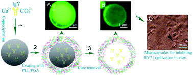
To save egg yolk immunoglobulin (IgY) from damage by protease digestion in oral delivery, IgY specific to enterovirus 71 (EV71) was loaded into polypeptide microcapsules by the method of layer-by-layer encapsulation. Porous CaCO3 particles doped with IgY were used as templates which were coated with multilayer poly (L-lysine) (PLL) and poly (L-glutamic acid) (PGA). Then, IgY-loaded polypeptide microcapsules were fabricated after the removal of CaCO3 templates. The alternating adsorption was confirmed by the zeta potential and the successful formation of a multilayer polypeptide (PLL/PGA) shell was visualized by SEM. The influence of the PLL/PGA layer numbers on the IgY sustained release performance was studied in simulated gastric fluid. IgY release could be accurately controlled by adjusting the layer number of the polypeptide shells and only 45% of the IgY was released from 5-bilayer microcapsules in the initial 2 hours. The biological activity of the encapsulated IgY was investigated both in vitro and in vivo. Encapsulated EV71-specific IgY remained highly active and capable of neutralizing EV71 after multilayer polypeptide encapsulation, which provided a promising method to protect EV71-specific IgY in the gastric environment and to specifically release IgY at the target intestinal site for the prevention and control of EV71 infectious diseases.
1. Introduction
Human enterovirus 71 (EV71), which belongs to the enterovirus genus of the Picornaviridae family, has been associated with sporadic cases or outbreaks of a wide spectrum of diseases, including hand-foot-and-mouth disease, herpangina and poliomyelitis-like syndrome in neonates and infants.1,2 In recent years, EV71 infections have become one of the serious public health issues in China and the Western Pacific region.3,4 Several approaches have been attempted to combat EV71 infection. Some antiviral chemicals have been reported to be anti-EV71 active in vitro5,6 and candidate EV71 vaccines have been evaluated in animal models.7,8 However, there are no effective antiviral drugs or vaccines against EV71 in the clinic presently.9 Neutralizing antibody provides protection against EV71 because it is able to bind the virus to reduce its replication.10–12 In addition, IgY antibodies from hens immunized with EV71 are demonstrated to significantly reduce the morbidity and mortality in EV71-infected mouse pups.13
IgY is an inexpensive antibody which is convenient, highly stable, safe and has been used to combat and treat bacterial and viral infections in animals.14 Specially, IgY which can be administered orally, has been proven to be effective against intestinal pathogens,15 enterovirus16 and influenza virus.17 Therefore, it is a promising antiviral infection medicine for passive immunotherapy application and has attracted considerable attention recently. However, IgY has its own disadvantage that the activity of IgY may be reduced or destroyed under gastric conditions, and particularly it is sensitive to pepsin and low pH digestion.18 Therefore, it is necessary to take measures to keep the therapeutic value of IgY in the gastric environment and improve its sustained release in the intestinal target site. Microencapsulation techniques have been previously studied to load IgY from pepsin digestion, such as chitosan–alginate microcapsulation,19 feed containers,20 polymer coatings21 and multiple emulsions.22 However, all these methods have some defects, such as complexity, low effectiveness or great loss of activity.
Recently, layer-by-layer (LBL) microcapsules have been designed for protein delivery,23–25 which are one of the most promising and simple methods due to the controllability of the wall thickness, permeability and size distribution.26 A variety of slow-release drugs have been effectively loaded in this way. Multilayer polyelectrolyte encapsulation technology is based on oppositely charged polymers being adsorbed on colloid-sized templates, followed by selective template removal.27 Charged polyelectrolyte has a great impact on the quality of the microcapsules because of its electric adsorption capacity, solubility and cross linking properties. Moreover, this polyelectrolyte film should have some properties of drug delivery vehicles, such as biocompatibility, permeability and biodegradability.28 The template is another key factor in preparing LBL microcapsules. Some colloidal particles can be employed as the microcapsule core, such as mesoporous silica,29 melamine formaldehyde30 and manganese carbonate.31 In particular, as a biocompatible and nontoxic template, calcium carbonate has been widely used for the encapsulation of proteins in polyelectrolyte capsules recently.24,32 Moreover, protein molecules can be pre-loaded in CaCO3 cores by co-precipitation and still keep a relatively higher bioactivity.33
This paper proposes the design of multilayer microcapsules for EV71-specific IgY loading and oral delivery to achieve the protection and sustained release of IgY for inhibiting enterovirus 71 replication. We attempted to pre-load EV71-specific IgY into CaCO3 templates via co-precipitation and adsorb multilayer poly (L-lysine) (PLL) and poly (L-glutamic acid) (PGA) (PLL/PGA) on the surface of the templates. It has been previously demonstrated that these multilayer polypeptide films have good cell adhesion properties which aid in targeting the release of the payload at the intestinal wall.34,35 This study is focused on the encapsulation of EV71-specific IgY antibodies by multilayer polypeptide microcapsules and its bioactivity for inhibiting virus after sustained release. The physical characteristics, encapsulation efficiency and release performance of the IgY-loaded microcapsules have been examined. While the biological activity of the encapsulated EV71-specific IgY for inhibiting enterovirus 71 replication have been investigated both in vitro and in vivo.
2. Materials and methods
2.1. Materials
Poly (L-lysine) (PLL) (molecular weight 70
000–150
000) and poly (L-glutamic acid) (PGA) (molecular weight 50
000–100
000) were obtained from Sigma-Aldrich (Munich, Germany). 4-(2-hydroxyethyl) piperazine-1-ethanesulfonic acid (HEPES) and Coomassie brilliant blue G-250 were obtained from Invitrogen (USA). Dulbecco's phosphate-buffered saline (DPBS) and fluorescein isothiocyanate (FITC) were purchased from Tianjin LianXing Biotechnology Co. Ltd. (Tianjin, China). The Bradford protein assay kit and chicken immunoglobulin Y (IgY) ELISA kit were purchased from the Beyotime Institute of Biotechnology (Shanghai, China). The EV71 real-time PCR (RT-PCR) kit was purchased from Huaruian Biology (Guangzhou, China). All of the other reagents used in this study were of analytical grade. FITC-labeled IgY (or PLL) was prepared by overnight incubation under ambient conditions of their mixtures (0.5 M carbonate buffer, pH 9.5, [IgY or PLL]/[FITC] = 25) followed with an exhaustive dialysis at 4 °C to remove the unconjugated FITC. The dialyzed solution was freeze-dried for 24 h when there was no fluorescence detected in the supernatant.
The specific anti-EV71 IgY antibody was prepared and purified in our laboratory as described previously.36 The antibody titer tested by ELISA was 20480 and the purity of IgY was 94.86%. Pregnant ICR mice were obtained from the experimental animal center of Guangdong Province (Foshan, China) and were maintained in a standard rearing room. The EV71 virus strain and Vero cells were provided by Landbiology (Guangzhou, China).
2.2. Ethics statement
The experiments involving animal procedures in this study were approved by the Animal Ethical and Welfare Committee of Sun Yat-sen University (Project Approval no. 201300132). The animal experiments were conducted in strict accordance with the recommendations in the Guide for the Care and Use of Laboratory Animals of the National Institute of Health. To minimize suffering after viral challenge, the mice were monitored each day and the mice that met end point criteria were humanely euthanized by inhalation of CO2.
2.3. Preparation of the IgY-loaded calcium carbonate templates
The preparation process was based on a previous study with some modifications.32 CaCO3 particles were prepared by mixing Na2CO3 (0.04 M) and Ca(NO3)2 (0.04 M) solutions with equal volumes under vigorous stirring and the CaCO3 templates obtained were stored after centrifugation three times. To obtain calcium carbonate particles loaded with IgY, abbreviated as CaCO3(IgY), 1.0 mg lyophilized powder of IgY was reconstituted with DPBS buffer (1.0 mL, pH ∼ 7.0) as the IgY loading solution, followed by this the solution was added into an equal volume of Ca(NO3)2 solution prior to its mixing with the Na2CO3 solution. The process resulted in IgY entrapped within growing CaCO3 particles via co-precipitation.25
2.4. Preparation of multilayer polypeptide microcapsules
Multilayered microcapsules were fabricated by immersion of the CaCO3(IgY) cores into oppositely-charged polypeptide solutions. In detail, the CaCO3(IgY) particles were alternately dispersed in 1.0 mg mL−1 PLL and PGA solutions containing 10 mM HEPES buffer (pH ∼ 7.05). Each adsorption was performed on a shaking table which maintains gentle agitation at 4 °C for 10 min, followed by three consecutive washes with distilled water to separate the bound polypeptides from excess polypeptides at 2000 × g centrifugation for 3 min. The desired number of polypeptide layers was achieved by the repeated deposition of PLL and PGA. The calcium carbonate core was then dissolved with 0.2 M EDTA solution (pH ∼ 7.0) under shaking (100 r min−1 for 30 min) and centrifugation of the suspension at 5000 × g for 10 min. With this routine, the multilayer IgY-loaded polypeptide microcapsules were successfully obtained after being washed in water and stored at 4 °C before further study. In particular, the amount of IgY entrapped in CaCO3 was not affected by the number of layers.
2.5. Characteristics of the IgY-loaded microcapsules
The surface morphology and size of the CaCO3(IgY) templates, multilayer polypeptides assembly and core-dissolved microcapsules were examined by scanning electron microscopy (SEM) (S3400N, Hitach, Japan). The suspended sample was dropped on the glass sheet and sputtered with gold after being dried and the imaging was carried out at an accelerating voltage of 15.0 kV. Fluorescent FITC-labeled IgY trapped in CaCO3 cores and FITC-labeled PLL adsorbed on the template surface were prepared for observation with a fluorescence microscope (BX51, Olympus, Japan). Images were taken with an MShot Digital Imaging System and the excitation wavelength λex = 488 nm and emission wavelength λem = 525 nm were used. The size distribution of the CaCO3(IgY) templates was assessed with a Mastersizer 2000 laser particle analyzer. The sample was prepared under the optimal process conditions and was mixed homogenously with deionized water. The formula for the average particle diameter was as follows:
where Dn represents the average diameter, ni refers to the number of microcapsules, and di the diameter of a single microcapsule.
To verify the assembly of the multilayer polypeptides, the zeta potential of the particles was measured at each adsorption step by a Zeta Potential Analyzer (Zetasizer 3000 HS, Malvern, England) in distilled water (pH ∼ 7.0). The potential value is the average of three consecutive measurements.
2.6. Determination of the IgY encapsulation efficiency and loading percentage
To analyze the IgY content incorporated into the microcapsules, the CaCO3(IgY) was prepared by varying the initial concentration of IgY and the amount of unbound IgY was measured by Bradford protein assay.24,29 This is a widely accepted method for the quantitative determination of proteins. Generally, the clear supernatant containing free IgY was separated by centrifugation at 2500×g for 5 min (Hettich, Germany). Afterwards, the supernatant was added into 96-well ELISA plates according to the Bradford protein assay kit instructions and the value of IgY content was measured with a standard enzyme reader at 570 nm (Tecan's Infinite 200, Switzerland). For standard curves, the dilution series of IgY was prepared in the concentration range of 50–600 μg mL−1 of PBS buffer solution. The IgY encapsulation efficiency (EE) and loading capacity (LC) of the CaCO3 templates were calculated as described by the following two equations:
EE% = (total IgY − free IgY)/total IgY × 100% |
LE% = (total IgY − free IgY)/CaCO3(IgY) × 100% |
The quality changes of the CaCO3(IgY) templates and core–shell microcapsules were measured by a Pyris 6 thermogravimetric analyzer (TGA) (Norwalk, CT, Perkin-Elmer, USA) at a heating rate of 10 °C min−1 from 30 °C to 800 °C. The samples were treated at 100 °C before TGA analysis.
2.7. IgY release studies
The in vitro IgY release profiles were carried out at a constant temperature on a shaking table containing simulated gastric fluid (SGF).37 In detail, the stored 6, 8 and 10 layers microcapsules, abbreviated as IgY/(PLL/PGA)n, containing 3.5 mg IgY were re-dispersed in 4 mL simulated gastric fluid (3.2 mg mL−1 pepsin, 0.03 M NaCl, pH ∼ 1.2) at 150 r/min, 37 °C. 100 μL of the supernatant was taken out from SGF and replaced with the same amount of fresh medium at intervals of 30, 60, 90 and 120 min. After 2 h, the microcapsules were filtered and transferred to 4 mL simulated intestinal fluid (SIF, 0.05 M KH2PO4, 10 mg mL−1 pancreatin pH 6.8) and incubated in the same conditions for 2 h.
The amount of free IgY from the microcapsules was determined by the chicken immunoglobulin Y (IgY) ELISA kit (96 wells).33 The kit uses a double-antibody sandwich enzyme-linked immunosorbent assay for the concentration of IgY. In brief, the standards and samples were added into the microplate wells which was pre-coated with IgY monoclonal antibody and all of the IgY was bound by the immobilized antibody during incubation. After washing, the enzyme-linked monoclonal antibody specific for IgY was pipetted into the well. Following another wash to remove unbound enzyme, color developed along with the addition of substrate solution to the wells. Absorbance was read at 450 nm by the microplate reader (Tecan Spectrafluor Plus). The accumulative release percentage was calculated and the data represented the mean of three measurements in three different trials.
2.8. Inhibiting EV71 replication in vitro
The capacity of the encapsulated EV71-specific IgY to inhibit EV71 replication in vitro was assessed by the cytopathic effect (CPE) of Vero cells caused by EV71 infection. Specifically, a 100 μL Vero cell suspension (2 × 10⁵ cells) was seeded into 96-well plates at 37 °C in a humid atmosphere of 5% CO2 and 95% air incubator. The cell culture medium consisted of DMEM with 10% FBS, penicillin (50 U mL−1), and streptomycin (0.05 mg mL−1). After 24 hours, the adherent Vero cells were then infected with 50 TCID50 per well EV71, followed by 20 μL microcapsules containing 0.1 mg EV71-specific IgY which were added into each well in the experimental group. As a control group, the equal free IgY and PBS were added into the Vero cells after being infected with EV71 respectively. After incubation at 37 °C for 24 h, the effect of the IgY microcapsules on inhibiting EV71 replication was directly observed by optical microscopy. Images of the cell morphology were captured at different wells of each sample by an Olympus IX51 microscope in a bright-field mode.
2.9. Anti-EV71 study in vivo
For the EV71 challenge, groups of 24 one day-old ICR pups were orally inoculated with 50 μL EV71 (1 × 10⁶ TCID50 per mouse) after being fasted for 8 h. The mice were divided randomly into three groups which were treated orally with PBS (50 μL per mouse), 50 μL IgY (1.0 mg mL−1) and equivalent doses of IgY-loaded microcapsules 2 h and 26 h post-infection respectively. The mice were killed by the inhalation of CO2 on day 5 of infection. Their skeletal muscle and small intestine were collected to analyze the existence of EV71 and to observe the histopathology. The obtained tissues were fixed in 10% buffered formalin for 24 h, embedded in paraffin and stained with hematoxylin and eosin (H&E). As with the evaluation standard, the histopathology of the skeletal muscle was evaluated based on muscle fiber degeneration and necrosis, while the evaluation of the small intestine was carried out in the same way on the intestinal villus interstitial edema and epithelial cell vacuolar degeneration by pathologists.38
Relative expression of the virus in the small intestine was detected by real-time PCR which was performed in a simplified two-step procedure: a reverse-transcription step leading to the synthesis of cDNA from the total RNA and quantitative real-time PCR step leading to the relative quantitation of EV71 mRNA expression levels among the samples. Viral RNA in the small intestine taken from each group was detected by using an EV71 real-time PCR kit according to the manufacturers' instructions. The specificity of the real-time RT-PCR assay was analyzed with the LightCycler (Roche Molecular Biochemicals, Germany).
3. Results and discussion
3.1. Characterization of the CaCO3(IgY) microspheres
The activity of IgY was relatively stable without general reduction at room temperature for one month,39 so there was no need for any special processing in the process of preparing IgY-loaded CaCO3 microparticles. IgY was directly added into the reaction before the mixing of Ca(NO3)2 and Na2CO3 solutions for controlling the growth of spherical CaCO3 microparticles and CaCO3(IgY) were formed from the co-precipitation.40 The morphology and size of these microparticles can be roughly detected by the SEM micrograph. The CaCO3(IgY) microparticles were spherical and porous with a mean size of about 5 μm (Fig. 1a and b), but the CaCO3 microparticles without IgY presented almost rhombohedral morphologies (Fig. 1e). The results with laser particle size analysis (Fig. 1f) demonstrated that the CaCO3(IgY) microspheres had a relatively homogeneous distribution and a particle size of 2–10 μm. In order to confirm IgY was loaded into CaCO3 templates, fluorescence microscopy was used to detect the loading result of the FITC-IgY microcarriers. IgY was labeled by FITC and then precipitated with the formation of CaCO3. As shown in Fig. 2a, the FITC-IgY was successfully encapsulated inside the microspheres, which was based on the green fluorescence light that was emitted from the cores. It was found earlier that some other proteins, such as BSA,41 HSA and IgG,24 could be incorporated into CaCO3. The reason for the successful incorporation was that the protein additives were absorbed on the surface of primary nanocrystallites which gradually formed the superstructure of porous carbonate microparticles.25 Some previous studies have noticed that electrostatic interactions contributed greatly to the affinity of the protein additives to the carbonate surface.24,42
 |
| Fig. 1 SEM surface images of (a) complete CaCO3(IgY) microspheres, (c) CaCO3(IgY)/(PLL/PGA)5 microspheres and (e) pure CaCO3. Enlarged view of (b) a single CaCO3(IgY) microsphere and (d) a single coating microsphere. (f) The particle size distribution of the CaCO3(IgY) microspheres was assayed with a Mastersizer 2000 laser particle analyzer. | |
 |
| Fig. 2 Fluorescence microscope images of (a) CaCO3(FITC-IgY) cores and (b) CaCO3(FITC-IgY) covered with 5-bilayer FITC-PLL/PGA. The insets show single enlarged microspheres. (c) Zeta potential of the CaCO3(IgY) cores coated with a PLL/PGA multilayer as a function of the layer number. | |
3.2. Characterization of the multilayer core–shell structure and microcapsules
The doped CaCO3 microparticles were coated with multilayered PLL/PGA films via LBL deposition. The process of stepwise polypeptide adsorption on the CaCO3 templates was monitored by measuring the surface potential after the deposition of each polyelectrolyte layer. The zeta potential of the CaCO3(IgY) cores at pH 7.0 was −10.11 mV under the influence of the more negatively charged IgY, whereas it switched to +9.01 mV after the first deposition of PLL (pI ∼ 9) (Fig. 2c). After the adsorption of PGA (pI ∼ 3.5), the zeta potential completely reversed and exhibited a negative value of −18.53 mV. Regular surface charge reversal after each new PLL and PGA adsorption indicated that the oppositely charged polypeptides were deposited on the microparticles successfully. Previous research also reported charge reversal after adsorption of the PGA layer and the PLL layer, which keeps a stable transformation with increasing numbers of layers.43
The microsphere covered with five PLL/PGA bilayers (abbreviated: CaCO3(IgY)/(PLL/PGA)5) was characterized by SEM. As Fig. 1 shows, the surface of CaCO3(IgY)/(PLL/PGA)5 (Fig. 1d), compared with the uncovered ones (Fig. 1b), became flocculent and rough. Also, the particles had almost the same size before and after the layer-by-layer self-assembly. The SEM images proved that the multilayer polypeptide layers could be adsorbed onto the template surface and form a stable core–shell structure successfully. Fluorescence microscopy was also performed to verify the successful fabrication of core–shell capsules. FITC-labeled IgY was doped in CaCO3 templates which were enveloped in the multilayer capsule shells and the PLL layer was also labeled with FITC. As could be seen from Fig. 2b, the cores were surrounded by a layer of light green shadow (Fig. 2a just showing some green cores), indicating that the multilayer polypeptide was packaged on the template wall by thin shells and IgY was still retained in the core. This multilayer core–shell structure containing IgY was fabricated via a polyelectrolyte adsorption method under mild conditions. This manufacturing method was more convenient and effective than the traditional multiple emulsification method in which homogenizer and a heat gelation process were necessary.44
The mass of adsorbed 5-bilayers polypeptide was measured by TGA. It could be observed in Fig. 4 that the weight loss was 4.2% to 6.2% at the temperature of 600 °C when the IgY and polypeptides were burning out for CaCO3(IgY) and CaCO3(IgY)/(PLL/PGA)5, respectively. In detail, the weight loss at temperatures lower than 200 °C represented the absorbed water content in the microcapsules, while the weight loss in the temperature range of 200–600 °C corresponded to IgY and polypeptide decomposition (including the combined water decomposition). The TGA results indicated that the mass of the deposited polypeptide membrane was 2.0%, consistent with a previous report.45
After the core–shell microparticles were obtained, the carbonate core was removed with EDTA at pH 7.0 under the standard procedure.46 In this way, CaCO3 could be moderately dissolved and the IgY-filled microcapsules were formed. In Fig. 3a, the drying 5-bilayers PLL/PGA capsules without cores in a collapsed state could be seen by SEM imaging. Compared with the spherical core–shell microparticles (Fig. 1c), the microcapsules collapsing into a flat shape indicated that the calcium carbonate cores were nearly completely dissolved. Fig. 3d shows a fluorescence microscopy image of hollow 5-bilayer capsules, which were fluorescently labeled by the adsorption of FITC-PLL. As was shown in Fig. 3d, the green fluorescence light of the microcapsule walls was higher than that from the interior of the microcapsules (the enlarged view of a single microcapsule showed this more clearly). This confirmed the successful formation of PLL/PGA multilayer shells after removal of the cores. Under bright field imaging it could be observed that the original shape and size of the CaCO3 templates was maintained after core removal (Fig. 3c). However, this would not mean that the capsules were absolutely “hollow” because the IgY molecules were encapsulated within the microcapsules by being doped into the porous CaCO3 templates with a molecular-sized confinement.37 Microcapsules without shrinkage and breakage indicated that the 5-bilayer PLL-PGA film is rather stable under the calcium carbonate core dissolution condition.
 |
| Fig. 3 Morphology of the IgY microcapsules. (a) SEM observation of the surface morphology for hollow IgY-load microcapsules, (b) an enlarged view of the microcapsules, (c) fluorescence microscope images of hollow microcapsules in bright-field and (d) in fluorescent-field. The inset shows a single enlarged microcapsule. | |
3.3. IgY encapsulation efficiency and loading capacity of the microparticles
The amount of IgY incorporated into the CaCO3 cores was affected by the initial loading concentration of the IgY in the process of co-precipitation. As demonstrated in Fig. 5, the increasing concentration of IgY (from 0.5 mg mL−1 to 2.0 mg mL−1) did affect the IgY encapsulation efficiency, which was observed to gradually decrease from 37% to 16%. On the other hand, the loading capacity of CaCO3 templates obviously increased with an increase of the initial loading concentration of IgY from 0.5 mg mL−1 to 2.0 mg mL−1. Meanwhile, it slowly decreased to reach an equilibrium (about 7%) when the initial concentration was greater than 1.0 mg mL−1. A possible explanation for this result could be that the amount of IgY doping into the CaCO3 cores was limited by co-precipitation. Some studies suggested that the loading amount related to spontaneous deposition is driven by electrostatic attraction.28,40 It was also observed that the EE% was 29% and LC% was 6.7% at the 1.0 mg mL−1 loading concentration which provided data for the release experiments.
The IgY incorporated into the CaCO3 microspheres was also quantified by TGA with which the CaCO3(IgY) particles were prepared under the initial concentration of 0.5 mg mL−1. From Fig. 4A it could be seen that the weight loss of pure CaCO3 and doped CaCO3 was greatly different in the temperature range from 200 to 600 °C. A considerably large amount (4.2%) of mass was lost for the CaCO3(IgY) particles compared with pure CaCO3. Specifically, the weight loss in the temperature range of 100–240 °C should represent the evaporation of water; while in the temperature range of 240–600 °C the weight loss relates to the decomposition of IgY. It could be concluded that the loading capacity of the templates was about 4.2%, consistent with the results displayed in Fig. 5.
 |
| Fig. 4 TGA degradation profiles. (a) TGA degradation profiles for CaCO3(IgY) templates and pure CaCO3. (b) TGA degradation profile of CaCO3(IgY) templates coated with 5-bilayers PLL/PGA and CaCO3(IgY) templates. | |
 |
| Fig. 5 Encapsulation efficiency and loading capacity. Effect of IgY initial loading concentration on the encapsulation efficiency and loading capacity of the CaCO3 microparticles. The data points represent the mean ± standard deviation of data from three independent samples. | |
3.4. Release studies
According to the above sections, IgY was encapsulated into the polypeptide microcapsules successfully. The ultimate goal of our study was not only to protect IgY from gastric inactivation, but also to estimate its sustained release in the intestine targets by oral administration. The release property of IgY from multilayer microcapsules was evaluated under simulated gastric fluid conditions for 2 h and simulated intestinal fluid for 4 h. From Fig. 6, a relatively large amount of release at the initial stage can be seen followed by a sustained release and the total release quantity of IgY (6–10 layers) was approximately 75% for 6 h. A small number of microcapsules were considered to see if leakage had occurred in the process of preparation. A short release with a high rate at the early stage was quite normal. The cumulative release reached 68% from the 3-bilayer microcapsules for 2 h, whereas only 45% of the IgY had been released from the 5-bilayer microcapsules, which indicated that the IgY release was slowed down as the number of layers increased. The release mechanism in the simulated gastric passage could be attributed to IgY diffusion and erosion of the PLL/PGA microcapsules.47 The amount of IgY released displayed a nearly linear relation with time in SGF, which indicated that the drug release behavior was mainly governed by a diffusion mechanism. On the other hand, although the strong electrostatic interaction among the layers ensured stability of the capsules at a low pH value, the polypeptide microcapsules were slowly broken down for the large concentration gradient between the capsule interior and the bulk.40 As a result of the thicker walls with 5-bilayers and a lower permeability, the microcapsules were more resistant to release in 2 h. It was also known from the release experiment that more than 28% of IgY was released when the 5-bilayered microcapsules were exposed to simulated intestinal fluid for 4 h, which meant that 28% of the IgY could be safely transported in the detrimental gastric environment with the protection of polypeptide microcapsules and could be released in the intestine.
 |
| Fig. 6 IgY cumulative release profiles. IgY cumulative release profiles from the polypeptide microcapsules having different numbers of layers. IgY microcapsules were suspended in simulated gastric fluid for 0–120 min followed by a suspension in simulated intestinal fluid for 2 h. The data points represent mean ± standard deviation of the data from three independent samples. | |
3.5. Sustained release of IgY from microcapsules for inhibiting enterovirus 71 replication
Specific anti-EV71 IgY has been confirmed to neutralize EV71 in vitro.13 In order to validate the activity of IgY not being destroyed after microencapsulation, the cytopathic effect (CPE) of EV71 on Vero cells was observed after IgY microcapsules, free IgY and PBS treatment respectively. The ability of EV71 to infect and cause CPE has been demonstrated in Vero cells previously.48 As Fig. 7 shows that both encapsulated (Fig. 7d) and free IgY (Fig. 7c) exhibited a high degree of integrity of cellular structure, which was almost the same as the untreated control (Fig. 6a), while the PBS treated group (Fig. 7b) after infection by EV71 showed signs of cell damage and death. This demonstrated that the cytopathic effects of EV71 on the Vero cells were noticeably inhibited by the encapsulated anti-EV71 IgY. The results further proved that the activity of IgY was not damaged after the process of polypeptide LBL microencapsulation, so that it was capable of inhibiting EV71 replication after release from the microcapsules. The main reason that microencapsulated IgY retained the majority of its activity was likely to be that the IgY was relatively stable and the preparation conditions were quite mild.
 |
| Fig. 7 The inhibition of EV71 replication in Vero cells culture. (a) untreated control (b) the trans-infection of EV71 on Vero cells showing the CPE (c) free anti-EV71 IgY and EV71 mixed into Vero cells showed slight CPE (d) anti-EV71 IgY microcapsules (white rectangle) and EV71 mixed into Vero cells showed no CPE, indicating virus neutralization. Scale 100 μm at 100×. | |
3.6. Anti-EV71 study in vivo
Several attempts have been made to control virus infection in animal models by oral IgY administration.14,17 In our study, the protective effect of IgY-loaded microcapsules against EV71 infection was evaluated in newborn ICR mice. Considering the small intestine and skeletal muscle as notable target sites of EV71 replication in mice,49 the pathologies of both parts were compared to evaluate the protective effect of IgY-loaded microcapsules and the relative expression of virus in the small intestine was detected by RT-PCR.50 Compared with the uninfected control, muscle fiber degeneration, necrosis and damage of the intestinal villus occurred in the EV71 infected control (Fig. 8). In the experimental group, the tissues from the free IgY group showed moderate necrosis and the IgY-loaded microcapsules group was observed without virus locus in the skeletal muscle and small intestine of the mice, compared with the EV71 infected control. The relative inhibition of expression of the virus in the small intestine was determined to be 19% and 85% for the free IgY and IgY-loaded microcapsules respectively (Fig. 9). The result demonstrated that both free IgY and IgY-loaded microcapsules could inhibit virus replication, which was similar to the previous research that the morbidity and mortality were significantly reduced after specific IgY was orally fed with infected EV71.13 However, it was noteworthy that the IgY-loaded microcapsules were significantly more effective than free IgY. This could be attributed to the ability of the polypeptide microcapsules to protect IgY from pepsin enzyme destruction in the gastric environment. Hence, greater quantities of active IgY were delivered to the intestine and could more effectively inhibit EV71 growth.
 |
| Fig. 8 Histopathology observation of tissues of EV71 infected neonatal mice. Tissues of uninfected 6 day old mice were used as the uninfected control. Both the skeletal muscle and small intestine were sampled for 6 days from being infected. Tissues from the infected EV71 mice orally treated with PBS, free IgY and IgY-loaded microcapsules are also shown. The lesion sites are denoted by black arrows. | |
 |
| Fig. 9 Real-time quantitative PCR analyses for the expression of EV71 in the small intestine. The results are reported as the fold change or relative quantitation of the target mRNA expression (2−ΔΔCT Ct method). Error bars represent the standard error of the mean associated with the ΔΔCt value. | |
Conclusion
We have demonstrated that IgY can successfully be loaded in multilayer polypeptide microcapsules and the delayed release finely adjusted by the layer number of the shells. Fabrication of the IgY capsules was performed by a simple co-precipitation method to form IgY-loaded CaCO3 microparticles, followed by PLL/PGA alternate assembly and dissociation of the inorganic template. The bioactivity of IgY has not been damaged in the encapsulation process because of its high stability and mild preparation conditions. These capsules show a strong ability for the encapsulation and controlled release of IgY. Results have shown that only 45% of IgY is released from the 5-bilayer capsules within two hours, which implies that the remaining IgY remained protected while passing through the detrimental gastric environment. Here the LBL assembled microcapsules as oral protein delivery carriers are applied to protect EV71-specific IgY from gastric inactivation and their effectiveness in inhibiting enterovirus 71 replication is confirmed both by in vitro cell culture and in vivo animal experiments. The encapsulated IgY has demonstrated a better ability to neutralize the EV71 than the same dosage of free IgY in EV71-infection newborn ICR mice. Thus, oral administration of multilayer polypeptide microencapsulated specific IgY may provide an effective means of controlling intestinal infection disease. Meanwhile, these findings are valuable to inspire more research to combine various therapeutic proteins drugs with multilayer polypeptide microcapsules for oral therapy. Further investigations into this issue are currently ongoing.
Acknowledgements
The authors are grateful to the combination research projects of the Guangdong Province and Ministry of Education for the financial support (project no. 2011B090400279 and 2012B091100188). We thank Guangzhou Landbiology Company for investigating biological activity of encapsulation IgY both in vitro and in vivo and Cheng Gong for their assistance with the determination of IgY. We also thank the anonymous reviewers for their valuable comments.
References
- L. Y. Chang, T. Y. Lin, K. H. Hsu, Y. C. Huang, K. L. Lin, C. Hsueh, S. R. Shih, H. C. Ning, M. S. Hwang, H. S. Wang and C. Y. Lee, Lancet, 1999, 354, 1682–1686 CrossRef CAS.
- N. J. Schmidt, E. H. Lennette and H. H. Ho, J. Infect. Dis., 1974, 129, 304–309 CrossRef CAS PubMed.
- Z. Liang, Q. Mao, F. Gao and J. Wang, Front. Med., 2013, 7, 111–121 CrossRef PubMed.
- P. C. McMinn, Curr. Opin. Virol., 2012, 2, 199–205 CrossRef CAS PubMed.
- Y. Yang, L. Zhang, X. Fan, C. Qin and J. Liu, Bioorg. Med. Chem. Lett., 2012, 22, 2209–2211 CrossRef CAS PubMed.
- G. Zhang, F. Zhou, B. Gu, C. Ding, D. Feng, F. Xie, J. Wang, C. Zhang, Q. Cao, Y. Deng, W. Hu and K. Yao, Arch. Virol., 2012, 157, 669–679 CrossRef CAS PubMed.
- E. J. Bek, K. M. Hussain, P. Phuektes, C. C. Kok, Q. Gao, F. Cai, Z. Gao and P. C. McMinn, Vaccine, 2011, 29, 4829–4838 CrossRef CAS PubMed.
- B. Premanand, M. Prabakaran, T. K. Kiener and J. Kwang, PLoS One, 2013, 8, e55536 CAS.
- X. Tian, X. Su, X. Li, H. Li, T. Li, Z. Zhou, T. Zhong and R. Zhou, PLoS One, 2012, 7, e41381 CAS.
- C. K. Yu, C. C. Chen, C. L. Chen, J. R. Wang, C. C. Liu, J. J. Yan and I. J. Su, J. Biomed. Sci., 2000, 7, 523–528 CrossRef CAS.
- G. H. Chang, Y. J. Luo, X. Y. Wu, B. Y. Si, L. Lin and Q. Y. Zhu, Virol. J., 2010, 7, 106 CrossRef PubMed.
- X. F. Lim, Q. Jia, W. X. Khong, B. Yan, B. Premanand, S. Alonso, V. T. Chow and J. Kwang, PLoS One, 2012, 7, e29751 CAS.
- J. F. Liou, C. W. Chang, J. J. Tailiu, C. K. Yu, H. Y. Lei, L. R. Chen and C. Tai, Vaccine, 2010, 28, 8189–8196 CrossRef CAS PubMed.
- Y. C. Dai, X. F. Zhang, M. Tan, P. Huang, W. Lei, H. Fang, W. Zhong and X. Jiang, Antiviral Res., 2013, 97, 293–300 CrossRef CAS PubMed.
- Z. V. Malekshahi, S. L. Gargari, I. Rasooli and W. Ebrahimizadeh, Microb. Pathog., 2011, 51, 366–372 CrossRef CAS PubMed.
- C. G. Vega, M. Bok, A. N. Vlasova, K. S. Chattha, F. M. Fernandez, A. Wigdorovitz, V. G. Parreno and L. J. Saif, PLoS One, 2012, 7, e42788 CAS.
- J. Wen, S. Zhao, D. He, Y. Yang, Y. Li and S. Zhu, Antiviral Res., 2012, 93, 154–159 CrossRef CAS PubMed.
- H. Hatta, K. Tsuda, S. Akachi, M. Kim, T. Yamamoto and T. Ebina, Biosci., Biotechnol., Biochem., 1993, 57, 1077–1081 CrossRef CAS.
- X. Y. Li, L. J. Jin, T. A. McAllister, K. Stanford, J. Y. Xu, Y. N. Lu, Y. H. Zhen, Y. X. Sun and Y. P. Xu, J. Agric. Food Chem., 2007, 55, 2911–2917 CrossRef CAS PubMed.
- C. J. Wu, H. Wang, Y. L. Chan and T. L. Li, Fish Shellfish Immunol., 2011, 30, 1042–1048 CrossRef CAS PubMed.
- J. Kovacs-Nolan and Y. Mine, J. Immunol. Methods, 2005, 296, 199–209 CrossRef CAS PubMed.
- M. Shimizu and Y. Nakane, Biosci., Biotechnol., Biochem., 1995, 59, 492–496 CrossRef CAS.
- S. De Koker, B. N. Lambrecht, M. A. Willart, Y. van Kooyk, J. Grooten, C. Vervaet, J. P. Remon and B. G. De Geest, Chem. Soc. Rev., 2011, 40, 320–339 RSC.
- M. L. De Temmerman, J. Demeester, F. De Vos and S. C. De Smedt, Biomacromolecules, 2011, 12, 1283–1289 CrossRef CAS PubMed.
- A. I. Petrov, D. V. Volodkin and G. B. Sukhorukov, Biotechnol. Prog., 2005, 21, 918–925 CrossRef CAS PubMed.
- I. Marchenko, A. Yashchenok, T. Borodina, T. Bukreeva, M. Konrad, H. Mohwald and A. Skirtach, J. Controlled Release, 2012, 162, 599–605 CrossRef CAS PubMed.
- W. J. Tong, X. X. Song and C. Y. Gao, Chem. Soc. Rev., 2012, 41, 6103–6124 RSC.
- J. Tripathy and A. M. Raichur, Colloids Surf., B, 2013, 101, 487–492 CrossRef CAS PubMed.
- S. J. Shu, C. Y. Sun, X. G. Zhang, Z. M. Wu, Z. Wang and C. X. Li, Acta Biomater., 2010, 6, 210–217 CrossRef CAS PubMed.
- S. Manju and K. Sreenivasan, Colloids Surf., B, 2011, 82, 588–593 CrossRef CAS PubMed.
- Q. R. Wei, H. Ai and Z. W. Gu, Colloids Surf., B, 2011, 85, 63–72 CrossRef CAS PubMed.
- D. V. Volodkin, N. I. Larionova and G. B. Sukhorukov, Biomacromolecules, 2004, 5, 1962–1972 CrossRef CAS PubMed.
- Z. She, C. X. Wang, J. Li, G. B. Sukhorukov and M. N. Antipina, Biomacromolecules, 2012, 13, 2174–2180 CrossRef CAS PubMed.
- A. M. Yu, I. R. Gentle and G. Q. Lu, J. Colloid Interface Sci., 2009, 333, 341–345 CrossRef CAS PubMed.
- N. Palath, S. Bhad, R. Montazeri, C. A. Guidry and D. T. Haynie, J. Biomed. Mater. Res., Part B, 2007, 81, 261–268 CrossRef PubMed.
- S. S. Zhu, S. Q. Zhao, Y. M. Li and J. L. Lin, Chinese Journal of Immunology, 2011, 10, 913–916 Search PubMed.
- Q. H. Zhao, B. S. Han, Z. H. Wang, C. Y. Gao, C. H. Peng and J. C. Shen, Nanomed.: Nanotechnol., Biol. Med., 2007, 3, 63–74 CrossRef CAS PubMed.
- J. N. Liu, W. Wang, J. Y. Duo, Y. Hao, C. M. Ma, W. B. Li, S. Z. Lin, X. Z. Gao, X. L. Liu, Y. F. Xu, W. B. Xu, C. Qin and L. F. Zhang, Vaccine, 2010, 28, 7444–7451 CrossRef CAS PubMed.
- E. Nilsson, J. Stalberg and A. Larsson, Br. Poult. Sci., 2012, 53, 42–46 CrossRef CAS PubMed.
- H. J. Shen, H. Shi, K. Ma, M. Xie, L. L. Tang, S. Shen, B. Li, X. S. Wang and Y. Jin, Acta Biomater., 2013, 9, 6123–6133 CrossRef CAS PubMed.
- J. Coleman and A. Lowman, J. Biomater. Sci., Polym. Ed., 2012, 23, 1129–1151 CAS.
- G. B. Sukhorukov, D. V. Volodkin, A. M. Gunther, A. I. Petrov, D. B. Shenoy and H. Mohwald, J. Mater. Chem., 2004, 14, 2073–2081 RSC.
- A. M. Diez-Pascual and J. E. Wong, J. Colloid Interface Sci., 2010, 347, 79–89 CrossRef CAS PubMed.
- Y. H. Cho, J. J. Lee, I. B. Park, C. S. Huh, Y. J. Baek and J. Park, J. Food Sci., 2005, 70, E148–E151 CrossRef CAS.
- J. Y. Tan, Y. R. Ren and S. J. Yao, Carbohydr. Polym., 2011, 84, 351–357 CrossRef CAS PubMed.
- J. Flemke, M. Maywald and V. Sieber, Biomacromolecules, 2013, 14, 207–214 CrossRef CAS PubMed.
- S. Yan, S. Rao, J. Zhu, Z. Wang, Y. Zhang, Y. Duan, X. Chen and J. Yin, Int. J. Pharm., 2012, 427, 443–451 CrossRef CAS PubMed.
- Q. C. Zhu, Y. Wang, Y. P. Liu, R. Q. Zhang, X. Li, W. H. Su, F. Long, X. D. Luo and T. Peng, Eur. J. Pharm. Sci., 2011, 44, 392–398 CrossRef CAS PubMed.
- J. N. Liu, W. Wang, J. Y. Duo, Y. Hao, C. M. Ma, W. B. Li, S. Z. Lin, X. Z. Gao, X. L. Liu, Y. F. Xu, W. B. Xu, C. Qin and L. F. Zhang, Vaccine, 2010, 28, 7444–7451 CrossRef CAS PubMed.
- R. Garimella, S. E. Tague, J. Zhang, F. Belibi, N. Nahar, B. H. Sun, K. Insogna, J. Wang and H. C. Anderson, J. Histochem. Cytochem., 2008, 56, 569–577 CrossRef CAS PubMed.
|
This journal is © The Royal Society of Chemistry 2014 |
Click here to see how this site uses Cookies. View our privacy policy here.