Guanghong Zeng‡
a,
Lei Liu‡ab,
Dan Xiaa,
Qiang Lia,
Zhuo Xina,
JianGuo Wangd,
Flemming Besenbacherac,
Troels Skrydstrupa and
Mingdong Dong*a
aInterdisciplinary Nanoscience Center (iNANO), Aarhus University, DK-8000 Aarhus C, Denmark. E-mail: dong@inano.au.dk
bInstitute For Advanced Materials, JiangSu University, China
cDepartment of Physics and Astronomy, Aarhus University, DK-8000, Aarhus C, Denmark
dCollege of Chemical Engineering and Materials Science, Zhejiang University of Technology, Hangzhou 310032, China
First published on 3rd January 2014
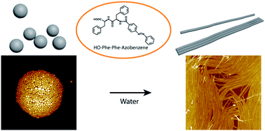
Self-assembled structures from aromatic dipeptides have attracted a lot of attention. It is highly desirable to produce dipeptide assemblies which undergo structural transitions in response to external stimuli. In this paper, solid nanospheres were successful produced from the self-assembly of chemically modified diphenylalanine in hexafluoroisopropanol (HFIP), a highly polar solvent. Interestingly, after treatment with water, the nanospheres were transformed into nanofibers. The intermediate transition state of nanospheres embedded along the nanofibers was captured by atomic force microscopy (AFM) imaging. In addition, AFM-based nanomechanical measurement revealed the increased stiffness after the transition, suggesting enhanced molecular packing due to favoured intermolecular interactions in water. This study presents a new method to fabricate novel dipeptide structures and provides new information for understanding the mechanism of dipeptide self-assembly driving by intermolecular interactions.
Among other methods, chemical modification and solvent induction have been proved to be powerful in tuning the structure and properties of dipeptide assemblies from FF. Vertically aligned nanoforests of FF nanotubes have been fabricated by solution evaporation22 and vapor deposition.23 Modification of Fmoc (9-fluorenylmethoxycarbonyl) on the N-terminus of FF resulted in fibrils which formed macroscopic hydrogels.24–26 Nanospheres are formed from diphenylglycine and cysteine–diphenylalanine,11 and peptide quantum dot (QD) structures have been fabricated by diluting tertbutoxycarbonyl-Phe-Phe-OH (Boc-FF) in 1:
1 water–ethanol solution.9 Nanotubes formed from D-Phe–D-Phe27 and H–Phe–Phe–NH2·HCl10 were able to rearrange to vesicles by diluting their solution with water.
In this paper, we report our attempt to use solvent exchange to achieve the transition of nanostructures from a chemically modified aromatic dipeptide. We designed a molecular building block by attaching an azobenzene group to the N-terminus of the FF peptide. The modified dipeptide has a structure of HO-Phe-Phe-Azobenzene (PPA). 1,1,1,3,3,3-Hexafluoro-2-propanol (hexafluoroisopropanol, HFIP), a commonly used peptide solvent, was used to dissolve PPA. Solid nanospheres were readily formed after solvent evaporation. Intriguingly, after treatment with water, the nanospheres were converted to nanofibers. The morphologies, chemical structures and nanomechanical properties of the nanospheres and nanofibers were investigated in order to reveal the molecular mechanism behind the solvent induced structural transition.
In an attempt to investigate the influence of solvent on the structure of PPA assemblies, water was introduced as a second solvent. A drop of water was added to the PPA nanospheres and incubated for 5 min before it was left to evaporate. The transition of from 0D nanospheres to 1D nanofibers occurred and the intermediates of the transition were captured by AFM imaging (Fig. 2a). The nanospheres were dissolved and a film composed of fibers covered the substrate. Fortunately, a partially dissolved nanosphere clusters were observed (Fig. 2b). Compared to untreated ones, the height of the nanospheres dropped significantly from more than 70 nm to less than 30 nm. In the high resolution images (Fig. 2d and e), the half-converted nanospheres were visible along the nanofibers.
After extending the incubation of water to 10 min, all PPA nanospheres were fully converted to mature PPA nanofibers. These nanofibers have a strong tendency to aggregate and align in parallel, forming nanofiber films on mica surface (Fig. 3a and b). The height histogram and line profile (Fig. 3c and d) show that the nanofibers fabricated from 0.5 mg mL−1 solution measure about 4 nm in height and 40 nm in diameter. Nanofibers fabricated from half concentration were 2 nm in height and 20 nm in diameter (Fig. 3e–g), indicating that their sizes are dependent on the concentration of the PPA solution. It is noteworthy that a layer of underlying thin film was observed in Fig. 3e, measuring 0.5 nm in height. Considering the small height of the film, it is probably composed of peptide monomers adsorbed on mica surface. It is also clearly observed that some of the nanofibers lie on top of the thin film, having no direct contact with the substrate (Fig. 3e). This indicates that the formation and alignment of the PPA nanofibers are independent of the substrate. In order to further exclude the possible substrate effect, the same fabrication process of PPA nanofibers was carried out on carbon film suspended on TEM grids, and TEM images were taken (Fig. S4†). It was observed that PPA formed bundles of solid nanofibers, confirming the formation of fiber was not induced by substrate. Interestingly, in the AFM image, several fibers were broken due to geometrical constraint, as indicated by the arrows in Fig. 3b. This observation implies that the PPA nanofibers are rigid fibers with a strong tendency to remain straight. This is confirmed in Fig. 3e inset, in which the Fourier transform analysis of the height signal clearly shows the directionality of the nanofibers. This rigid property also explains why the nanofibers tend to arrange in parallel to form fiber films. If the fibers had certain flexibility, they would tend to form interweaving networks.
To obtain an insight into the transition of PPA self-assembled nanomaterials, the mechanical properties of nanospheres and nanofibers were explored. Quantitative nanomechanical mapping28,29 was performed to detect the stiffness of nanospheres and nanofibers. In order to determine the optimal conditions to measure the stiffness, cantilevers with different spring constant were tested at different imaging forces (Fig. S5†). The 26.2 N m−1 cantilever with 4 nN moderate imaging force was used in the following measurements. The Young's moduli of spheres (Fig. 4c) and fibers (Fig. 4d) were fitted by Gaussian distribution, and the values were determined to be 2.77 ± 0.66 GPa and 6.42 ± 1.33 GPa, respectively. These values are lower than 19 GPa of the FF nanotubes,18 but comparable to those of the amyloid fibrils which varies from hundreds of MPa to several GPa,30–32 falling in the high region. The Young's modulus of PPA nanofibers is higher than that of the nanospheres, i.e., the nanofibers are more rigid than the nanospheres. PPA nanofibers were imaged with increasing peak forces to test their response under mechanical pressure (Fig. 4e–i). The morphologies of the nanofibers remained unchanged under forces up to 8 nN. However, at 16 nN force, ∼0.5 nm irreversible deformation was observed (Fig. 4j). Therefore, the PPA nanofibers can at least withstand forces up to 8 nN in vertical direction.
The chemical structures of the nanospheres and nanofibers were studied by Fourier transform infrared (FT-IR) spectroscopy, and it was proved that they have similar secondary structures of peptide in the assemblies. The IR spectra of the nanospheres and nanofibers are similar (Fig. S6†). In both spectra, two peaks at 1630 cm−1 and 1534 cm−1 were observed, which were identified as amide I and amide II absorption bands, respectively. The position of amide I band indicates the presence of β-sheets, and the absence of distinct absorption band at 1670–1695 cm−1 suggests the β-sheets are parallel.33,34 Therefore, in both nanospheres and nanofibers, intermolecular hydrogen-bonding exists in the form of parallel β-sheets.
By combining the morphological observation, nanomechanical measurement, and chemical characterization, we propose that the transition from nanospheres to nanofibers is attributed to solvent interactions. The diphenylalanine motif in PPA has a strong tendency to form hydrogen-bonding networks of β-sheets. In HFIP, as the solvent has strong hydrogen bonding capability, large area network of hydrogen bonds is not stable and is possibly disrupted to small segments under solvent interactions. Upon the evaporation of the solvent, zero-dimensional nanospheres were formed from the small segments. In water, the solvent has weaker hydrogen bonding capability compared to HFIP. As a result, the ability to form long-range 1D fibers by the stacking of β-sheets is restored. Furthermore, due to the amphiphilic structure of PPA, the peptide backbone and aromatic groups, which are hydrophobic parts, are embedded inside and protected from solvent interactions in water. As a result, long-range hydrogen bonding networks and aromatic stacking, which are favorable for the formation of 1D nanostructure, can be sustained in water. Similar mechanism has been proposed for the formation of FF peptide nanotubes in water.35
Molecular dynamics (MD) simulation of PPA dimers in the two solvents supported the above argument (Fig. S7, S8 and Table S1†). Compared to the parallel conformation in water, PPA dimers showed a more disordered conformation in HFIP, suggesting stronger disturbance from solvent interaction by HFIP. Nanomechanical measurements by AFM also supported the above argument. It is known that high packing density of molecules in molecular assemblies results in high stiffness.36 Due to the higher level of order and directionality in the nanofibers, it is reasonable that the Young's modulus of the nanofibers is increased compared to nanospheres. Besides, it is also noteworthy to highlight the role of azobenzene group in PPA. After using UV irradiation to switch the conformation of azobenzene, no regular structure was observed from HFIP solution (Fig. S9†). Therefore, extra π–π stacking interaction by azobenzene contributes significantly to the formation of novel dipeptide nanospheres and nanofibers thereafter. To sum up, the possible mechanism of PPA nanostructure transition is proposed (Fig. S10†).
From the discussion above, the chemical modification of FF is the key to this novel behavior. The addition of an azobenzene group to FF not only added extra π–π stacking, but also disturbed the overall network of β-sheets, making it more vulnerable to solvent interactions, resulting in novel nanostructures as well as the solvent responsive behaviour. The transition from nanospheres to nanofibers is intriguing, as it can be utilized to fabricate smart dipeptide materials, such as forming a surface coating of nanofibers immediately after contact with water, and some drug release application from nanospheres to nanofibers. Given the biocompatibility of dipeptides, this material will probably find more opportunities in novel biomedical applications.
Footnotes |
† Electronic supplementary information (ESI) available: Details on methods, synthesis, additional AFM and TEM images, FT-IR and DLS spectra, results of MD simulations. See DOI: 10.1039/c3ra46718j |
‡ These authors contributed equally. |
This journal is © The Royal Society of Chemistry 2014 |