Nanofibers to nanocuboids of polyaniline by lead nitrate: hierarchical self-assembly with lead ions

Plawan Kumar Jha, Barun Dhara and Nirmalya Ballav*
Department of Chemistry, Indian Institute of Science Education and Research (IISER), Dr Homi Bhabha Road, Pune – 411 008, India. E-mail: nballav@iiserpune.ac.in

Received 14th November 2013 , Accepted 28th January 2014

First published on 28th January 2014


Abstract

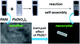
By mixing the solutions of polyaniline base and lead nitrate together an oxidized form of polyaniline was gradually precipitated-out together with toxic lead ions under ambient conditions. A remarkable morphological transformation of polyaniline nanofibers to nanocuboids with lead ions suggested a spontaneous and hierarchical self-assembly. We have attributed such in situ formation of supramolecular nanomaterial to the specific influence of relativistic inert-pair effects of the lead ions. Our exploration of one of the relativistic quantum effects in generating supramolecular nanomaterials are thought-provoking and pave the way toward designing a new type of self-assembled materials through which toxic heavy metal ions are automatically removed from aqueous solution.


An elegant example of molecular self-assembly1,2 is the crystallization process where mass transfers of solute particles from usually liquid (or gas) phase into solid material take place. Like natural zeolites, artificial crystallization could also result in the formation of porous crystalline materials, so called coordination polymers or metal-organic frameworks.3,4 The spontaneous organization of various components, specifically, metal ions and organic ligands, into nanocrystals is thus emerging in the view of understanding distinctive self-assembly processes at the nanometer length scale and at the same time triggering the bottom-up approach towards fabrication of functional nanomaterials.1 In case of the heavier metal atoms/ions, relativistic variation of the mass of the electron becomes crucial, thereby causing a significant change in the spatial distribution of the orbitals (contraction) which leads to the relativistic quantum effects.5 Some of the typical periodic discrepancies like liquidity in metal mercury, color of gold and cesium, aurophilicity, and inert electron pair have been successfully explained with the help of the relativistic effect.6,7

While in a technical report IUPAC recommended the term heavy metal as meaningless,8 we are still very much accustomed to it non-technically in our social life (for example, heavy metal ions poisoning). In isoelectronic Tl(I), Pb(II), and Bi(III) ions there is an electron pair in the 6s orbital which resists further oxidation due to a relativistic contraction of the orbital and behaving as if those electrons are chemically inert – so called inert-pair effect.9–12 We wanted to explore a bottom-up approach where the relativistic inert-pair effect of Pb(II) ions could in principle play an important role, possibly through self-assembled complex formation with multi-dented polymeric ligand, and if so, it could potentially shed new light on an important environmental concern by removing Pb(II) ions from aqueous solution. Aqueous Pb(II) ions are known to be toxic and poisonous, and about 20% of total lead-exposure are coming only from drinking water in USA.13

Herein we have simply mixed the solutions of commercially available nitrogen containing π-conjugated polyaniline (PANI) base (which could play the role of a poly-dented nitrogen-donor ligand to various metal ions) and lead nitrate together at ambient conditions whereby an oxidized form of polyaniline was gradually precipitated-out together with Pb(II) ions as metal-polymer nanocrystals. PANI is one of the most-studied, inexpensive and intrinsically conducting polymers which exhibits three characteristic π-conjugated redox forms, leucoemeraldine base (LB, all amines), emeraldine base (EB, amines–imines), and pernigraniline base (PB, all imines).14,15 The key advantages of PANI over other conducting polymers are the proton-induced conversion from insulator to metallic phase without changing the number of electrons and its solubility in organic polar aprotic solvents.

In case of the fabrication of PANI nanostructures, the self-assembled monomers are so far encoded in a specific spatial arrangement (or in a template) in the initial step followed by the polymerization (decoding step) reaction resulting in the formation of the desired structures like nanotube, nanorod, nanosphere, and nanoplate.16–22 To our knowledge, post-assembly of PANI units into well-defined nanostructures is not explored and at the same time supramolecular chemistry of Pb(II) ions influenced by the relativistic inert-pair effect remained limited to academic interest only.23,24

Field-emission scanning electron microscopy (FESEM) images of EB and the precipitate (here onwards referred as EB–Pb) are presented in Fig. 1a. The morphology of EB was mostly detected as nanofibers (see ESI, Fig. S1) however the EB–Pb predominantly exhibited very uniform, discrete, and well-defined nanocuboid structures (Fig. 1b and see also Fig. S2). Such a transformation of morphology, starting from nanofibrous material and finally to nanocuboids, is rather surprising and resembles previous observations on the shape transformation of nanowires to nanocubes in the case of metal–organic coordination polymer particles.25 Notably, in case of EB–Pb we could not detect a single nanofiber. In order to investigate the uniqueness of Pb(II) ions generating the nanocuboid structures, we have performed similar experiments of EB with other water soluble divalent nitrates containing metal ions representing s-, p- and d-block elements e.g., Mg(II), Ca(II), Cu(II) and Zn(II). FESEM images (Fig. 1c and d) of the so called the EB–Mg and EB–Cu systems are entirely different from that of EB–Pb system – in both cases only globular polymeric moieties revealing a non-uniform morphological pattern could be detected.


image file: c3ra46691d-f1.tif
Fig. 1 (a and b) FESEM images of EB showing non-uniform nanofibers with average diameter below 50 nm and from EDX analysis elemental ratio between nitrogen and carbon was estimated to be ∼1[thin space (1/6-em)]:[thin space (1/6-em)]6 which is characteristic of PANI. (c–f) FESEM images of the EB–Pb system, additional zoom-in images clearly representing discrete nanocuboids and EDX analysis showed elemental ratio of ∼1[thin space (1/6-em)]:[thin space (1/6-em)]6 for N and C in addition to the characteristic signature of Pb. (g) FESEM images of the precipitate obtained after reaction of EB with magnesium nitrate. Notably no EDX signature of Mg was detected. (h) FESEM images of the precipitate obtained after reaction of EB with copper nitrate and from EDX little amount of Cu was detected.

FESEM data is supported by the powder X-ray diffraction (PXRD) patterns recorded at room temperature (Fig. 2). In line with previously reported data, a broad PXRD peak of EB in the 2θ range of 15–40 was observed.26 Such PXRD pattern suggests the truly non-crystalline nature of EB, as one would have expected from the fibrous nature of the material. On the contrary, PXRD pattern of the EB–Pb consistently exhibited (see also Fig. S3) very sharps peaks starting from 2θ values of ∼20 and beyond, thereby revealing a significant structural order in the material. Specifically, the highest intensity peak at 2θ ∼ 27 suggests that in the EB–Pb system d001 planes are placed ∼3.3 Å apart and are possibly interconnected by the Pb(II) ions resulting into the self-assembled supramolecular nanocuboids like various common Pb(II) salts. Notably, no PXRD peak(s) characteristic of Pb(NO3)2 is found to be visible in the EB–Pb system (Fig. S3).


image file: c3ra46691d-f2.tif
Fig. 2 Distinctive PXRD patterns of EB and the precipitates obtained after reaction with various divalent metal nitrates. Sharp and crystalline peaks were observed only for the EB + Pb(NO3)2 system.

In case of other nitrates, although gradual precipitation took place, all the precipitated materials were found to be non-crystalline in nature as evidenced from the respective PXRD pattern (i.e. practically remained almost unchanged as compared to EB). Thus, either those nitrates did not react with EB or even if they reacted the respective reaction did not lead to the formation of supramolecular nanocrystals likewise Pb(II) ions. Our blank experiment suggested that without any metal ion there was no precipitate from the EB solution (Fig. S4). Overall, a distinctive interaction scenario of Pb(II) ions with EB is clearly reflected in the PXRD patterns.

To elucidate further the in situ formation of such nanocuboid structures, we have performed high-resolution transmission electron microscopy (HRTEM) studies (Fig. 3a). In general, well-defined structure of the EB–Pb system was visible with sub-nanometer resolution (see also Fig. S5). A spacing of ∼3.0 Å noted in <001> plane (c.f. Fig. 3b) , which is in excellent agreement with the PXRD peak at 2θ ∼ 29.9 (d-spacing of ∼2.9 Å), could be due to inter-chain distance among PANI moieties. A selected area electron diffraction (SAED) pattern (Fig. 3c) fully supported the HRTEM observation by showing the bright spots. In line with the previously reported monoclinic crystal lattice of Pb(II) ions with polymeric ethylene glycol ligand23 we would be in favour of assigning crystal lattice of the EB–Pb system as monoclinic. Such SAED pattern was consistently observed for individual EB–Pb nanocrystals (Fig. S6). We would like to attribute the occurrence of such unique nanocuboids to (i) the cleavage of nanofibers into different lengths during oxidation of EB; (ii) subsequent self-organization of nanofibers by non-covalent (typically H-bonding) interaction into 1D-nanosheets; and (iii) finally self-sorting and the assembly of 1D-nanosheets into well-defined 3D-nanocuboids through specific coordination to Pb(II) ions influenced by the relativistic inert pair effect (see the scheme in Fig. 4a).27,28


image file: c3ra46691d-f3.tif
Fig. 3 (a) HRTEM image taken on EB–Pb system (inset: nanocrystal). (b) Zoomed-in HRTEM image clearly showing the spacing between white lines is ∼ 3.0 Å in the <001> plane. (c) SAED pattern on EB–Pb system on which monoclinic crystal lattice is marked with small black dotted circles and white lines (inset: nanocrystal).

image file: c3ra46691d-f4.tif
Fig. 4 (a) Schematic representation of the formation of self-assembled PANI nanocuboids. (b) FTIR spectra of EB (blue) and the EB–Pb system (red). (c) N 1s XPS of EB. (d) N 1s XPS of EB–Pb. (e) Pb 4f XPS of EB–Pb and the binding energy clearly show presence of Pb(II) ions in the system.

The relative intensity of the peaks at ∼1495 cm−1 and ∼1585/∼1160 cm−1 (characteristic of benzenoid and quinonoid moieties in polyaniline, respectively) in the Fourier transformed infra-red (FTIR) spectra (Fig. 4b) suggests an oxidation reaction in the system converting EB to PB.19,29 Raman spectroscopic data also indicated the oxidation of EB moieties to PB and supported the FTIR observation. Specifically, the intensity ratio of the Raman signals at ∼1475 cm−1 (νC[double bond, length as m-dash]N of the quinoid units) and ∼1590 cm−1 (νC[double bond, length as m-dash]C of the quinoid rings)30 was much higher for the EB–Pb system than EB (Fig. S7). 1H nuclear magnetic resonance (NMR) data complemented the FTIR/Raman data by revealing a significant reduction in the intensity of the signal at δ = 6.0–7.5 ppm and the absent of signal at δ = 7.5–9.0 ppm which are characteristic proton signatures in the backbone of polyaniline base (Fig. S8).31

The chemical identity of the EB–Pb system is further probed by X-ray photoelectron spectroscopy (XPS). One would expect two chemically non-equivalent N atoms (amine and imine) of equal amount in EB and only one type of N (imine) atoms in PB. Indeed, N 1s XPS shows two different signals, one at ∼399.0 eV and the other at ∼398.0 eV (Fig. 4c) which are characteristic binding energies (BEs) of respective amine and imine N atoms in EB.32 In case of the EB–Pb system, we observed only one major N1s photoemission signal at ∼399.0 eV which we would like to assign to imine N atoms (Fig. 4d). While there was no detectable signature of Pb 4f photoemission in EB (as expected), a clear Pb 4f photoemission signature showing the 4f7/2 peak at ∼139.0 eV and the 4f5/2 peak at ∼145.0 eV, due to spin–orbit coupling (Fig. 4e), was detected which confirmed the incorporation of Pb with +2 oxidation state (presence of inert 6s2 pair of electrons) in the EB–Pb system.33 The relative shift in the N 1s photoemission signal towards higher binding energy indicates coordination of imine N atoms to Pb(II) ions. The negligible N 1s photoemission signals at ∼395.6 eV and ∼402.2 eV (for EB) and at ∼396.0 eV and ∼402.0 eV (for EB–Pb) could be related to X-ray photoelectron diffraction and/or the satellite features in π-conjugated systems.34,35 The additional small peak at ∼406.0 eV observed only for the EB–Pb contributing to ∼3% weight in the spectrum is due to presence of NO3 anions in the supramolecular coordination sphere of Pb(II) ion. Thus overall, XPS analysis together with the FTIR/Raman and NMR data confirmed oxidation of EB to PB and a predominant coordination of PB (as polymeric nitrogen-donor ligand) to Pb(II) ions forming such metal-polymer nanocrystals.36

The primary question remained is what was responsible for the in situ oxidation of EB to PB? Lead nitrate is known to be an oxidizing agent, however, depending on the reaction conditions, neutral or acidic, either Pb(II) (neutral) or NO3 ions (acidic) plays the role, respectively.9–11 First of all, we used lead nitrate in much excess compared to EB (see Experimental section) and secondly, we observed separately that upon addition of EB, the change in the pH value of the lead nitrate solution was negligible (pH ∼ 4.4 to pH ∼ 5.0) thereby meaning that the constituents' mixture was rather in acidic condition. Thus NO3 anions acted as the oxidizing species. To investigate the loading of lead in the nanocrystals, we have performed gravimetric analysis and from the difference in the weight loss (initial amount and the final amount after evaporation of the filtrate) ∼55% (weight) loading was estimated. Our combined XPS and energy-dispersive X-ray (EDX) spectroscopy analyses (see Table S1 and S2) suggested elemental ratio of ∼10[thin space (1/6-em)]:[thin space (1/6-em)]1 for N[thin space (1/6-em)]:[thin space (1/6-em)]Pb which directs towards supramolecular arrangements of PANI around Pb(II) ion.23 Furthermore, in case of the so called EB–Mg system we could not detect any EDX signal of Mg (Table S3) and even if little EDX signature of Cu could be detected in the EB–Cu system (Table S4) the morphologies were entirely different in comparison to EB–Pb system (cf. Fig. 1).

Thermo-gravimetric analysis (TGA) revealed that almost complete weight loss of EB took place at ∼700 °C whereas the EB–Pb system remained very much stable and only about ∼30% weight loss could be detected in that temperature range (Fig. S9). Temperature dependent current–voltage (IV) plots indicated non-linear behaviour and a monotonically increasing dc conductivity value of the EB–Pb system with increasing temperature up to 150 °C (Fig. S10) was noted. Furthermore, by showing the same IV characteristic upon cooling down the sample to the same temperature, say for example ∼60 °C suggested that the heating did not modify or damage the nanocrystals thereby clearly complementing the high-thermal stability.

Our argument in establishing the formation such PANI-based nanocuboids influenced by the relativistic inert-pair effect of Pb(II) ions is rather simple. We have used five different water soluble divalent nitrates overall representing typical non-transition s-block (Mg(II) and Ca(II)), transition d-block (Cu(II) and Zn(II)) and p-block (Pb(II)) metal ions. Out of those, it is only the Pb(II) ion which is behaving in a significantly different way, although in all the cases, NO3 ions played the similar role in oxidizing EB to PB as clearly visible in the FTIR spectra (Fig. S11). Even taking the soft-hard acid-base (SHAB) theory into account one can realize that Mg(II) and Ca(II) are hard-acids; Cu(II), Zn(II) and Pb(II) are boarder-line acids (neither soft nor hard) while PANI (which resembles aromatic primary or secondary amines) behaves like a boarder-line base. Respective electronegativity values of Mg, Ca, Cu, Zn, Pb and N are 1.31, 1.00, 1.90, 1.65, 1.80 and 3.04 (using Pauling scale)10 and the difference alone is clearly not enough to explain the noteworthy bonding scenario. Furthermore, there exist innumerable complexes of nitrogen-donor ligands with Cu(II) and Zn(II) ions which are stable at ambient conditions, whereas similar stable complexes of Pb(II) ion are really very rare. The paradoxical observation goes beyond the Pearson-Pauling paradox (soft–hard acid–base versus electronegativity difference)37 and directly pointing towards the decisive role of the relativistic inert-pair electron in imposing specific stereo-electronic influence on the formation of such supramolecular nanocrystals.

One of the important aspects of our self-assembly-induced removal of aqueous Pb(II) ions by PANI in the form of nanocuboids is the regeneration of starting material EB. The EB–Pb system was simply immersed into dilute nitric acid (HNO3) for overnight and filtered; and upon addition of aqueous solution of sodium sulfate or carbonate, white precipitation occurred confirming the formation of lead sulfate or carbonate, respectively. Washing with HNO3 was continued until no white precipitate appeared and the dark colored mass, namely EB-salt, was found to dissolve in solvents like DMSO, NMP and DMF. Furthermore, EB-salt showed high dc conductivity value (in the range of ∼1.5 S cm−1) as expected. In a separate experiment, we added dilute HNO3 to a EB/(DMSO + MeOH) solution which resulted in the formation of a EB + HNO3 salt, as expected. Notably, the FTIR and UV-vis spectra (cf. Fig. 5a and b) of EB-salt and EB + HNO3, taken in DMSO, are almost identical and showed an additional peak at ∼445 nm (polaron to π* inter band transition) which is absent in EB. The morphology of EB-salt (cf. Fig. 5c) was found to be globular and is different from that of EB nanofibers. Such non-crystalline morphological features are complemented by the PXRD patterns (cf. Fig. 5d). To regenerate EB, one just needs to treat the EB-salt with ammonium hydroxide solution (deprotonation) and we expect its re-usability for further extraction of Pb(II) ions in a cyclic manner. Another aspect in the present study is the adopted method i.e. precipitation which is known to be a widely used industrial technique.38


image file: c3ra46691d-f5.tif
Fig. 5 (a) FTIR spectra of EB-salt (black) obtained after extraction of the nanocuboids with HNO3 (namely EB-salt) and the precipitate resulted upon addition of HNO3 to EB solution, namely EB + HNO3 (dark grey). (b) UV-vis spectra of EB (grey), EB-salt (black) and EB + HNO3 (dark grey). (c) FESEM image of EB-salt (inset: powder sample). (d) PXRD patterns of EB-salt (black) and EB + HNO3 (dark grey).

In summary, we have presented in situ generation of self-assembled nanocuboids of polyaniline with lead ions. The elemental ratio between nitrogen and lead suggested supramolecular coordination of polyaniline to lead ions forming the monoclinic lattice of the nanocuboids. Our exploration of the relativistic inert-pair effect of lead ions in generating new type of supramolecular nanomaterials in one hand is stimulating and on the other hand it can provide a very simple and better mean of addressing an important environmental concern. We expect that our self-assembly approach can also be employed to remove other heavy metal ions (e.g., mercury) from aqueous solutions and a spatiotemporal evolution of such polymer-based nanocrystals would be fundamentally very much useful in engineering nanoscale polymeric crystal growth for various other applications.

Acknowledgements

N.B. gratefully acknowledges the financial support from IISER Pune (India) through start-up research grant, DST Nano Mission (India, project no. SR/NM/NS-42), DAE-BRNS (India, project no. 2011/20/37C/17/BRNS) and personally thanks Prof. K. N. Ganesh (IISER Pune) for the encouragement. P.K.J. and B.D. thank CSIR (India) for providing Junior Research Fellowships. The authors thank SPECS GmbH, Berlin for XPS analysis.

Notes and references

  1. J.-M. Lehn, Science, 2002, 295, 2400–2403 CAS.
  2. J.-M. Lehn, Angew. Chem., Int. Ed., 1990, 29, 1304–1319 Search PubMed.
  3. E. R. Parnham and R. E. Morris, Acc. Chem. Res., 2007, 40, 1005–1013 CAS.
  4. D. J. Tranchemontagne, J. L. Mendoza-Cortes, M. O'Keeffe and O. M. Yaghi, Chem. Soc. Rev., 2009, 38, 1257–1283 CAS.
  5. U. Kaldor and S. Wilson, Theoretical Chemistry and Physics of Heavy and Superheavy Elements, Springer, 2003 Search PubMed.
  6. P. Pyykko and J. P. Desclaux, Acc. Chem. Res., 1979, 12, 276–281 CAS.
  7. L. J. Norrby, J. Chem. Educ., 1991, 68, 110 CAS.
  8. J. H. Duffus, Pure Appl. Chem., 2002, 74, 793–807 CrossRef CAS.
  9. A. Earnshaw and N. Greenwood, Chemistry of the Elements, Elsevier Science, 1997 Search PubMed.
  10. J. E. Huheey, E. A. Keiter, R. L. Keiter and O. K. Medhi, Inorganic Chemistry: Principles of Structure and Reactivity, Pearson Education, 2006 Search PubMed.
  11. F. A. Cotton, G. Wilkinson, C. A. Murillo and M. Bochmann, Advanced Inorganic Chemistry, Wiley India Pvt. Limited, 2007 Search PubMed.
  12. G. L. Miessler, Inorganic Chemistry, Pearson Education, 2008 Search PubMed.
  13. R. P. Maas, S. C. Patch, D. M. Morgan and T. J. Pandolfo, Publ. Health Rep., 2005, 120, 316–321 Search PubMed.
  14. A. J. Heeger, Rev. Mod. Phys., 2001, 73, 681–700 CAS.
  15. A. G. MacDiarmid, Angew. Chem., Int. Ed., 2001, 40, 2581–2590 CAS.
  16. G. Ćirić-Marjanović, in Nanostructured Conductive Polymers, John Wiley & Sons, Ltd., 2010, pp. 19–98 Search PubMed.
  17. L. J. Pan, L. Pu, Y. Shi, S. Y. Song, Z. Xu, R. Zhang and Y. D. Zheng, Adv. Mater., 2007, 19, 461–464 CAS.
  18. M. Wan, Adv. Mater., 2008, 20, 2926–2932 CAS.
  19. M. Trchová and J. Stejskal, Pure Appl. Chem., 2011, 83, 1803–1817 Search PubMed.
  20. H. W. Park, T. Kim, J. Huh, M. Kang, J. E. Lee and H. Yoon, ACS Nano, 2012, 6, 7624–7633 CAS.
  21. L. Zhang and M. Wan, Adv. Funct. Mater., 2003, 13, 815–820 CAS.
  22. J. Fei, Y. Cui, X. Yan, Y. Yang, K. Wang and J. Li, ACS Nano, 2009, 3, 3714–3718 CAS.
  23. R. D. Rogers, A. H. Bond and D. M. Roden, Inorg. Chem., 1996, 35, 6964–6973 CAS.
  24. S.-Y. Wan, J. Fan, T. Okamura, H.-F. Zhu, X.-M. Ouyang, W.-Y. Sun and N. Ueyama, Chem. Commun., 2002, 2520–2521 CAS.
  25. S. Jung and M. Oh, Angew. Chem., Int. Ed., 2008, 47, 2049–2051 CAS.
  26. J. P. Pouget, M. E. Jozefowicz, A. J. Epstein, X. Tang and A. G. MacDiarmid, Macromolecules, 1991, 24, 779–789 CAS.
  27. Y. Wang, J. Liu, H. D. Tran, M. Mecklenburg, X. N. Guan, A. Z. Stieg, B. C. Regan, D. C. Martin and R. B. Kaner, J. Am. Chem. Soc., 2012, 134, 9251–9262 CAS.
  28. P. Pyykko, Chem. Rev., 1988, 88, 563–594 CAS.
  29. Y. Sun, A. G. MacDiarmid and A. J. Epstein, J. Chem. Soc., Chem. Commun., 1990, 529–531 CAS.
  30. S. H. Domingues, R. V. Salvatierra, M. M. Oliveira and A. J. G. Zarbin, Chem. Commun., 2011, 47, 2592–2594 CAS.
  31. S. Mu and Y. Yang, J. Phys. Chem. B, 2008, 112, 11558–11563 CrossRef CAS PubMed.
  32. K. L. Tan, B. T. G. Tan, E. T. Kang and K. G. Neoh, Phys. Rev. B: Condens. Matter Mater. Phys., 1989, 39, 8070–8073 CAS.
  33. J. F. Moulder and J. Chastain, Handbook of X-ray photoelectron spectroscopy: a reference book of standard spectra for identification and interpretation of XPS data, Physical Electronics Division, Perkin-Elmer Corporation, 1995 Search PubMed.
  34. S. Hüfner, Photoelectron Spectroscopy: Principles and Applications, Springer, 2003 Search PubMed.
  35. C. Wackerlin, C. Iacovita, D. Chylarecka, P. Fesser, T. A. Jung and N. Ballav, Chem. Commun., 2011, 47, 9146–9148 RSC.
  36. M. Tan, Y. N. Sum, J. Y. Ying and Y. Zhang, Energy Environ. Sci., 2013, 6, 3254–3259 CAS.
  37. G. Wulfsberg, Inorganic Chemistry, University Science Books, 2000 Search PubMed.
  38. Y. Seida and Y. Nakano, Water Res., 2000, 34, 1487–1494 CAS.

Footnote

Electronic supplementary information (ESI) available: Experimental details and additional data. See DOI: 10.1039/c3ra46691d

This journal is © The Royal Society of Chemistry 2014
Click here to see how this site uses Cookies. View our privacy policy here.