Anti-inflammatory terpenoids from Boswellia ovalifoliolata†‡
Renu Chiba,
Manjeet Kumara,
Masood Rizvib,
Simmi Sharmaa,
Anjali Pandeya,
Sarang Bania,
Samar S. Andotraa,
Subhash C. Tanejaa and
Bhahwal A. Shah*a
aNatural Product Microbes, CSIR-Indian Institute of Integrative Medicine, Canal Road, Jammu-Tawi, India
bDepartment of Chemistry, University of Kashmir, Hazaratbal, J&K, India. E-mail: bashah@iiim.ac.in; Fax: +91-191-25693331; Tel: +91-191-25692217
First published on 2nd December 2013
Abstract
Ten compounds, including three new compounds (1–3, one sesquiterpenoid and two triterpenoids), were isolated and characterized from the ethanolic extract of oleo-gum-resin from Boswellia ovalifoliolata. It is noteworthy that, for the first time, compound 4 has been reported from any of Boswellia species, and 5–9 from B. ovalifoliolata. These isolated compounds were screened for TNF-α inhibition and the results showed that 2 and 5 significantly inhibited the expression of TNF-α at 1 μM, whereas inhibition by rolipram (a standard TNF-α inhibitor) occurred at 100 μM. Furthermore, 2 and 5 were found to considerably reduce levels of interleukin (IL-6 and IL-8) and nitric oxide (NO) production, which suggests they have anti-inflammatory potential and possible therapeutic uses.
Introduction
Extracts of Boswellia sp. are known for their effectiveness in the treatment of rheumatoid arthritis and chronic inflammation, mainly due to their boswellic acid content.1,2 The biological activities of boswellic acids, against ulcerative colitis,3 chronic colitis,4 asthma,5 and their anti-complimentary activities, are also well documented.6 Furthermore, the cytotoxicity and anti-cancer studies carried out on boswellic acids in the last few years have provided an additional dimension to their bioactivity profile.7,8 In view of the importance of the compounds from Boswellia sp., we undertook detailed investigations into the gum resin of Boswellia ovalifoliolata, which has been reported to contain molecules such as α-amyrin (10),9 as well as macrocyclic diaryl ether i.e., ovalifoliolatins A and B,10 acetogenin C11and sitost-4-en-3-one.12 Therefore, as a continuation of our efforts towards the study of the chemical diversity13 and medicinal potential of Boswellia sp. and their constituents,14 herein we report the isolation and characterization of ten compounds (1–10), including three new compounds (1–3), from the ethanolic extract of the oleo-gum-resin of B. ovalifoliolata (Fig. 1). Notably, for the first time, 1β,3β,11α-trihydroxy-urs-12-ene (4)15 has been reported from a Boswellia sp., whereas, 3α-hydroxy-tirucall-8,24-dien-21-oic acid (5), 3α-acetoxy-tirucall-8,24-dien-21-oic acid (6), serratol (7),16 neoilexonol (8),17 3-hydroxy-urs-9,11-dien-24-oic acid (9)18 from B. ovalifoliolata. In an attempt to evaluate their anti-inflammatory potential, the isolated pure compounds were screened for TNF-α inhibitory activity. Compounds 2 and 5 were found to significantly inhibit TNF-α expression and also considerably reduce levels of interleukin, IL-8 and IL-6, and nitric oxide (NO) production, warranting further investigation into their development as useful moieties in therapeutic applications.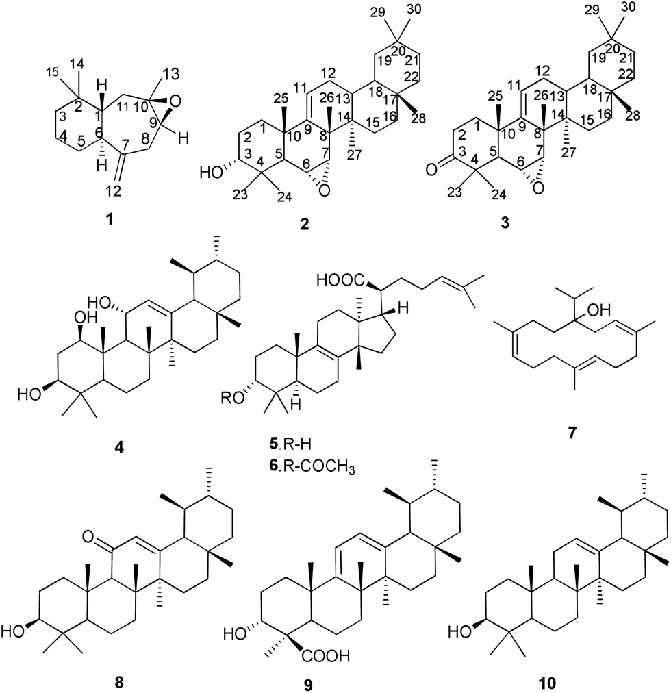 | ||
| Fig. 1 Structures of isolated compounds 1–10. | ||
Results and discussion
Purification of the ethanolic extract of oleo-gum resin of B. ovalifoliolata, by repeated column chromatography with hexane–ethyl acetate, resulted in the isolation of compounds 1–10. Their structures were assigned by 1H, 13C NMR and two-dimensional spectroscopy (COSY, HSQC, HMBC and NOESY).Compound 1 was isolated as an aromatic white semisolid. Its molecular formula, C15H24O, was established by high-resolution positive-ion-mode electrospray ionization mass spectrometry (HRESIMS) at m/z 221.3510 [M + H]+ (calcd, 221.3578) with four double-bond equivalents (DBEs). The IR spectrum displayed absorbances at 1121.4 and 1402.4 cm−1, indicating the presence of epoxy and alkene respectively. 13C NMR displayed 15 carbon resonances classified as 3 methyls, 6 methylenes, 3 methines (1 epoxy) and 3 quaternary carbons (1 olefinic, 1 epoxy) (Table 1). In accordance with these functionalities the compound is bicyclic which corresponds to 2 DBEs leading to a total of 4 DBEs including 2 DBEs for epoxy and alkene double bond groups. 1H and 13C NMR data analysis suggests that compound 1 is sesquiterpene. 1H NMR spectra indicate the presence of a tertiary methyl group at δH 1.13 positioned at C13 from HMBC correlation with C9 and C11. The peak at δH 4.79 reveals the presence of an exocyclic double bond which shows correlation with C6 and C8; further confirmed by HSQC. Furthermore, two singlets at δH 0.90 and 0.93, located at C14 and C15, confirm the presence of two gem methyl groups showing a correlation with each other in HMBC spectroscopy. Furthermore, the linkages from 3 → 6 → 11 were also confirmed by 1H–1H COSY, showing correlations between H3 → H6 → H11. 1H–1H COSY also revealed the correlation of H2-8 (δH 2.15–2.20) with H9 (δH 2.85). The downfield signal of H9 (δH 2.85) indicates its attachment to the epoxy group which was positioned by HMBC correlations with C8 and C13. The large coupling constant (J = 10.0 Hz) between H1 and H6 indicates that the rings in 1 are transfused.19 The absence of any correlation between the two centres in the NOESY experiment further supports this. Additionally, the chemical shift of H3-13 (δH 1.13) indicates that its stereochemistry is alpha, suggesting the configuration of epoxide to be β.20 Thus, the structure of compound 1 was elucidated as (1R,6S,9S,10R)-2,2,10-trimethyl-7-methylenedecahydro-10H-benzo[4,5]cyclo hepta[9,10]oxirene.
| Compound 1 | |||
|---|---|---|---|
| Position | 13C NMR | 1H NMR (J in Hz) | HMBC |
| 1 | 51.5, CH | 1.69, t, (10.0) | 3, 5, 14 |
| 2 | 34.7, C | — | 4, 14, 15 |
| 3 | 27.9, CH2 | 1.34, 1.55 m | 1, 4, 14 |
| 4 | 30.5, CH2 | 1.99–2.07; 2.24–2.29, m | 3, 6 |
| 5 | 40.5, CH2 | 1.52–1.60, m | 1, 3 |
| 6 | 49.4, CH | 2.53, dd, (8.8) | 4, 8, 12 |
| 7 | 152.5, C | — | 1, 12 |
| 8 | 30.9, CH2 | 2.15–2.20, m | 6, 10, 12 |
| 9 | 64.4, CH | 2.85, dd, (4.0, 6.4) | 8, 13 |
| 10 | 60.5, C | — | 1, 8, 13 |
| 11 | 39.8, CH2 | 1.99–2.07, m | 6, 13 |
| 12 | 113.5, CH2 | 4.79, d, (0.8); 4.90, d, (1.2) | 6, 8 |
| 13 | 30.6, CH3 | 1.13, s | 9, 11 |
| 14 | 22.3, CH3 | 0.90, s | 1, 3, 15 |
| 15 | 17.7, CH3 | 0.93, s | 1, 3, 14 |
Compound 2 was isolated as a white amorphous powder. Its molecular formula, C30H48O2, was established by HRESIMS at m/z 441.3657 [M + H]+ (calcd, 441.3727) with seven double-bond equivalents (DBEs). The IR spectrum displayed absorbances at 1220.9, 1402.4 and 3143.2 cm−1 indicating the presence of epoxy, alkene and hydroxyl groups respectively. 13C NMR displayed 30 carbon resonances classified as 8 methyls, 8 methylenes, 7 methines (1 hydroxylated, 2 epoxy and 1 olefinic) and 7 quaternary carbons (1 olefinic) (Table 2). In accordance to these functionalities the compound should be pentacyclic which corresponds to 5 DBEs leading to a total of 7 DBEs including 2 DBEs for the epoxy and alkene double bond groups. The 1H and 13C NMR data analysis suggests that compound 2 was an oleanane-type terpenoid.21 The 1H NMR spectrum (Table 2) indicates the presence of 8 tertiary methyl groups (δH 0.83, 0.86, 0.88, 0.96, 0.97, 1.00, 1.08, 1.09) each a singlet of 3 protons, an olefinic proton (δH 5.54) and an oxymethine proton at (δH 3.45). Extensive comparison of the 2D spectra indicates the presence of one hydroxyl group having δC 76.1 in 13C NMR with δH 3.45 (H-3α) fixed at C3 by HMBC correlation with H2-1 (δH 0.90), H2-2 (δH 1.60, 2.04) and H3-23 (δH 1.00) respectively. 1H and 13C NMR spectra show the presence of a double bond between C9 (δC 157.3) and C11 (δC 118.8 and δH 5.54) which are positioned on the basis of HMBC correlation with H7 (δH 2.81), H12 (δH 1.73, 2.01), H3-25 (δH 1.09) and H3-26 (δH 0.88). The two typical downfield chemical shifts at C6 and H6 (δC 52.0, δH 3.12) and, C7 and H7 (δC 58.2, δH 2.81) show the presence of an epoxy group further confirmed by the literature of already known compounds of this class.22 The position of the epoxy group was established to be C6 and C7 by HMBC correlations with C4, C5, C7 and C5, C9, C14, respectively. 1H–1H COSY revealed the direct linkage between H11→H13; between H5→H7. The position of the two tertiary methyls H3-29 (δ 29.9) and H3-30 (δ 28.1) at C20 was confirmed by HMBC correlation with H19 (δH 1.36 and 2.05) and H21 (δH 1.58) respectively. The small coupling constant (J6,7 = 4.6 Hz) indicates that H6 and H7 are cis configured,19 having R-configuration. Thus, the structure of 2 was assigned as 6R,7R-epoxy-1-oleanen-3-ol.
| Compound 2 | Compound 3 | |||||
|---|---|---|---|---|---|---|
| Position | 13C NMR | 1H NMR (J in Hz) | HMBC | 13C NMR | 1H NMR (J in Hz) | HMBC |
| 1 | 33.2, CH2 | 0.90, 1.35, m | 2, 5, 25 | 39.8, CH2 | 1.34, 2.15, m | 5, 25 |
| 2 | 25.0, CH2 | 1.60, 2.04, m | 1, 4, 10 | 33.9, CH2 | 2.4, 2.67, m | 1, 4, 10 |
| 3 | 76.1, CH | 3.45, s | 1, 2, 23 | 216.8, C | — | 1, 2, 23 |
| 4 | 36.7, C | — | 2, 23, 24 | 47.3, C | — | 5, 23 |
| 5 | 53.3, CH | 1.12,s | 6, 24, 25 | 52.9, CH | 1.31, m | 1, 6, 24, 25 |
| 6 | 52.0, CH | 3.12, t, (5.1) | 4, 5, 7 | 51.7, CH | 3.14, t, (5.1) | 7, 8, 10 |
| 7 | 58.2, CH | 2.81, d, (4.6) | 5, 9, 14 | 58.2, CH | 2.82, d, (4.4) | 5, 9 |
| 8 | 39.1, C | — | 11, 13 | 38.9, C | — | 11, 13 |
| 9 | 157.3, C | — | 7, 12, 25, 26 | 156.6, C | — | 7, 12, 25, 26 |
| 10 | 35.4, C | — | 11, 25 | 35,3, C | — | 5, 11, 25 |
| 11 | 118.8, CH | 5.54, dd, (3.3, 8.1) | 12 | 119.2, CH | 5.61, dd, (3.2, 8.1) | 12 |
| 12 | 38.2, CH2 | 1.73, 2.01,m | 9, 11, 13 | 38.2, CH2 | 1.72, 2.13, m | 11, 13 |
| 13 | 48.3, CH | 1.28–1.29, m | 12, 18 | 54.7, CH | 1.05, d, (4.4) | 17, 18 |
| 14 | 37.5, C | — | 13, 26, 27 | 37.9, C | — | 13, 16, 27 |
| 15 | 18.8, CH2 | 1.44–1.56, m | 16, 27 | 20.1, CH2 | 1.23–1.41, m | 16 |
| 16 | 35.2, CH2 | 1.11, 1.44, m | 18, 28 | 35.2, CH2 | 1.17, 1.56, m | 15, 18, 28 |
| 17 | 37.2, C | — | 13, 15 | 36.36, C | — | 13, 15, 21, 28 |
| 18 | 48.1, CH | 1.37, m | 12, 22, 28 | 48.1, CH | 1.22, s | 12, 16, 22 |
| 19 | 40.4, CH2 | 1.36, 2.05, m | 29, 30 | 38.6, CH2 | 1.64, 2.11, m | 13, 21, 29 |
| 20 | 28.7, C | — | 18, 22, 29, 30 | 26.7, C | — | 18, 22, 29, 30 |
| 21 | 32.9, CH2 | 1.58, m | 22, 29, 30 | 33.1, CH2 | 1.38, 1.50, m | 22, 29, 30 |
| 22 | 36.6, CH2 | 1.18, 1.48, m | 18, 29, 30 | 36.57, CH2 | 1.57, 1.19, m | 29, 30 |
| 23 | 33.7, CH3 | 1.00, s | 5, 24 | 21.6, CH3 | 1.01, s | 5, 24 |
| 24 | 27.2, CH3 | 1.08, s | 5, 23 | 28.3, CH3 | 1.15, s | 5, 23 |
| 25 | 16.8, CH3 | 1.09, s | 5, 9 | 16.2, CH3 | 1.24, s | 1, 9 |
| 26 | 22.1, CH3 | 0.88, s | 7, 9, 14 | 30.2, CH3 | 0.86, s | 9, 14 |
| 27 | 19.5, CH3 | 0.83, s | 15 | 19.6, CH3 | 0.82, s | 13, 15 |
| 28 | 30.2, CH3 | 0.86, s | 16, 22 | 30.2, CH3 | 1.08, s | 16, 21, 22 |
| 29 | 29.9, CH3 | 0.97, s | 19, 21 | 33.7, CH3 | 1.0, s | 19, 21 |
| 30 | 28.1, CH3 | 0.96, s | 19, 21 | 29.9, CH3 | 0.96, s | 19, 21, 29 |
Compound 3 was isolated as a white, amorphous powder with a core structure similar to 2, deduced by analysis of the NMR spectrometric data (Table 2). C3 in 3 was conspicuously absent from the 1H NMR spectrum but a chemical shift at δC 216.3 was present in the 13C NMR spectrum indicating the presence of a carbonyl group, further confirmed by a downfield shift in C2 and C4 (δC 8.9 and 10.6 respectively). The HRESIMS showed an [M + H]+ ion at m/z 439.6887 confirming the molecular formula as C30H46O2 (calcd, [M + H]+ 439.6924) with two protons less than 2, indicating the presence of a carbonyl group at C3, positioned by multiple HMBC correlations with H1 (δH 2.13), H2 (δH 2.4 and 2.6, H-1) and H3-23 (δH 1.01). The structure of 3 was therefore elucidated to be 6R,7R-epoxy-1-oleanen-3-one.
Cytokines are regulators of host responses to infection, immune responses and inflammation. Attention has been focused on blocking cytokines, which can be harmful to the host, particularly during overwhelming inflammation. Tumor necrosis factor-alpha (TNF-α) is a central regulator of inflammation, and TNF-α antagonists may be effective for treating inflammatory disorders in which it plays an important pathogenic role.23
Flow cytometric studies were carried out to determine the effects of the isolated compounds on intracellular TNF-α expression in murine neutrophils, separated from whole blood by a histopaque gradient.24 From the results shown in Table 3, it is clear that compounds 2 and 5 displayed a maximum inhibitory effect on TNF-α secretion at 1 μM i.e., 55.01 and 58.73% respectively, when compared to that of an LPS control and a standard, rolipram, at 100 μM. These findings demonstrate that compounds 2 and 5 have potent TNF-α inhibitory activity, indicating their anti-inflammatory potential and possible development as moieties for use in therapeutic applications.
| Sample | Conc (μM) | TNF-α expression (mean ± SE) | % activity against LPS control |
|---|---|---|---|
| LPS Control | — | 2.69 ± 0.08 | — |
| 1 | 1 | 1.47 ± 0.04* | 45.35↓ |
| 2 | 1 | 1.21 ± 0.08* | 55.01↓ |
| 3 | 1 | 1.55 ± 0.05* | 42.37↓ |
| 4 | 1 | 1.44 ± 0.08* | 46.46↓ |
| 5 | 1 | 1.11 ± 0.05* | 58.73↓ |
| 6 | 1 | 1.40 ± 0.07* | 47.95↓ |
| 7 | 1 | 1.32 ± 0.07* | 50.92↓ |
| 8 | 1 | 1.48 ± 0.21* | 44.98↓ |
| 9 | 1 | 1.56 ± 0.11* | 42.00↓ |
| 10 | 1 | 1.57 ± 0.04* | 41.63↓ |
| Rolipram | 100 | 0.73 ± 0.08* | 72.86↓ |
Compounds 2 and 5 were then subjected to in vitro studies for the estimation of intracellular interleukins, IL-8 and IL-6, as well as the extracellular estimation of nitric oxide (NO), by an ELISA technique. IL-6 is produced at the site of inflammation and plays a key role in the acute phase response of a host, as defined by a variety of clinical and biological features such as the production of acute phase proteins.25 IL-8 is a polypeptide able to induce the responses observed in chemotactically stimulated neutrophils, i.e. activation of directional migration, expression of surface adhesion molecules, release of lysosomal enzymes, and production of reactive oxygen intermediates.26 Neutrophil collection and measurement revealed a significant reduction in the levels of intracellular IL-6 and IL-8 by 2 and 5 which suggests a broad multi-pronged efficacy in the control of the inflammatory response (Fig. 2). Together, this data suggests a regulatory role of compounds 2 and 5 in response to increased LPS concentration, not only in the level of TNF-α production, but also in the reduction of IL-6 and IL-8 levels.
 | ||
| Fig. 2 Flow cytometric quadrant plots showing representative values for the expression of intracellular IL-6 and IL-8 in LPS activated murine neutrophils. | ||
NO is a signaling molecule that plays a key role in the pathogenesis of inflammation. The undesired effects of NO are due to its impaired production and in short include, vasoconstriction, inflammation and tissue damage.27 Treatment with NOS inhibitors or NO quenchers suppresses NO production and effectively abrogates inflammation. Compounds 2 and 5 suppress the increased levels of nitric oxide and show a marked inhibition resulting in a decreasing level of NO from 237.74 μmol ml−1 in the LPS control group to 139.66 μmol ml−1 and 112.56 μmol ml−1 in 2 and 5 treated samples respectively (Fig. 3). To check whether the NO inhibition by compounds 2 and 5 is due to their cytotoxicity, we analyzed their cytotoxic effects in three different human cancer cell lines viz., THP1, COLO-205 and A549. Interestingly, neither of these compounds showed any cytotoxic activities in any of these cell lines at 1 and 10 μM, however, compound 2 displayed significant growth inhibition at 100 μM, 77 and 81% in THP1 and A549 cell lines respectively. Similarly, compound 5 was toxic in A549 with 67% growth inhibition at 100 μM (please see Table 1, ESI†). The lack of cytotoxicity at concentrations of 1 and 10 μM suggests that the NO inhibition shown by these compounds does not emanate from their cytotoxicity.
 | ||
| Fig. 3 Effect of test samples on the expression of extracellular nitric oxide (NO) in LPS activated murine neutrophils. | ||
Conclusions
In conclusion, ten compounds were isolated and characterized from the ethanolic extract of oleo-gum-resin of Boswellia ovalifoliolata. Compounds 2 and 5 significantly inhibited the expression of TNF-α in murine neutrophils and also reduced the interleukin (IL-6 and IL-8) and NO production levels. Further studies are underway to isolate other metabolites from B. ovalifoliolata. Additional studies on 2 and 5 aim to elucidate further details on their molecular mode of action.Experimental
General experimental procedures
1H and 13C NMR spectra in CDCl3 were recorded on 400 MHz spectrometers with TMS as an internal standard. Chemical shifts are expressed in parts per million (δ ppm); J values are given in Hertz. The reagents and solvents used were mostly AR grade. Silica gel aluminum plates were used for TLC. Optical rotations were measured on a polarimeter at 25 °C using sodium D light. HRMS were recorded on UHD LC/MS Q-TOF.Extraction and isolation
The dried oleo-gum-resin (100 g) of B. ovalifoliolata was extracted with ethanol (3 × 1.5 L). The successive extraction after maceration was achieved with fresh solvent and the resulting extract was subjected to vacuum drying, resulting in a dark brown sticky mass (30 g). This was then subjected to repeat column chromatography with hexane–ethyl acetate to retrieve isolates 1 (0.0013%), 2 (0.043%), 3 (0.0043%), 1β,3β,11α-trihydroxy-urs-12-ene (4), 3α-hydroxy-tirucall-8,24-dien-21-oic acid (5), 3α-acetoxy-tirucall-8,24-dien-21-oic acid (6), serratol (7), neoilexonol (8), 3-hydroxy-urs-9,11-dien-24-oic acid (9) and α-amyrin (10). The structures of compounds 1–3 were assigned by 1H and 13C NMR spectroscopy with connectivity established by two-dimensional techniques (COSY, HSQC, HMBC and NOESY).Biological activities
![[thin space (1/6-em)]](https://www.rsc.org/images/entities/char_2009.gif) 000 events). Rolipram at 100 μM was used as the standard inhibitor of TNF-α in this study. Fluorescence compensation, data analysis, and data presentation were performed using Cell Quest Pro software (Becton Dickinson).29000 events).
000 events). Rolipram at 100 μM was used as the standard inhibitor of TNF-α in this study. Fluorescence compensation, data analysis, and data presentation were performed using Cell Quest Pro software (Becton Dickinson).29000 events).Acknowledgements
We thank CSIR, New Delhi for financial assistance and Ajay Mahajan for cytotoxic studies. RC and MK would also like to thank CSIR for the award of Senior Research Fellowships.References
- (a) H. Safayhi, T. Mack, J. Sabieraj, M. I. Anazodo, L. R. Subramanian and H. P. Ammon, J. Pharmacol. Exp. Ther., 1992, 261, 1143 CAS; (b) H. Safayhi, E. R. Sailer and H. P. Ammon, Mol. Pharmacol., 1995, 47, 1212 CAS; (c) S. Schweizer, A. F. Von Brocke, S. E. Boden, E. Bayer, H. P. Ammon and H. Safayhi, J. Nat. Prod., 2000, 63, 1058 CrossRef CAS PubMed.
- (a) A. O. Tucker, Econ. Bot., 1986, 40, 425 CrossRef CAS; (b) J. J. W. Coppen, Flavours and Fragrances of Plant Origin, Non-wood Forest Products 1, FAO, Rome, 1995, vol. 81 Search PubMed; (c) B. Mahajan, V. K. Sethi, S. C. Taneja and K. L. Dhar, Phytochemistry, 1995, 39, 453 CrossRef CAS.
- I. Gupta, A. Parihar, P. Malhotra, G. B. Singh, R. Ludtke, H. Safayhi and H. P. Ammon, Eur. J. Med. Res., 1997, 2, 37 CAS.
- I. Gupta, A. Parihar, P. Malhotra, S. Gupta, R. Ludtke, H. Safayhi and H. P. Ammon, Planta Med., 2001, 67, 391 CrossRef CAS PubMed.
- A. Gupta, V. Gupta, A. Parihar, S. Gupta, R. Ludtke, H. Safayhi and H. P. Ammon, Eur J. Med. Res., 1998, 3, 511 Search PubMed.
- A. Kapil and N. Moza, Int. J. Immunopharmacol., 1992, 14, 1139 CrossRef CAS.
- R. Han, Stem Cells, 1994, 12, 53 CrossRef CAS PubMed.
- (a) B. Shashi, A. Kumar, F. Malik, S. S. Andotra, V. K. Sethi, I. Kaur, S. C. Taneja, G. N. Qazi and J. Singh, Apoptosis, 2007, 12, 1911 CrossRef PubMed; (b) T. Glaser, S. Winter, P. Groscurth, H. Safayhi, E. R. Sailer and H. P. Ammon, Br. J. Cancer, 1999, 80, 756 CrossRef CAS PubMed; (c) G. Jannsen, U. Bode, H. Breu, B. Dohm, V. Engelbrecht and U. Gobel, Klin. Paediatr., 2000, 212, 189 CrossRef PubMed; (d) M. Winkling, S. Sarikaya, A. Rahmanian, A. Jodicke and D. K. Boker, Neurooncology, 2000, 46, 97 CrossRef; (e) R. F. Hoernlein, T. Orlikowsky, C. Zehrer, D. Niethammer, E. R. Sailer, T. Simmet, G. E. Dannecker and H. P. Ammon, J. Pharmacol. Exp. Ther., 1999, 288, 613 CAS; (f) Y. Jing, S. Nakajo, L. Xia, K. Nakaya, Q. Fang, S. Waxman and R. Han, Leuk. Res., 1999, 23, 43 CrossRef CAS; (g) Y. Shao, C. T. Ho, C. K. Chin, V. Badmaev, W. Ma and M. T. Huang, Planta Med., 1998, 64, 328 CrossRef CAS PubMed.
- (a) D. E. U. Ekong and J. I. Okogun, Phytochemistry, 1969, 8, 669 CrossRef CAS; (b) U. V. Ahmad and A. Rahman, in Handbook of Natural Products Data, Pentacyclic Triterpenoids, Elsevier, Amsterdam, The Netherlands, 1994, 2, 718 Search PubMed.
- V. L. N. Reddy, K. Ravinder, M. T. Srinivasulu, V. Goud, S. M. Reddy, D. Srujankumar, T. P. Rao, U. S. Murty and Y. Venkateswarlu, Chem. Pharm. Bull., 2003, 51, 1081 CrossRef CAS.
- (a) M. Kubo, M. Nagai and T. Inoue, Chem. Pharm. Bull., 1983, 31, 1917 CrossRef CAS; (b) G. I. Gonzalez and J. J. Zhu, Org. Chem., 1997, 62, 7544 CrossRef CAS.
- K. C. Joshi, R. K. Bansal and P. Singh, Indian J. Chem., Sect. B: Org. Chem. Incl. Med. Chem., 1974, 12, 903 CAS.
- (a) B. A. Shah, R. Chib, P. Gupta, V. K. Sethi, S. Koul, S. S. Andotra, A. Nargotra, S. Sharma, A. Pandey, S. Bani, B. Purnima and S. C. Taneja, Org. Biomol. Chem., 2009, 7, 3230 RSC; (b) M. Kumar, M. Qadri, P. R. Sharma, A. Kumar, S. S. Andotra, T. Kaur, K. Kapoor, V. K. Gupta, R. Kant, A. Hamid, S. Johri, S. C. Taneja, R. A. Vishwakarma, S. U. Hassan and B. A. Shah, J. Nat. Prod., 2013, 76, 194 CrossRef CAS PubMed.
- (a) B. A. Shah, A. Kumar, P. Gupta, V. K. Sethi, A. K. Saxena, J. Singh, G. N. Qazi and S. C. Taneja, Bioorg. Med. Chem. Lett., 2007, 17, 6411 CrossRef CAS PubMed; (b) A. Kumar, B. A. Shah, S. Singh, A. Hamid, S. K. Singh, V. K. Sethi, A. K. Saxena, J. Singh and S. C. Taneja, Bioorg. Med. Chem. Lett., 2012, 22, 431 CrossRef CAS PubMed; (c) S. Khan, R. Kaur, B. A. Shah, F. Malik, A. Kumar, S. Bhushan, S. K. Jain, S. C. Taneja and J. Singh, Mol. Carcinog., 2011, 51, 679 CrossRef PubMed; (d) S. Khan, R. Chib, B. A. Shah, Z. A. Wani, N. Dhar, D. M. Mondhe, S. Lattoo, S. K. Jain, S. C. Taneja and J. Singh, Eur. J. Pharm., 2011, 660, 241 CrossRef CAS PubMed; (e) R. Kaur, S. Khan, R. Chib, T. Kaur, P. R. Sharma, J. Singh, B. A. Shah and S. C. Taneja, Eur. J. Med. Chem., 2011, 46, 1356 CrossRef CAS PubMed.
- Y. H. Xiao, X. T. Ming, W. Qiang, C. Yong and S. W. Heng, J. Chin. Pharm. Sci., 2008, 17, 144 Search PubMed.
- (a) R. S. Pardhy and S. C. Bhattacharya, Indian J. Chem., Sect. B: Org. Chem. Incl. Med. Chem., 1978, 16, 174 Search PubMed; (b) R. S. Pardhy and S. C. Bhattacharya, Indian J. Chem., Sect. B: Org. Chem. Incl. Med. Chem., 1978, 16, 176 Search PubMed.
- W. M. Bandaranayake, Phytochemistry, 1980, 19, 255 CrossRef CAS.
- B. A. Shah, G. N. Qazi and S. C. Taneja, Nat. Prod. Rep., 2009, 26, 72 RSC.
- B. Maurer, A. Hauser and G. Ohloff, Helv. Chim. Acta, 1986, 69, 2026 CrossRef CAS.
- (a) E. Lassaba, H. El. Jamili, A. Chekroun, A. Benharref, A. Chiaroni, C. Riche and J.-P. Lavergne, Synth. Commun., 1998, 28, 2641 CrossRef CAS; (b) A. P. S. Narula and S. Dev, Tetrahedron, 1977, 33, 813 CrossRef CAS.
- H. Z. Xue, Z. Z. Lu, C. Konno, D. D. Soejarto, G. A. Cordell, H. H. S. Fong and W. Hodgson, Phytochemistry, 1988, 27, 233 CrossRef CAS.
- H. L. Chiu, J. H. Wu, Y. T. Tung, T. H. Lee, S. C. Chien and Y. H. Kuo, J. Nat. Prod., 2008, 71, 1829 CrossRef CAS PubMed.
- T. Oda, M. Katori, K. Hatanaka and S. Yamashina, Mediators Inflammation, 1992, 1, 403 CrossRef CAS PubMed.
- D. L. Spector, R. D. Goldman and L. A. Leinwand, Cells: A Laboratory Manual, Cold Spring Harbor Laboratory Press, New York, USA, 1998 Search PubMed.
- C. Gabay, Arthritis Res. Ther., 2006, 8, 2:S3 Search PubMed.
- A. Harada, N. Sekido, T. Akahoshi, T. Wada, N. Mukaida and K. J. Matsushima, J. Leukocyte Biol., 1994, 56, 559 CAS.
- J. N. Sharma, A. Al-Omran and S. S. Parvathy, Inflammopharmacology, 2007, 15, 252 CrossRef CAS PubMed.
- B. Clara, R. C. Arancha, G. M. Andres, P. Atansio, A. Julie and O. Alberto, J. Immunol. Methods, 2002, 264, 77 CrossRef.
- K. A. Bhat, B. A. Shah, K. K. Gupta, A. Pandey, S. Bani and S. C. Taneja, Bioorg. Med. Chem. Lett., 2009, 19, 1939 CrossRef CAS PubMed.
- (a) R. E. Weir, A. R. Morgan, W. J. Britton, C. R. Butlin and H. M. Dockrell, J. Immunol. Methods, 1994, 176, 93 CrossRef CAS; (b) R. Hussain, A. Kaleem, F. Shahid, M. Dojki, B. Jamil, H. Mehmood, G. Dawood and H. Dockrell, J. Immunol. Methods, 2002, 264, 95 CrossRef CAS.
Footnotes |
| † CSIR-IIIM Communication No. IIIM/1560/2013 |
| ‡ Electronic supplementary information (ESI) available: NMR spectral data (1H, 13C, 1H–1H COSY, HSQC, HMBC and NOESY) of compounds (1–3) are available in the supporting information. See DOI: 10.1039/c3ra46412a |
| This journal is © The Royal Society of Chemistry 2014 |