Preparation of CeO2 nanoparticles supported on 1-D silica nanostructures for room temperature selective oxidation of styrene†
Bipul Sarkar,
Rajib Kumar Singha,
Ritesh Tiwari,
Shilpi Ghosh,
Shankha Shubhra Acharyya,
Chandrashekar Pendem,
L. N. Sivakumar Konathala and
Rajaram Bal*
Catalytic Conversion & Processes Division, Indian Institute of Petroleum (Council of Industrial & Scientific Research), Dehradun 248005, India. E-mail: raja@iip.res.in; Fax: +91 135 2660202; Tel: +91 135 2525797
First published on 10th December 2013
Abstract
CeO2 nanoparticles of 2–5 nm size supported on 1-D silica nanostructure with diameter of ∼25–40 nm and a length of ∼1–4 μm were synthesized hydrothermally and it was found that the catalyst is very active for selective oxidation of styrene to styrene oxide at room temperature.
Cerium oxide (ceria) is a very important rare earth metal oxide and has received much attention due to its unique properties such as high mechanical strength, high oxygen storage capacity and oxygen ion conductivity.1 It is also used in different applications such as fuel cells, ultraviolet absorbents for cosmetics and glass windows, polishing agents for the chemical mechanical planarization processes, in metal oxide semiconductor devices, as three way catalysts for redox reactions in order to clean the exhaust of automobiles and fluid catalytic cracking.1,2 Controlled synthesis of nanoparticles is of both fundamental and technical interest due to their size, shape dependent physical properties and diverse applications. Therefore simple preparation method is required for easy manipulation of the nucleation, growth kinetics and for tunable size and shape of the nanoparticle.2 The silicon is the most common element in the earth surface after oxygen and have application in semiconductors, ceramics, plastics, electronics, resins, mesoporous molecular sieves, and catalysis, optic fiber and coating, insulators and many others.
One dimensional (1-D) inorganic nanostructure like nanotube, nanorod, and nanowire have gain attention due to their potential application in optics, mechanical and electronic devices.3–5 1-D nanostructure material are often used in sensors and catalysis.5–7 Silica nanostructure can be prepared by using different type of anisotropic templates (soft and hard). Due to the difficulty to choose proper template, the wide use of silica nanostructure is very limited. Moreover, the fine tuning of the size, shape and large scale production of those nanostructure materials still remain the common problem which have to addressed. As a result the facial synthesis of silica nanostructure with tuneable size and shape or a large scale production is desirable for fully explore their practical application. Silica nanostructure base material are very attractive material for their chemical inertness, corrosion resistance and mechanical and thermal stability.
Although various nanoparticles supported on silica or alumina have been successfully exploited by post synthesis method but the preparation of nanoparticle supported on silica in a single preparation method is rarely reported, and this is most probably due to the difficulty in choosing the proper synthesis precursor and auxiliary reagents.8 So a general strategy is required for the preparation of nanostructured metal oxides. To the best of our knowledge there is no report where in a simple preparation method CeO2 nanoparticles can be prepared on a silica nanosheet. Nanostructure, materials are the building unit for fabricating more complicated hierarchical structure. The nanostructure materials can also be converted to a composite structure by functionalization. So the modification of nanostructure materials has the great potential for making material with novel properties.
Fossilized hydrocarbon to high value chemicals is a basic technology which is mainly based on efficient selective epoxidation.9 Epoxidation of higher terminal cycloalkanes to cyclic oxide is a important application of this reaction normally carried out over a Pd(II) catalyst using either molecular oxygen or hydrogen peroxide as oxidant.10 Several research groups have investigated the selective oxidation of styrene to styrene oxide.11 But still the development of a simpler, efficient, low cost and eco-friendly material for the selective oxidation of styrene to styrene oxide is desirable. Herein, we report a novel and effective way to prepare CeO2 nanoparticles supported on silica nanostructure and studied their catalytic activity in the selective oxidation of styrene to styrene oxide at room temperature.
The preparation of CeO2 nanoparticles supported on Si nanostructures is described as follows. 6.58 g (31.6 mmol) tetraethyl orthosilicate (TEOS), 0.28 g (0.63 mmol) Ce(NO3)3·6H2O was dissolved in 100 ml ethanol under continuous stirring. The pH of the resultant mixture was adjusted to ∼9.5 by adding 1 M ammonium hydroxide solution and mixed it properly at 323 K to get a homogeneous solution. After the solution was stirred for 1 h; a solution of 11.6 g cationic copolymer poly(diallyldimethylammonium chloride) (PDD-AM) in 250 ml ethanol along with 1.26 g stabilizer polyvinylpyrrolidone (PVP), was added dropwise under vigorous stirring. The solution was maintained at 373 K and finally the resultant gel was hydrothermally treated at 458 K for 7 days in a Teflon-lined stainless steel autoclave vessel under an autogenous pressure. The product was washed with distilled water and ethanol, and dried at 373 K for 12 h, followed by calcination in air at 823 K. The obtained sample was denoted as CeO2-SiO2. CeO2 supported on SiO2 was also prepared as reference by a conventional impregnation method using Ce(NO3)3·6H2O and fumed SiO2 and denoted as CeO2-SiO2imp.
The schematic illustration of the possible formation process of the CeO2 nanoparticles supported on SiO2 nanostructure is presented in Scheme 1. Si(OEt)4 will hydrolyzed first in the alkaline medium to form Si(OH)4. Surfactant poly(diallyldimethylammonium chloride) (PDDAM) will from (PDDAM)+ in the solution and a co-operative self-assembly between ionic part of the surfactant and the lone pair of the oxygen atom. When the surfactant concentration become high (greater than critical micelle concentration), surfactant poly(diallyldimethylammonium) chloride molecule will start adsorbing into the surface of Si and form spherical micelles. It is well established phenomenon that the curvature of an ionic micelle can be turned from spherical to rod like micelle by adding certain additive.12 In our preparation method, the spherical micelle Si–O-PDDAM were transformed into sheet like micelle as a result of attractive electronegative interaction between PDDAM stabilized negatively charged silicate ions and positively charged Ce4+ ions, where Ce4+ induced symmetrically breaking anisotropic growth through selective adsorption into particular crystal plates of silica. Thus the decrease in surface energy of the silica sheet crystal in one direction results the formation of 1-D sheet like micellar structure and this will act as a nucleating agent for the growth of nanosheets. It was also observed that there is no nanostructure formation in the absence of Ce. We believe, PVP coordinates easily with Ce3+ with its carbonyl end and prevents agglomeration of Ce particles. Earlier reports also revealed the PVP act as capping agent13 and control the size of nanoparticles. In absence of PVP we could not observe any formation of such CeO2 nanoparticles (Fig. S1 ESI†). The association of poly(diallyldimethylammonium) and PVP with the Ce4+ crystals were confirmed by FTIR analysis. The FTIR spectra of the uncalcined sample exhibit the preparation of organic–inorganic hybrid material. The vibrational band at 1633.9 cm−1 may be assigned to C![[double bond, length as m-dash]](https://www.rsc.org/images/entities/char_e001.gif) O stretching which is in consistent with presence of PVP.14 The sharp peak at 1408.2 cm−1 may be attributed to the N+–CH3 bond of cationic polymer (Fig. S2 ESI†).15
O stretching which is in consistent with presence of PVP.14 The sharp peak at 1408.2 cm−1 may be attributed to the N+–CH3 bond of cationic polymer (Fig. S2 ESI†).15
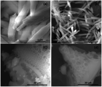
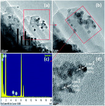
The as prepared CeO2 nanoparticle supported on SiO2 were characterized by X-ray diffraction, SEM, TEM, FTIR and XPS. Fig. 1 shows the SEM images with high and low magnification of the SiO2/CeO2 nanostructure. The SEM images show the formation of uniform rod like structures (Fig. 1 and S3 ESI†). The TEM images of the samples are shown in Fig. 2. TEM shows the presence of CeO2 nanoparticles on the SiO2 nanostructures (Fig. 2 and S4 ESI†). The typical diameters of these SiO2 nanostructure are around 25–40 nm and length are ∼1–4 μm in length. As clearly shown by the TEM images, ceria nanoparticle exhibits as dark particles of size 2–5 nm dispersed over silica nanostructure. The lattice fringes with a d-spacing of 2.93 Å (±0.02) are shown in Fig. 2d and this corresponds to the (h, k, l) values (111) of CeO2. The EDS is recorded and shown in Fig. 2c.
 | ||
| Fig. 1 SEM images of (a and b) CeO2-SiO2 catalyst and (c and d) SiO2 prepared in absence of Ce by following same preparation method. | ||
 | ||
| Fig. 2 (a), (b) and (d) represents the HR-TEM images; whereas (c) represent the EDS spectra of the CeO2-SiO2 catalyst. Partial size distribution histogram (inset 2a). | ||
The XPS spectra (Fig. S5 ESI†) for the CeO2 nanoparticles supported on SiO2 nanosheet (CeO2-SiO2) shows three different energy ranges containing Si 2p, Ce 3d and O 1s (carbon was a ubiquitous impurity). The peak at 284.8 eV indicated the presence of the C 1s. The Ce 3d5/2 binding energy values of 881.2 eV can be assigned for Ce4+ and the peak of Ce 3d5/2 at 883.8 eV is for Ce3+.16 CeO2-SiO2 showed no sharp XRD peaks for crystalline SiO2 or ceria (Fig. S6 ESI†). Due to the small size of CeO2 particles, no peak for CeO2 was detected in XRD. Whereas the XRD pattern shows the presence of amorphous SiO2. The amorphous SiO2 was also characterized by a Si 2p peak of 103.4 eV and an O1s peak of 532.9 eV (Fig. S5 ESI†).17 In accordance with the EDAX analysis (Fig. S7 ESI†) and XPS analysis, we found that there is no impurity presence in CeO2 nanoparticles supported silica nanostructure. The amount of CeO2 present in the silica nanostructures was estimated by ICP-AES and it was 0.98%. The BET surface area estimated by nitrogen adsorption of CeO2-SiO2 was 135 m2 g−1.
The FTIR spectrum of CeO2-SiO2 sample shows three characteristic bands, identified as SiO2 (Fig. S2 ESI†). The characteristic band at 1090 and 808 cm−1 corresponds to the stretching and bending of Si–O bonds, respectively. The position and the shape of Si–O vibrational bands at 1090 cm−1 reflects a stoichiometric silicon dioxide structure.18 Also, the band at 1634 cm−1 and 3429 cm−1 corresponds to O–H bending and H–O–H stretching mode indicating the hydrated SiO2.
The reactivity of the different CeO2-SiO2 catalyst for the selective oxidation of styrene are shown in Table. 1. All the experiments were carried out at room temperature. Commercial SiO2 (entry 1) and CeO2-SiO2imp (prepared by impregnation method) (entry 8) showed no activity, 0.98% CeO2-SiO2 shows excellent activity toward the selective conversion of styrene to styrene oxide. The catalyst shows 42.9% conversion with 92.4% styrene epoxide selectivity (entry 3) with a turn over frequency (TOF) values of 20.7 min−1 was achieved after 6 h reaction time and the remaining 7.6% was benzaldehyde. It was also noticed that with increasing reaction time, although the conversion was increasing but the selectivity for styrene epoxide decreasing, favouring the formation of benzaldehyde. The TOF values remain constant after 3 reuse, which is the advantage of our catalyst. The catalyst shows 21.4% conversion with 98.9% styrene oxide selectivity after 3 h (entry 2) and the conversion increases to 72.1% with 72.1% styrene oxide selectivity after 12 h (entry 4). With increasing catalyst to styrene ratio the conversion increases but the styrene oxide selectivity decreases (entry 5 & 6). We also observed the increase in conversion from 21.4 to 81.5 when the temperature was increased to 323 K (entry 7). When a radical scavenger (2, 6-di-tert-butyl-4-methyl-phenol) was added initially before the reaction, at room temperature; no reaction takes place. The result strongly suggest that the formation of styrene epoxide from styrene proceeds through free radical pathway, where Ce nanoparticle act as an initiator in the homolytic cleavage of H2O2 into hydroperoxyl (˙O–OH) radical in room temperature. Dissociation of H2O2 is catalyzed by the transition metal oxides are already reported in the literature.19 The redox reaction has been taking place, where Ce3+ is oxidized by H2O2 in to relatively strong one electron oxidant Ce4+. Where, that Ce4+ initiates the radical to attack on styrene which been converted into styrene epoxide subsequently.
| Entry no | Catalyst | Time (h) | XS (%) | Rca | TOFb (min−1) | SSO (%) | SB (%) |
|---|---|---|---|---|---|---|---|
a Rc: rate of styrene consumption in mole gcat−1 h−1.b TOF: moles of styrene oxide produced per moles of CeO2 per sec.c 2% catalyst (0.02 g) with respect to styrene.d Temperature = 323 K. Styrene 1.0 g (10 mmol) in 10 mL CH3CN, catalyst = 0.01 s g (1% with respect to styrene), room temperature, styrene![[thin space (1/6-em)]](https://www.rsc.org/images/entities/char_2009.gif) : :![[thin space (1/6-em)]](https://www.rsc.org/images/entities/char_2009.gif) H2O2 (50 wt%) = 1 H2O2 (50 wt%) = 1![[thin space (1/6-em)]](https://www.rsc.org/images/entities/char_2009.gif) : :![[thin space (1/6-em)]](https://www.rsc.org/images/entities/char_2009.gif) 5 (mole ratio). XS: conversion of styrene. SSo: selectivity of styrene oxide. SB: selectivity of benzaldehyde. 5 (mole ratio). XS: conversion of styrene. SSo: selectivity of styrene oxide. SB: selectivity of benzaldehyde. |
|||||||
| 1 | SiO2 | 6 | — | — | — | — | - |
| 2 | 0.98% CeO2-SiO2 | 3 | 21.4 | 71.3 | 20.6 | 98.9 | 1.1 |
| 3 | 0.98% CeO2-SiO2 | 6 | 42.9 | 71.5 | 20.7 | 92.4 | 7.6 |
| 4 | 0.98% CeO2-SiO2 | 12 | 72.1 | 60.1 | 17.4 | 82.1 | 17.9 |
| 5 | 0.98% CeO2-SiO2c | 6 | 68.2 | 56.8 | 16.4 | 90.9 | 7.1 |
| 6 | 0.98% CeO2-SiO2c | 12 | 80.1 | 66.7 | 9.6 | 72.7 | 27.3 |
| 7 | 0.98% CeO2-SiO2d | 3 | 81.4 | 271.3 | 78.6 | 92.8 | 7.2 |
| 8 | 1% CeO2-SiO2imp | 6 | — | — | — | — | — |
| 9 | 0.98% CeO2-SiO2 | 6 | 40.1 | 66.8 | 19.3 | 97.9 | 3.1 |
| 10 | 1.97% CeO2-SiO2 | 6 | 42.9 | 71.5 | 8.4 | 86.4 | 13.6 |
The reusability of the catalyst CeO2-SiO2 was studied without any regeneration. It was observed that the catalyst showed no change in its activity (conversion and selectivity) after 3 successive runs (Table 1, entry 9). The amount of Ce present in the catalyst (0.98% CeO2-SiO2) after 3 run is almost same as the fresh catalyst (estimated by ICP-AES), confirming that the heterogeneity of the catalyst.
Conclusions
In conclusion, we have found a surfactant mediated new strategy for the preparation of ceria nanoparticles supported on SiO2 nanosheet for the first time. The hydrothermal treatment of a mixture of Ce-precursors, tetraethyl orthosilicate (TEOS), poly(diallyldimethylammonium chloride), polyvinylpyrrolidone (PVP-40) produced ceria nanoparticles with the size of 2–5 nm supported on SiO2 nanosheet with the diameter of 25–40 nm and a length of 1–4 μm and characterized by XRD, SEM, TEM, XPS, ICP-AES, EDAX, BET-surface area. The supported ceria nanoparticles were very active for selective oxidation of styrene to styrene oxide at room temperature.Acknowledgements
B. S., R. K. S., S. G. thanks UGC, India and S. S. A. thanks CSIR, India for the fellowship. The Director, CSIR-IIP, is acknowledged for his support. The authors thank Analytical Science Division, Indian Institute of Petroleum for analytical services.Notes and references
- J. Kaspar, P. Fornasiero and N. Hickey, Catal. Today, 2003, 77, 419–449 CrossRef CAS.
- N. Shang, P. Papakonstantinou, P. Wang, A. Zakharov, U. Palnitkar, l. Lin, M. Chu and A. Stamboulis, ACS Nano, 2009, 3, 1032–1038 CrossRef CAS PubMed; S. Wu, G. Han, D. J. Milliron, S. Aloni, V. Altoe, D. V. Talapin, B. E. Cohen and P. J. Schuck, PNAS, 2009, 106, 10917–10921 CrossRef PubMed; J. Zhuang, A. D. Shaller, J. Lynch, H. Wu, O. Chen, A. D. Q. Li and Y. C. Cao, J. Am. Chem. Soc., 2009, 131, 6084–6085 CrossRef PubMed.
- H. Okamoto, Y. Kumai, Y. Sugiyama, T. Mitsuoka, K. Nakanishi, T. Ohta, H. Nozaki, S. Yamaguchi, S. Shirai and H. Nakano, J. Am. Chem. Soc., 2010, 132, 2710–2718 CrossRef CAS PubMed.
- H. Nakano, T. Mitsuoka, M. Harada, K. Horibuchi, H. Nozaki, N. Takahashi, T. Nonaka, Y. Seno and H. Nakamura, Angew. Chem., Int. Ed., 2006, 45, 6303–6306 CrossRef CAS PubMed.
- T. Sasaki, M. Watanabe, H. Hashizume, H. Yamada and H. Nakazawa, J. Am. Chem. Soc., 1996, 118, 8329–8335 CrossRef CAS; N. Miyamoto, H. Yamamoto, R. Kaito and K. Kuroda, Chem. Commun., 2002, 20, 2378–2379 RSC; N. Yamamoto, T. Okuhara and T. Nakato, J. Mater. Chem., 2001, 11, 1858–1863 RSC.
- H. Nakano, M. Ishii and H. Nakamura, Chem. Commun., 2005, 23, 2945–2947 RSC; A. B. Smith, C. M. Adams, S. A. L. Barbosa and A. P. Degnan, J. Am. Chem. Soc., 2003, 125, 350–351 CrossRef CAS PubMed.
- R. Ferrando, J. Jellinek and R. L. Johnston, Chem. Rev., 2008, 108, 845–910 CrossRef CAS PubMed.
- H. M. Chen and R. S. Liu, J. Phys. Chem. C, 2011, 115, 3513–3527 CAS.
- X. Deng and C. M. Friend, J. Am. Chem. Soc., 2005, 127, 17178–17179 CrossRef CAS PubMed.
- M. Roussel and H. Mimoun, J. Org. Chem., 1980, 45, 5387–5390 CrossRef CAS.
- R. J. Chimentao, I. Kirm, F. Medina, X. Rodriguez, Y. Cesteros, P. Salagreb and J. E. Sueiras, Chem. Commun., 2004, 7, 846–847 RSC; D. H. Zhang, H. B. Li, G. D. Li and J. S. Chen, Dalton Trans., 2009, 10527–10533 RSC; W. Zhan, Y. Guo, Y. Wang, Y. Guo, X. Liu, Y. Wang, Z. Zhang and G. Lu, J. Phys. Chem. C, 2009, 113, 7181–7185 Search PubMed; S. C. Laha and R. Kumar, J. Catal., 2001, 204, 64–70 CrossRef CAS.
- P. Fleming, S. Ramirez, J. D. Holmes and M. A. Morris, Chem. Phys. Lett., 2011, 509, 51–57 CrossRef CAS PubMed.
- Y. Borodko, S. E. Habas, M. Koebel, P. Yang, H. Frei and G. A. Somorjai, J. Phys. Chem. B, 2006, 110, 23052–23059 CrossRef CAS PubMed.
- H. Sun, J. He, J. Wang, S. Y. Zhang, C. Liu, T. Sritharan, S. Mhaisalkar, M. Y. Han, D. Wang and H. Chen, J. Am. Chem. Soc., 2013, 135, 9099–9110 CrossRef CAS PubMed.
- T. K. Sau and C. J. Murphy, Langmuir, 2005, 21, 2923–2929 CrossRef CAS PubMed.
- B. V. Crist, Handbook of The Elements and Native Oxides, XPS International, LLC, 1999 Search PubMed.
- A. E. Gonzalez, A. L. M. Hernandez, C. A. Chavez, V. M. Castano and C. V. Santos, Nanoscale Res. Lett., 2010, 5, 1408–1417 CrossRef PubMed.
- R. E. Lamont and W. A. Ducker, J. Am. Chem. Soc., 1998, 120, 7602–7607 CrossRef CAS.
- B. Cornils and W. A. Herrmann, J. Catal., 2003, 216, 23–31 CrossRef CAS.
Footnote |
| † Electronic supplementary information (ESI) available: Material characterization, experimental detail, SEM, TEM images, XPS, XRD, FTIR patterns and EDAX analysis. See DOI: 10.1039/c3ra46179c |
| This journal is © The Royal Society of Chemistry 2014 |

