Folate and biotin based bifunctional quantum dots as fluorescent cell labels

Atanu Chakraborty, Amit Ranjan Maity and Nikhil R. Jana*
Centre for Advanced Materials, Indian Association for the Cultivation of Science, Kolkata, 700032, India. E-mail: camnrj@iacs.res.in; Fax: +91-33-24732805; Tel: +91-33-24734971

Received 24th October 2013 , Accepted 19th December 2013

First published on 20th December 2013


Abstract

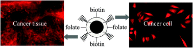
Although nanoprobes functionalized with one type of affinity molecule are commonly used as bioimaging probes, biolabeling studies of nanoprobes functionalized with more than one type of affinity molecule remains an unexplored area of research. Here we show that the labeling specificity of a nanoprobe can be enhanced if they are functionalized with more than one type of affinity molecule. We have synthesized functional quantum dots (QDs) of 20–30 nm hydrodynamic diameter having both folate and biotin on their surface. This dual functional nanoprobe has an enhanced labeling specificity to cancer cells/tissue as compared to folate or biotin based monofunctional QDs.


1. Introduction

Nanoparticle based biological probes are emerging as an alternative to molecular probes, as they are brighter and have more stable optical properties.1–14 These nanoprobes are usually functionalized with affinity molecules that enhance the labeling specificity of the probes. For example, nanoparticles have been functionalized with antibodies,1 peptides,2,3 vitamins,4–10 carbohydrates11,12 and aptamers13 for an enhanced labeling specificity of cells/tissues. These results show that functionalization generally induces the labeling specificity, although other factors such as size, shape, surface charge and the density of the affinity molecules also play a significant role. Extensive research is ongoing to optimize the physical and chemical properties of the nanoprobes, and bioconjugation chemistry is being used to achieve efficient linking of the affinity molecules and to search for new affinity molecules. However, most of the currently available approaches conjugate a single affinity molecule with the nanoparticle for use in different biomedical applications. In reality, cellular interactions with a nanoscale object occur via more than one type of receptor, and thus functionalization of a nanoprobe with more than one type of affinity molecule may further influence its labeling specificity. Although such types of multifunctional nanoparticles have been used previously to enhance detection sensitivity15,16 and to enhance the performance of nanoparticle based drug delivery,8,17–19 they are rarely used as imaging probes. Here we have synthesized folate and biotin based bifunctional nanoprobes, and showed that dual functionalization offers enhanced labeling specificity for cancer cells and tissues.

We have selected folate and biotin for nanoparticle functionalization as these two vitamins are often overexpressed in tumor cells, and it is well known that tumor cells have receptors for both vitamins.4–10 It was found that folic acid and biotin functionalization can increase the cancer-targeting efficiency of drug loaded nanoparticles.4–10 Thus it is expected that folate and biotin based bifunctional nanoprobes may have a higher interaction with tumor cells than biotin or folate based monofunctional nanoprobes. In addition, these bifunctional nanoprobes can be useful for targeting cells that have either biotin or folate receptors. Quantum dots (QDs) have been chosen for the synthesis of bifunctional nanoprobes due to their size-dependent tunable emission and the fact that they have been widely used as bioimaging probes.1–3 Besides, a range of functionalization strategies are currently available for QDs.20–22

2. Materials and methods

Chemicals

Folic acid, N,N′-dicyclohexylcarbodiimide (DCC), N-hydroxysuccinimide (NHS), poly(ethylene glycol) methacrylate, 3-sulfopropyl methacrylate, N-(3-aminopropyl) methacrylamide hydrochloride and N,N-methylenebisacrylamide, biotin N-hydroxy succinimide ester were purchased from Sigma-Aldrich and used as received. N-(3-Aminopropyl) methacrylamide hydrochloride was purchased from Polyscience and used as received. Folate-free RPMI-1640 medium was purchased from Invitrogen and folate-containing DMEM was purchased from Sigma-Aldrich for cell culture experiments.

Synthesis of folic acid and biotin functionalized QDs

High quality CdSe/ZnS based QDs were synthesised by a previously reported method.23 First, red emissive CdSe nanocrystals were synthesised at 280 °C in octadecene solvent and then they were used for ZnS shelling at 220 °C.23 The hydrophobic QDs were converted into primary amine-terminated water soluble QDs by a polyacrylate coating approach, as reported previously.21,22 Folate–NHS was prepared using DCC based coupling chemistry, as reported elsewhere,10 and 10 mM of fresh solution was prepared in dimethylformamide. Similarly, 10 mM of a fresh solution of biotin–NHS was prepared in dimethylformamide.

Next, three different sets of one mL of primary amine-terminated QD solution were placed in a borate buffer solution of pH 9. In one vial 30 μL folic–NHS solution was mixed, in a second vial 30 μL biotin–NHS solution was mixed and in a third vial 15 μL folic–NHS and 15 μL biotin–NHS were mixed. These three solutions were stirred overnight to achieve a completed reaction. Next, the solutions were dialysed against basic water and then against normal water to remove excess reagents. Finally, the solutions were preserved at 4 °C.

Estimation of the number of folate and biotin molecules per QD

The number of folate and biotin molecules per QD has been estimated following our earlier reported method.24 The concentration of QDs was calculated from a previously described equation.25 Next, the number of amine molecules per QD has been estimated by a fluorescamine based titration method, and was found to be around 150 per QD. Next, the functionalized QDs were treated with HCl to dissolve the QDs, followed by neutralization with NaOH. Next, the fluorescence properties of folate were used to estimate the folate concentration. The number of folate molecules per QD was estimated from the ratio of folate and QD concentrations. In monofunctional QD–biotin, the number of biotin molecules per QD was assumed to be the same as the number of primary amine molecules, as excess biotin–NHS had been used so that all the primary amines were reacted. Similarly, in the bifunctional QDs, the number of biotin molecules per QD was estimated after subtracting the number of folate molecules per QD from the total number of primary amine molecules per QD. In summary, the QD–folate has about 150 folate molecules per QD, QD–biotin has about 150 biotin molecules per QD and folate–QD–biotin has about 100 folate and 50 biotin molecules per QD.

In vitro cell labeling

Cells were cultured in a folate-free RPMI-1640 (Invitrogen) medium with 10% heat-activated fetal bovine serum (FBS), 1% penicillin–streptomycin, at 37 °C and in a 5% CO2 atmosphere. For fluorescence microscopy, cells were cultured overnight in a 24 well plate with 500 μL medium. Next, about 10–100 μL of the samples was added to the cell culture medium and incubated for 1–12 hours. After incubation, the cells were washed twice with phosphate-buffered saline (PBS) to remove the free unbound particles, and then the cells were fixed using paraformaldehyde and used for imaging.

Instrumentation

All UV-visible spectra and fluorescence spectra were measured with a Shimadzu UV-2550 UV-visible spectrophotometer and Fluoromax-4 spectrofluorometer (Horiba JobinYvon), respectively. Fourier transform infrared spectroscopy on KBr pellets was performed using a Shimadzu FT-IR 8400S instrument. Bright field and fluorescence images were taken using a Carl Zeiss Apotome Imager Z1 Microscope.

3. Results and discussion

Synthesis and characterization of bifunctional quantum dots

Scheme 1 demonstrates the synthesis approach for bifunctional quantum dots. Hydrophobic ZnS-capped CdSe QDs of a 2–5 nm size are synthesized first via a high temperature colloid-chemical method.23 Next, these are converted into water soluble and primary amine terminated QDs of a hydrodynamic diameter of 20–30 nm, via an established polyacrylate coating approach.21,22 Next, bifunctional QDs are synthesized by reacting them with a mixture of an N-hydroxysuccinimide derivative of folate (folate–NHS) and an NHS derivative of biotin (biotin–NHS). The high reactivity of NHS derivatives with primary amine groups ensures the efficient functionalization of the QDs with both folate10 and biotin.8 The advantage of the polyacrylate coating is that it offers the QDs high water solubility along with a wider opportunity for variations of the surface charge and functionality.21,22 In the present cases the polyacrylate coating has been optimized so that it provide an anionic surface charge and a polyethylene glycol functionality, that permits low non-specific binding of QDs during cellular interaction/labeling.22 The present scheme is also applicable to produce folate or biotin based monofunctional QDs when folate–NHS or biotin–NHS is reacted with amine-terminated QDs. Thus the present synthetic scheme has successfully produced folate-functionalized QDs (QD–folate), biotin functionalized QDs (QD–biotin) and folate and biotin based bifunctional QDs (folate–QD–biotin). (Fig. 1–3 and ESI, Table S1, Fig. S1–S6) All the monofunctional or bifunctional QDs have high colloidal stabilities in physiological conditions and have a hydrodynamic diameter of 20–30 nm.
image file: c3ra46085a-s1.tif
Scheme 1 The synthesis strategy for folate and biotin based bifunctional QDs.

image file: c3ra46085a-f1.tif
Fig. 1 (a) UV-visible spectra of polyacrylate coated QDs before and after functionalization, showing the appearance of a characteristic folate band after folate functionalization. (b) Emission spectra of the mono- and bifunctional QDs, showing the characteristic emission of folate after folate functionalization along with the fluorescence band of the QDs. ((i) QDs, (ii) QD–folate, (iii) folate–QD–biotin, (iv) QD–biotin) (c) transmission electron microscopy (TEM) and dynamic light scattering (DLS) based size determination of the bifunctional QDs. (d) Streptavidin (Stv) based aggregation of the bifunctional QDs (fol–QD–bio), showing evidence of biotin functionalization.

The presence of both folate and biotin in the synthesized bifunctional QDs has been confirmed from the optical properties and a biochemical activity test (Fig. 1). The functionalization of the folate has been established from its UV-visible and fluorescence spectra. The characteristic absorption band of folate appears in the 370 nm region and the characteristic emission band of folate appears in the 400–500 nm region. Biotin functionalization has been confirmed via a streptavidin based biochemical activity test. Streptavidin has four binding sites for biotin, and it initiates the crosslinking of the QDs.26 Upon the addition of streptavidin, the solution of bifunctional QDs becomes turbid within 2–3 minutes and the QDs get precipitated out from the solution within an hour. It has been ensured that no free folate or biotin is present in the QD solution, as the sample is extensively dialysed against distilled water.

FTIR spectral studies of the QDs before and after conjugation also provide further proof that the folate and biotin are covalently conjugated with the primary amine groups of the QDs (ESI, Fig. S1). The N–H bending vibration peak at 1632 cm−1 becomes weaker after folate and biotin conjugation. In addition, the symmetry-deformed vibration peak of NH3+ at 1522 cm−1 becomes absent after the conjugation of folate and biotin, and a new peak is generated at around 1192 cm−1, corresponding to the signature of C–N stretching vibrations. The size of the bifunctional QDs has been determined by transmission electron microscopy (TEM) and dynamic light scattering (DLS) based studies (Fig. 1). The TEM study shows the size of the 3–4 nm inorganic cores of CdSe–ZnS, and the DLS shows the 20–30 nm hydrodynamic size that includes the inorganic core and the polymeric shell (including the biotin and folate).

Bifunctional quantum dots as fluorescent probes

The advantage of bifunctional QDs as biological labels has been investigated using fluorescence based labeling and the imaging of cells and tissue. We have selected HeLa cells and cervical cancer tissue as they are known to have overexpressed receptors for both folate and biotin.7 When a functional QD probe is introduced into culture media, the growing HeLa cells have a preferential interaction/uptake for bifunctional folate–QD–biotin compared to monofunctional QD–folate or QD–biotin. This preferential interaction/uptake of bifunctional QDs is clearly observed from the fluorescence image of labelled HeLa cells (Fig. 2) and the quantitative estimation of QD uptake in labelled cells (ESI, Fig. S5). HeLa cells of similar confluency are incubated for four hours, separately with mono- and bifunctional QDs of similar concentrations. Next, the labelled cells are washed and imaged under a fluorescence microscope and quantified under a microplate reader. The results clearly show that under similar conditions the bifunctional QDs have a higher cellular interaction/uptake, as observed from the much more intense fluorescence. Further study is conducted with the HeLa cells with a different time incubation, which clearly shows that QDs enter cells in a 12 h timeframe, and that the concentration of the internalized QDs are much higher for the bifunctional QDs (Fig. 3 and ESI Fig. S3 and S4). The control labeling experiment with other cell lines (CHO and H9C2) shows that cells are not at all labelled by the monofunctional and bifunctional QDs, as those cell lines have low numbers of folate and biotin receptors (ESI, Fig. S6).
image file: c3ra46085a-f2.tif
Fig. 2 (a) A fluorescence image (FL) of HeLa cells labeled with monofunctional QDs and bifunctional QDs after 4 h of incubation, showing that the bifunctional QDs have an enhanced labeling and uptake compared to respective monofunctional QDs. (b) A fluorescence image of cervical cancer tissue after labeling with monofunctional QDs and bifunctional QDs, showing that only the bifunctional QDs effectively label the tissue. Blue-emitting Hoechst dye has been used to stain the cell nucleus.

image file: c3ra46085a-f3.tif
Fig. 3 The intracellular localization of the mono- and bifunctional QDs after a longer time period (12 h) of incubation. In each case, the blue fluorescence is due to the Hoechst dye used for nucleus staining and the red fluorescence is due to the QDs. From the images it is evident that the QDs are successfully internalized inside the cells within 12 h, and that bifunctional QDs have a higher internalization efficiency as compared to the monofunctional QDs.

The potential advantage of these bifunctional QDs is manifested by the specific labeling of cervical cancer tissue. Fig. 2b shows that these bifunctional QDs can label the cervical cancer tissue more efficiently, compared to respective monofunctional QDs. Earlier work shows that multiple affinity molecules on a nanoprobe surface can significantly enhance the labelling and uptake of the nanoprobe.24,27,28 Thus we expect that the presented bifunctional QDs, with multiple numbers of folate and biotin molecules, interact strongly with the cells/tissue which have both folate and biotin receptors, and induce a higher labelling efficiency.

4. Conclusion

In summary, we have synthesized folate and biotin based bifunctional QDs of a 20–30 nm hydrodynamic diameter. These nanoprobes produce stable colloidal solutions in water and act as much more efficient fluorescent biological labels compared to respective monofunctional nanoprobes. This concept can be extended in preparing various new nanoprobes by combining various bioaffinity biomolecules and exploring their enhanced labeling efficiency.

Acknowledgements

This work is supported by DST and CSIR, government of India. AC thanks CSIR-India for a research fellowship and Dr. Arun Roy (V. J. R. College, Kolkata) for the tissue imaging study, and ARM thanks DST-India for a research fellowship.

Notes and references

  1. L. L. Yang, H. Mao, Y. A. Wang, Z. H. Cao, X. H. Peng, X. X. Wang, H. W. Duan, Q. G. Yuan, G. Adams, M. Q. Smith, W. C. Wood, X. H. Gao and S. Nie, Small, 2009, 5, 235–243 CrossRef CAS PubMed.
  2. J. B. Delehanty, I. L. Medintz, T. Pons, P. E. Dawson, F. M. Brunel and H. Mattoussi, Bioconjugate Chem., 2006, 17, 920–927 CrossRef CAS PubMed.
  3. Y. F. Wei, N. R. Jana, S. J. Tan and J. Y. Ying, Bioconjugate Chem., 2009, 20, 1752–1758 CrossRef CAS PubMed.
  4. H. S. Yoo and T. G. Park, J. Controlled Release, 2004, 100, 247–256 CrossRef CAS PubMed.
  5. D. J. Bharali, D. W. Lucey, H. Jayakumar, H. E. Pudavar and P. N. Prasad, J. Am. Chem. Soc., 2005, 127, 11364–11371 CrossRef CAS PubMed.
  6. R. L. Schneider, F. Schmitt, C. Frochot, Y. Fort, N. Lourette, F. Guillemin, J. F. Muller and M. Barberi-Heyob, Bioorg. Med. Chem., 2005, 13, 2799–2808 CrossRef CAS PubMed.
  7. W. J. Yang, Y. Y. Cheng, T. W. Xu, X. Y. Wang and L. P. Wen, Eur. J. Med. Chem., 2009, 44, 862–868 CrossRef CAS PubMed.
  8. Y. B. Patil, U. S. Toti, A. Khdair, L. Ma and J. Panyam, Biomaterials, 2009, 30, 859–866 CrossRef CAS PubMed.
  9. D. N. Heo, D. H. Yang, H. J. Moon, J. B. Lee, M. S. Bae, S. C. Lee, W. J. Lee, I. C. Sun and I. K. Kwon, Biomaterials, 2012, 33, 856–866 CrossRef CAS PubMed.
  10. A. R. Maity, A. Saha, A. Roy and N. R. Jana, ChemPlusChem, 2013, 78, 259–267 CrossRef CAS.
  11. C. Tassa, S. Y. Shaw and R. Weissleder, Acc. Chem. Res., 2011, 44, 842–852 CrossRef CAS PubMed.
  12. S. Basiruddin, A. R. Maity and N. R. Jana, RSC Adv., 2012, 2, 11915–11921 RSC.
  13. W. J. Kang, J. R. Chae, Y. L. Cho, J. D. Lee and S. Kim, Small, 2009, 5, 2519–2522 CrossRef CAS PubMed.
  14. N. Erathodiyil and J. Y. Ying, Acc. Chem. Res., 2011, 44, 925–935 CrossRef CAS PubMed.
  15. P. Hazarika, B. Ceyhan and C. M. Niemeyer, Small, 2005, 1, 844–848 CrossRef CAS PubMed.
  16. D. G. Georganopoulou, L. Chang, J. M. Nam, C. S. Thaxton, E. J. Mufson, W. L. Klein and C. A. Mirkin, Proc. Natl. Acad. Sci. U. S. A., 2005, 102, 2273–2276 CrossRef CAS PubMed.
  17. M. Thomas, J. Clarhaut, P.-O. Strale, I. Tranoy-Opalinski, J. Roche and S. Papot, ChemMedChem, 2011, 6, 1006–1010 CrossRef CAS PubMed.
  18. H. Li, Y. Lu, L. Piao, J. Wu, X. Yang, V. Kondadasula, W. E. Carson and R. J. Lee, Bioconjugate Chem., 2010, 21, 961–968 CrossRef CAS PubMed.
  19. K. Zhang, L. Hao, S. J. Hurst and C. J. Mirkin, J. Am. Chem. Soc., 2012, 134, 16488–16491 CrossRef CAS PubMed.
  20. I. L. Medintz, H. T. Uyeda, E. R. Goldman and H. Mattoussi, Nat. Mater., 2005, 4, 435–446 CrossRef CAS PubMed.
  21. S. Basiruddin, A. Saha, N. Pradhan and N. R. Jana, J. Phys. Chem. C, 2010, 114, 11009–11017 CAS.
  22. N. R. Jana, Phys. Chem. Chem. Phys., 2011, 13, 385–396 RSC.
  23. J. J. Li, Y. A. Wang, W. Guo, J. C. Keay, T. D. Mishima, M. B. Johnson and X. Peng, J. Am. Chem. Soc., 2003, 125, 12567–12575 CrossRef CAS PubMed.
  24. A. Saha, S. K. Basiruddin, A. R. Maity and N. R. Jana, Langmuir, 2013, 29, 13917–13924 CrossRef CAS PubMed.
  25. W. W. Yu, L. H. Qu, W. Z. Guo and X. G. Peng, Chem. Mater., 2003, 15, 2854–2860 CrossRef CAS.
  26. A. Gole and C. J. Murphy, Langmuir, 2005, 21, 10756–10762 CrossRef CAS PubMed.
  27. P. M. Valencia, M. H. Hanewich-Hollatz, W. W. Gao, F. Karim, R. Langer, R. Karnik and O. C. Farokhzad, Biomaterials, 2011, 32, 6226–6233 CAS.
  28. T. P. Thomas, B. H. Huang, S. K. Choi, J. E. Silpe, A. Kotlyar, A. M. Desai, H. Zong, J. Gam, M. Joice and J. R. Baker, Mol. Pharmaceutics, 2012, 9, 2669–2676 CrossRef CAS PubMed.

Footnote

Electronic supplementary information (ESI) available: FTIR characterization of functional QDs and control labeling experiments. See DOI: 10.1039/c3ra46085a

This journal is © The Royal Society of Chemistry 2014
Click here to see how this site uses Cookies. View our privacy policy here.