Runsheng
Jiang
ab,
Yongjun
Li
*a,
Zhihong
Qin
ab,
Liang
Xu
ab,
Daoben
Zhu
a and
Yuliang
Li
*a
aBeijing National Laboratory for Molecular Sciences (BNLMS), CAS Key Laboratory of Organic Solids, Institute of Chemistry, Chinese Academy of Sciences, Beijing 100190, P. R. China. E-mail: liyj@iccas.ac.cn; ylli@iccas.ac.cn; Fax: +86-10-82616576; Tel: +86-10-62587552
bGraduate University of Chinese Academy of Sciences, Beijing 100190, P. R. China
First published on 14th November 2013
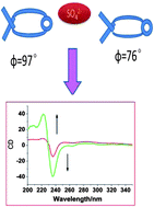
A novel macrocyclic binaphthalene derivative (L) containing amide and triazole units has been synthesized by “click” reaction. The binding behaviors of this receptor toward anions have been studied by 1H NMR titration, circular dichroism (CD) spectroscopy. The tetrahedral sulfate anion predominantly interacts with L through hydrogen bonds which could tune the dihedral angle between the two naphthalene rings and supply tunable CD output signals to form chiral receptor.
NMR, UV/vis or fluorescence spectroscopies were used to monitor anion binding processes.38,39 CD spectroscopy is a convenient method to monitor conformational changes of the supramolecular systems, which is a powerful tool for sensing anions when the supramolecular response detected from other spectroscopies is particularly weak.40
Molecular sensors for sulfate anions mainly based on triazolium, imidazolium, pyrrole, amide, urea and heterotopic combination of these binding motifs. Amide and urea NH groups have been employed to produce a wide range of sulfate receptors.41 For example, Das, Ganguly and co-workers have reported a tren based tris(urea) receptor which demonstrated selective binding with sulfate both in solution and solid states.42 Amide-based macrocyclic receptors are also found to be particularly well-suited for encapsulating sulfate anions.43 All these receptors prefer to bind sulfate over dihydrogenphosphate, acetate, nitrate and perchlorate. However, the construction of chiral receptors for sulfate anions is relatively rare.44 Several examples of chiroptical sensors that provide significantly differentiated CD signals upon binding of metal ions, neutral guests, or chiral polymers have been recently reported.45,46
Herein, we designed a molecule L (see Scheme 1) based on the chiral framework of binaphthalene,47 with 1,2,3-triazoles and amide units as the binding motifs. Anions can form strong intermolecular hydrogen bonds with the amide groups and triazole units which may result in the changes of the dihedral angle between the two naphthalene rings, thus the variation of the CD signals from the binaphthalene framework can be observed. This chiral molecular receptor shows an efficient sensor property with obvious CD signal changes in the presence of sulfate anion, however, it only exhibits weak CD signal changes when binding to other anions, such as H2PO4−, HSO4−, AcO− and Br−.
Sulfate is a tetrahedral anion which can form NH⋯X− hydrogen bonds through two oxygen atoms with amide groups, the remaining oxygen atoms can form hydrogen bonds with CH of the 1,2,3-triazole. 1H NMR titration was carried out to understand the binding mode of L with sulfate anion. Fig. 2b showed the 1H NMR spectra of L with different amounts of bis(tetrabutylammonium) sulfate (TBA2SO4) in [D6] acetonitrile. In general, π–π stacking results in upfield shift of protons, hydrogen-bonding interaction results in downfield shift of protons. Upon the addition of 1.0 equivalent of TBA2SO4, the signals from amide NH (ΔδHj = 3.35 ppm), triazole CH (ΔδHd = 2.13 ppm) and benzene CH (ΔδHa = 1.25 ppm) protons showed downfield shifts, due to the multiple hydrogen bonds between the sulfate and L. With the addition of 1.5 equivalents to 10.0 equivalents of TBA2SO4, the signals from the protons of triazole CH (Hd), amide NH (Hj), benzene CH (Ha) showed upfield shifts, and those from the protons Hm, Hh of the binaphthalene also showed slight upfield shifts.
As shown in Fig. 2c, there were strong NOE connections between the Hj and Ha (Hd–Hj, Hd–He, Hj–He) in the presence of 1.0 equivalent of sulfate. Ascribed to the unique configuration of the L, one amide NH and two triazoles CH of L could bind with one molecule of sulfate by multiple hydrogen bonds as 1:
1 host–guest complex. NOESY spectroscopy in the presence of 1.5 equivalents of sulfate (Fig. 2d) showed some new NOE connections between the benzene protons (Ha), triazole protons (Hd), and binaphthalene protons (Hf) (Hf–Hd, Ha–Hf, Ha–Hc). Due to the long distances between protons Hf and Hd, Ha and Hf, Ha and Hc in one molecule, intramolecular cross peaks in NOESY NMR could not be observed, these signals (Hf–Hd, Ha–Hf, Ha–Hc) must correspond to intermolecular contacts between different chains, indicating that two macrocyle L arranged in head to tail conformation in the final 2
:
3 complex which was stabilized by π–π stacking between the binaphthalene unit and benzene unit.
These results clearly indicated that the addition of sulfate to a solution of host L first led to the formation of a 1:
1 host–guest complex, two 1
:
1 complexes then sandwiched another sulfate with intermolecular hydrogen bonds, which resulted in the final 2
:
3 binding stoichiometry between L and sulfate (as shown on Fig. 2a). This configuration can explain the small upfield shift of the signals from triazole proton Hd, amide Hj, and even the up-field shift of those from the aromatic protons (Hm, Ha, Hh), which is due to the interaction of π–π stacking (upfield shift).
UV-vis spectral studies were carried out in acetonitrile to evaluate the binding properties of compound L (Fig. 3a). The absorption spectra of L exhibit an intense absorption band (λmax = 227 nm), assigned to the π–π* transition of the naphthalene chromophores. Upon addition of sulfate, the absorption at λmax = 227 nm decreased until one equivalent of sulfate was added. This absorption change was due to the destruction of the π–π conjugation within the binaphthaene rings through the binding of sulfate anions. The binding stoichiometry of the sulfate–L interactions was calculated to be 2:
3 from the Job's plot (Fig. S11†). The fluorescence intensity was quenched upon the addition of SO42− (Fig. S18†), due to the electron transfer from electron-rich sulfate anions to binaphthalene chromophore.
![]() | ||
Fig. 3 (a) UV-vis absorption spectra of L (1 × 10⁻⁵ mol L−1 in acetronitrile) upon addition of SO42− in acetronitrile, (b) CD spectra of L (1 × 10⁻⁵ mol L−1) upon addition of SO42− in acetronitrile. |
The multi-hydrogen bonding interaction can modulate the dihedral angle of naphthalene rings which leads to the changes of CD signals. As shown in Fig. 3b, the CD spectra of compound L (1.0 × 10⁻⁵ M) were changed upon the addition of SO42−. Compound L exhibits CD signals at 220 nm and 239 nm (Fig. 3b) due to the chiral binaphthane-rings. The intensity of CD band at 239 nm was increased upon the addition of SO42−, and it was increased about 200% (Fig. 3b) with 5.0 equiv. of SO42−, further addition of SO42− just increased CD signals slightly (Fig. S7†).
On the basis of previous studies,53,54 the enhancement of CD band intensity reflects that the dihedral angle of binaphthalene is reduced. As the energy-minimized structure of compound L and sulfate–L shown in Fig. S31,† the dihedral angle of binaphthalene ring in L was calculated to be 93°, for the sulfate–L complex, the dihedral angle of binaphthalene ring was calculated to be 76°.55 The dihedral angles of compound L and sulfate–L complex were respectively about 92° and 73° based on CD data according to the method described by Salvadori et al.56
The binding behaviors of L with other anions were also investigated with 1HNMR titration. As shown in Fig. S19–S24,† with the addition of I− (Fig. S20†), the signals from protons Hj (Δδ = 1.0 ppm), Ha (Δδ = 0.6 ppm), Hd (Δδ = 0.5 ppm) showed downfield shifts, which was due to the weak multiple hydrogen-bonding between compound L and I−. While in the presence of Cl− (as shown in Fig. S19†), only the signals from protons Hj (Δδ = 1.7 ppm) and Ha (Δδ = 2.0 ppm) showed downfield shifts, those from triazole Hd and other protons showed no chemical shifts. Similar phenomenon was observed for Br− titration (Fig. S19†). Upon the addition of H2PO4− (Fig. S20†), the signal of amide proton Hj (ΔδHj = 2.4 ppm) showed downfield shifts. Those from other protons such as triazole CH (Hd), aromatic proton (Ha) showed weak chemical-shifts. While in the presence of HSO4−, (as shown in Fig. S24†), the signals from protons Hj (Δδ = 2.2 ppm), Ha (Δδ = 0.5 ppm), Hd (Δδ = 0.4 ppm) showed downfield shifts. The spectra of receptor L with various amounts of AcO− anions were shown in Fig. S21,† the signal of amide (NH) shifted downfield more largely (ΔδHj = 2.8 ppm) than those of the triazole CH (ΔδHd = 0.5 ppm) and aromatic proton CH (ΔδHa = 1.0 ppm).
The binding stoichiometry of the L–anion (I−, Cl−, Br−, AcO−, H2PO4−) interaction was confirmed to be 1:
1 from the Job's plot (as shown in Fig. S12 and S13†). The stability constant of I− and AcO− with L were determined by the analysis of the signal shifts of benzene proton Ha, because of line broadening of the binding-site proton signals in the 1H NMR spectra. With the addition of H2PO4−, the signal from proton Hj shifted downfield (as shown on Fig. S20†). The stability constant of the H2PO4− was obtained by the analysis of the amide NH (Hj) signals (as shown in Fig. 4). The stability constant of the SO42− was calculated by fitting the chemical shifts of proton Hj (as shown on Fig. S27†). While the stability constant of the HSO4− was calculated by fitting the chemical shifts of proton Ha (as shown on Fig. S24†). The data were fitted with the WinEQNMR software to give K (Table 1).52 While the stability constant (K) of Br−, Cl− anions could not be determined by analysis of signal shifts of protons due to the nonlinear line broadening. Benesi–Hildebrand curve method47 was employed to calculate the stability constant K from spectropolarimetric titration data (Fig. S29 and 30†). The stability constant of SO42− is undoubtedly the biggest one among the anions studied.
Anions | Br−b | I−a | HSO4−a | Cl−b | SO42−a | H2PO4− | AcO− |
---|---|---|---|---|---|---|---|
a Anions as nBu4N+ salts in [D3] CH3CN, triazole protons Ha fitted with WinEQNMR,20 errors less than 10%. b Benesi–Hildebrand curve methods. c r = I0 − Ix/I0 (I0 is the CD intensity of L at 238 nm. Ix is the CD intensity of the L after addition of anions. r showed ratio of CD signals changes). | |||||||
K | 113 ± 1 | 108 ± 3 | 360 ± 4 | 120 ± 1.5 | 12970 ± 146 | 300 ± 5 | 140 ± 2 |
r c (%) | 28% | 30% | 60% | 28% | 28% | 28% | 28% |
As shown in Fig. 2a, compound L has three typical anion recognition sites: amide, triazole and benzene ring. Amide (NH) group is a typical strong hydrogen bond donor which can easily form hydrogen bonds as NH⋯X− complex. The strength of the hydrogen-bonding is influenced by the radius size and the basicity of X−.57,58 The smaller radius size and stronger basicity of X− can result in high strength hydrogen-bonding. From this theory, the anion binding constant with amide (NH) should be in the order of AcO− > H2PO4− ≫ Br−, but from the binding constants (K) (as shown in Table 1), L showed the binding order of H2PO4− > AcO− >Br−. This strange binding order is due to the specific structural compensation of the receptor and anions. For example, AcO− is a planar structure, in which two oxygen atoms preferentially bind with one amide and one triazole units or one benzene protons on the same side of the L through hydrogen-bonding, while SO42− with four binding sites could cooperatively bind with L. Due to the specific structure of compound L, L can bind with one sulfate as 1:
1 complex, then the complex binds with another sulfate as 2
:
3 host–guest complex, which shows large binding constants (K). We figure out its 1
:
1 complex binding constants (K1) by WinEQNMR software (as shown on Table 1). For tetrahedral H2PO4− and SO42−, H2PO4− should have bigger binding constants than SO42− based on basicity, while the size of SO42− is more suitable for the cavity of the compound L than that of H2PO4−, and the cooperative effect in the sulfate–L complex further increased the binding strength. On the other hand, the charge difference also contributed to binding affinity. Halogen anions are weak base anions with small size, which bind with compound L weakly.
The structural compensation between the receptor and anions also influenced the CD signals of this system. The trend of CD signal changes (Table 1) is consistent with the binding constants. Sulfate induced the largest CD signal increment (about 200%), while other anions caused weak or moderate CD signal changes (from 28% to 60%). In compound L, there are two pairs of amide and triazole units, anions which could form multiple hydrogen-bonds with both pairs of binding units of L could rotate the naphthane rings and tune the dihedral angle. Thus tetrahetral anions (SO42−, H2PO4−) induced more CD signal changes than the planar AcO− and sphere halides (Cl−, Br−, I−), and the smaller sulphate with two charges dragged both sides more strongly than H2PO4−, which showed the largest CD signal changes. Tetrahedral sulfate anions which could form multiple hydrogen bonds with both pairs of binding units of L could tune the CD signals of the binaphthalene framework.53,59
Footnote |
† Electronic supplementary information (ESI) available: 1H NMR and 13C NMR of compound L. UV-vis Spectra and CD Signals of compound L with various anions, See DOI: 10.1039/c3ra46049e |
This journal is © The Royal Society of Chemistry 2014 |