From (+)-epigallocatechin gallate to a simplified synthetic analogue as a cytoadherence inhibitor for P. falciparum

Sandra Gemmaabc, Simone Brogiabc, Pradeep R. Patilabc, Simone Giovaniabc, Stefania Lamponiabc, Andrea Cappelliabc, Ettore Novellinoad, Alan Browne, Matthew K. Higginsf, Khairul Mustafag, Tadge Szestak g, Alister G. Craigg, Giuseppe Campiani*abc, Stefania Butiniabc and Margherita Brindisiabc
aEuropean Research Centre for Drug Discovery and Development (NatSynDrugs), Università degli Studi di Siena, via Aldo Moro 2, 53100 Siena, Italy. E-mail: campiani@unisi.it; Tel: +39 0577 234172
bDipartimento di Biotecnologie, Chimica e Farmacia, Università degli Studi di Siena, via Aldo Moro 2, 53100 Siena, Italy
cCentro Interuniversitario di Ricerche sulla Malaria (CIRM), University of Perugia, Perugia, Italy
dDip. di Farmacia, University of Naples Federico II, Via D. Montesano 49, 80131 Naples, Italy
eDepartment of Biochemistry, University of Cambridge, Cambridge, CB2 1GA, UK
fDepartment of Biochemistry, University of Oxford, Oxford, OX1 3QU, UK
gLiverpool School of Tropical Medicine, Pembroke Place, Liverpool L3 5QA, UK

Received 18th October 2013 , Accepted 6th December 2013

First published on 9th December 2013


Abstract

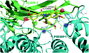
Parasite derived surface antigen PfEMP1 is a virulence factor of the human malaria parasite. PfEMP1 variants have been implicated in the cytoadherence of P. falciparum infected erythrocytes (iRBC) to several binding receptors on host vascular endothelium. Among them, binding to ICAM-1 seems to be related to severe manifestations of the disease such as cerebral malaria. The binding site for iRBC has been mapped to the BED-side of the N-terminal immunoglobulin-like domain of ICAM-1, and the DE-loop appears to be critical for binding. To date (+)-EGCG is the unique small molecule anti-cytoadherence inhibitor probably mimicking the DE-loop of ICAM-1. Here we report the discovery of a tetrahydroisoquinoline derivative, a prototype of a novel class of cytoadherence inhibitors, and an analogue of the natural compound characterized by a synthetically accessible scaffold. Molecular modeling analysis of (+)-EGCG and its synthetic tetrahydroisoquinoline analogue rationalized their binding mode to PfEMP1, confirming their ability to mimic the DE-loop.

Introduction

Malaria is a deadly disease killing millions of people worldwide.1 The disease is caused by infection from parasites of the genus Plasmodium. Several Plasmodium species affect humankind, e.g. P. falciparum, P. vivax, P. ovale, P. malariae, and P. knowlesi. Among them, P. falciparum is the most dangerous parasite and infection by P. falciparum can be lethal. The symptoms of uncomplicated malaria are normally mild and mainly comprise fever and headache. However, the symptoms of severe malaria can rapidly develop from uncomplicated malaria resulting in coma and death. Among the manifestations of severe malaria, cerebral malaria is responsible for a significant proportion of mortality. Although the coma that characterizes cerebral malaria is likely to have multifactorial etiology, post-mortem analysis reveals a common feature, namely sequestration of infected red blood cells (iRBCs) in the microvasculature of the brain.2–4 Cytoadherence properties of iRBCs are mainly mediated by interaction of the parasite antigenic protein PfEMP1 with specific host receptors; mainly CD36, ICAM-1, or CSA (for placental malaria) located on the endothelial cell surface,5,6 although endothelial protein C receptor has recently been identified as an important ligand in cerebral malaria.7 PfEMP1 is expressed on the surface of the iRBCs and is encoded by around 60 divergent var genes.8–10 This surface antigenic protein is responsible for both adhesion and immune evasion.10 Although still debated,11,12 several studies highlighted a role of ICAM-1 binding for the development of cerebral malaria.3,13–15 ICAM-1 is expressed on the surface of leukocytes and endothelial cells and its expression is increased following inflammation.3 ICAM-1 is composed of five Ig-like domains.16 The binding site for PfEMP1 has been located in the N-terminal Ig-like domain and mainly involves 3 β-strands (named B, D, and E: the BED domain).17 On the other hand, PfEMP1 proteins are composed by modular domains: Duffy binding like (DBL) domains (classified into α–ζ), and cysteine rich interdomain regions (CIDR).18–20 Although a crystallographic structure of an ICAM-1 binding PfEMP-1 has not been obtained yet, very recently a low resolution structure demonstrated a rigid and elongated arrangement of PfEMP1 domains.21 Moreover, in the same study it has been demonstrated that the binding of ICAM-1 to PfEMP1 is exclusively mediated through the DBLβ domain.21 As shown by mutagenesis studies,22 several regions within this domain are probably involved in the interaction with ICAM-1, potentially within a relatively small region of the DBLβ domain.23

Cytoadhesion represents a novel and unexplored target process for drug development and due to its role in severe malaria, the discovery of small molecules able to interfere with interaction between PfEMP1 and its cellular receptor would represent useful adjunct therapeutic approaches for the treatment of severe malaria symptoms. However, the development of small molecule inhibitors is difficult and extremely challenging. Recently, the anti-adhesion properties of (+)-epigallocatechin gallate (EGCG, 1, Fig. 1) have been reported.24,25 This molecule was discovered through a virtual screening approach and it was hypothesized (although not proved) to mimic the structural features of the DE-loop of ICAM-1 BED-domain.24 Its enantiomer (−)-EGCG was also tested and proved to be almost equally potent indicating a low degree of stereoselective interaction. Even considering recent progresses,26 the synthesis of this natural compound is still a challenge. Moreover, the presence of two chiral centres, and the low stability of this heterocyclic system, could prevent the implementation of a medicinal chemistry approach aimed at discovering optimized inhibitors. Here we report the conversion of the epigallocatechin scaffold of EGCG into a tetrahydroisoquinoline system and the synthesis of a small library of derivatives (Fig. 1 and Table 1, 2a–n) bearing different decoration patterns at the aromatic rings. In analogy to EGCG, also in this case the compounds were designed to target the DE-loop of ICAM-1 BED-domain. From these studies, we identified a suitable hit compound for further development and investigated its potential binding mode to PfEMP1 through molecular modeling studies. The isoquinoline 2n is the first inhibitor of cytoadhesion, easily accessible and designed to interfere with the protein–protein interaction between ICAM-1 and PfEMP1.


image file: c3ra45933k-f1.tif
Fig. 1 Chemical structures of reference (1) and title (2a–n) compounds (R1, X, and R2 are defined in Table 1).
Table 1 Inhibition of cytoadhesion (%) of compounds 2a–n and reference compound 1 measured for the ItG strain under static conditions25
Cpd X R1 R2 % Inhibitiona Fitnessc
R S
a Standard errors were all within 10% of the mean. The compounds have been dissolved in DMSO (8% DMSO final concentration in binding buffer).b Data from ref. 25.c Fitness is a score that represents how well the matching pharmacophore site of compounds (acceptors, donor, hydrophobic, etc.) align to those of the hypothesis.27
2a COO 4-OMe H 7 0.91 0.99
2b COO 4-OMe 4-OMe 15 1.18 1.19
2c COO 4-OMe 3,4-diOMe 13 1.16 1.15
2d COO 5-OMe H 16 1.16 1.20
2e COO 5-OMe 4-OMe 20 1.17 1.14
2f COO 5-OMe 3,4-diOMe 18 1.23 1.30
2g CONH 4-OMe 3-OMe 12 1.27 0.92
2h CONH 4-OMe 4-OMe 12 1.15 1.21
2i CONH 5-OMe 3-OMe 11 1.05 1.09
2j CONH 5-OMe 4-OMe 20 1.15 1.16
2k CO 4-OMe 4-OMe 18 1.18 1.26
2l CO 4-OMe 3,4,5-triOMe 21 1.20 1.27
2m CO 5-OMe 4-OMe 13 1.03 1.19
2n CO 5-OMe 3,4,5-triOMe 44 1.37 1.39
1 85b 2.13


Chemistry

The synthesis of compounds 2a–n is described in Scheme 1. Accordingly, aldehyde 3 was homologated to 4 through a two-step procedure. Firstly, the aldehyde group was converted into the corresponding vinyl ether through the Wittig reaction protocol; secondly, cleavage of the ether was accomplished under acidic conditions to afford 4. The aldehyde was then reacted with the Grignard reagent obtained by treatment of bromides 5a,b with Mg turnings. Mitsunobu reaction of the corresponding carbinols 6a,b afforded phthalimides 7a,b. The subsequent synthetic steps involved hydrazinolysis of the phthalimido group to release the free amines which were immediately treated with ethyl formate to afford formamides 8a,b. Treatment of these latter compounds with POCl3 afforded the corresponding dihydroisoquinolines, which were converted into 9a,b through reduction of the imine double bond using sodium borohydride in methanol. The tetrahydroisoquinoline core of 9a,b was further decorated by introducing appropriate methoxy-substituted aromatic rings linked to the heterocyclic nitrogen trough different tethers.
image file: c3ra45933k-s1.tif
Scheme 1 Synthetic procedure for the preparation of compounds 2a–n; Reagents and conditions: (a) PPh3CH2OMeCl, NaHMDS, THF, from 0 °C to 25 °C, 6 h, 90%; (b) 4 N HCl, acetone, 25 °C, 3–4 h, 100%; (c) (i) 5a,b, Mg, I2, THF, reflux, 1 h; (ii) 4, THF, reflux, 10 h, 47–50%; (d) phthalimide, PPh3, DIAD, THF, from 0 °C to 25 °C, 10 h, 58–61%; (e) hydrazine hydrate, EtOH, reflux, 8 h, 55–60%; (f) ethylformate, reflux, 12 h, 56–62%; (g) POCl3, toluene, 90 °C, 3 h, 94–98%; (h) NaBH4, MeOH, from 0 °C to 25 °C, 1 h, 83–89%; (i) TEA, THF, 55 °C, 10 h 33–49%; (j) THF, 50 °C, 6 h, 45–51%; (k) TEA, DCM, from 0 °C to 25 °C, 6 h, 19–25%.

Reaction of 9a,b with chloroformates 11a,b, isocyanates 12a,b, and acyl chlorides 13a,b, afforded the corresponding carbamates 2a–f, ureas 2g–j, and amides 2k–n, respectively.

Results and discussion

The biological investigation of the library was accomplished as previously described using recombinant ICAM-1-Fc protein as the cytoadhesion target.24,25 The screening was performed using the ICAM-1 binding parasite strain ItG, which is a strong ICAM-1 binder.25 Results are reported in Table 1. From the screening, compound 2n resulted the best inhibitor of cytoadhesion with an inhibition potency (44% at 50 μM) lower than the natural compound (+)-EGCG (85% at 50 μM) but still showing significant inhibition and being much more chemically accessible. The best compound of the series 2n was also evaluated to investigate its effects on cell viability in vitro. Acute toxicity assay was performed against murine fibroblast cell line NIH3T3. Regrettably, the hit 2n resulted endowed with an IC50 = 72 μM on NIH3T3, which is almost comparable with its cytoadhesion inhibitory activity.

In order to understand the structural features responsible for the ability of compound 2n to mimic the ICAM-1 DE-loop and to inhibit binding to PfEMP1, we undertook an in depth molecular modeling investigation. The in silico analysis was performed using the crystal structure of ICAM-1 (PDB ID: 1IAM) and a homology model of the DBLβ domain since it has recently been demonstrated that the binding of ICAM-1 to PfEMP1 is exclusively mediated through this domain.21 In order to build the DBLβ domain the information relative to the sequence to be modeled was extracted from a full length sequence of PfEMP1 IT4VAR13 (UniprotKB ID: A3R6S0; 3277AA) according to the recently published model.21 The selected domain was built applying a multiple template-based homology modeling approach successfully employed previously by means of MODELLER package (details concerning the generation of the model are reported in the Experimental section, while the homology model analysis is provided in Fig. S1).28 It is worthy of note that two parallel protein refinement protocols have been applied and their outcomes compared. In the first procedure, after structure optimization by using MacroModel (MM), the DBLβ domain homology model was submitted to Protein Preparation Wizard (PPW) (Method-1 MM-PPW). The second procedure envisaged the use of PPW in the first step of the homology model optimization procedure, with the MM minimization performed after a first run of PPW (Method-2 PPW-MM). Since no substantial changes were found between the two DBLβ domain homology models, for convenience's sake only the output obtained with the first methodology is described in the Main Text, while the output obtained by using the second methodology is provided in ESI together with the comparison of the two pharmacophores. The sequence concerning the ICAM-1D1D2 interacting domain was extracted from the crystal structure of this protein available from Protein Data Bank (PDB ID: 1IAM).21 The complex between the modeled DBLβ domain and ICAM-1D1D2 was built by HADDOCK (High Ambiguity Driven protein–protein DOCKing) web server.29 HADDOCK is an information-driven flexible docking approach for the modeling of biomolecular complexes.30

During the input steps, description of the interfaces that were involved in the protein–protein interaction was provided. The sequence of the DE-loop (L42-R49) of ICAM-1D1D2 was carefully chosen, while for the DBLβ domain cavity a larger interface was selected according to the binding site prediction performed by SiteMap (Fig. S3).31 The best complex obtained by means of HADDOCK web server was superimposed to the experimental SAXS (Small-Angle X-ray Scattering) data.21 The superposition between the SAXS data and the docked complex was performed by SUPCOMB.32 The complex was minimized taking into account constraint distances derived by experimental SAXS information using MacroModel.33

This series of computational approaches based on the experimental model allowed us to increase the reliability of the homology modeling and protein–protein docking simulation. The obtained complex between the DBLβ domain and ICAM-1D1D2 is depicted in Fig. 2, while the output of the second methodology is reported in Fig. S2. The two different procedures conveyed in no substantial changes in the final complex between DBLβ–ICAMD1D2 as demonstrated by their superimposition (Fig. S2). As shown in Fig. 3, the DE-loop of ICAMD1D2 showed a strong pattern of interaction with the cleft of DBLβ domain. In particular, a relevant number of residues located in the DE-loop are involved in polar contacts with residues of PfEMP1–DBLβ domain (Fig. 3). The L42–R49 loop interacts with the DBLβ domain by a series of polar contacts: the backbone carboxyl group of residue L43 produced a contact with K1005 belonging to PfEMP1; the side chain of R49 is involved in a hydrogen-bond with residue E1035 of PfEMP1, while the side chain of residue N47 produced a hydrogen-bond with the backbone of the residue Q1121 of PfEMP1. Moreover, the backbone NH and the side chain carbonyl group of N48 produced two hydrogen-bonds with Q1122 and H1125 belonging to the DBLβ domain, respectively (the numbering of PfEMP1 residues above mentioned is referred to the full length protein sequence as shown in Fig. S4). In the second step of our computational analysis, in order to evaluate the ability of compound 1 and 2n to interact with the DBLβ domain, mimicking the ICAM-1 DE-loop, a Structure-Based (SB) pharmacophore was developed, taking into account the interactions highlighted for the DE-loop reported in Fig. 3 and S2. For this purpose the e-Pharmacophore application implemented in Maestro molecular modeling environment was used.33,34 This innovative method for generating SB pharmacophores combines pharmacophore perception with protein ligand energetic terms computed by the Glide XP (extra precision) scoring function (details of the SB pharmacophore generation are provided in the Experimental section).35 Since the DBLβ domain homology model was minimized using two different methods, both resulting DBLβ–ICAMD1D2 complexes were used to generate a SB pharmacophore. The SB pharmacophore hypothesis (AADDHP) shown in Fig. 4 is referred to the Method-1 MM-PPW and consists of six features: two hydrogen-bond acceptors (A; represented by red vectors), two hydrogen-bond donors (D; represented by light blue vectors), one hydrophobic function (H; represented by a green sphere) and one positive ionizable centre (blue sphere). The SB pharmacophore obtained by using the complex deriving from DBLβ domain minimized with Method-2 PPW-MM and the comparison between the two pharmacophore hypotheses are provided in ESI (Fig. S5). The AADDHP SB pharmacophore was used to evaluate the fitness of compounds reported in this study in order to rationalize their inhibitory activity. The outcome of these calculations are reported in Tables 1 and S1, while the superposition between compounds 1 and 2n and AADDHP hypothesis is shown in Fig. 5A and B, respectively (the superposition between all compounds and AADDHP hypothesis is reported in Table S2 of the ESI). Both enantiomers of each compound were taken into consideration; no significant differences of fitness for each couple of enantiomers were observed.


image file: c3ra45933k-f2.tif
Fig. 2 Superposition between the DBLβ–ICAMD1D2 (cyan and green cartoon, respectively) by HADDOCK web server and the shorter enveloped derived by experimental SAXS data.21 The DE-loop is represented by spheres. The picture was generated by PyMOL (PyMOL is an OPEN SOURCE program distributed under the “Python” license. The PyMOL Molecular Graphics System, v1.6-alpha; Schrodinger LLC: New York, 2013). Nonpolar hydrogens were omitted for the sake of clarity.

image file: c3ra45933k-f3.tif
Fig. 3 Details of the DBLβ–ICAMD1D2 complex (cyan and green cartoons, respectively). Key residues of the ICAM-1 docked into PfEMP1 DBLβ binding domain are represented by green tubes and blue lines respectively. Hydrogen-bonds were reported as gray dotted lines. The numbering of PfEMP1 is referred to full length protein sequence (see Fig. S4 in the ESI for further details). The picture was generated by PyMOL. Nonpolar hydrogens were omitted for the sake of clarity.

image file: c3ra45933k-f4.tif
Fig. 4 AADDHP hypothesis generated by e-Pharmacophore. The DBLβ domain is represented by turquoise ribbons and the key residues are reported as ball and stick, while the DE-loop is represented by green ribbons and ball and stick. The hydrogen-bonds between the key residues of DE-loop and the DBLβ domain binding site are reported as gray dotted lines. Features are as follows: H-bond acceptors = A; red vector; hydrogen-bond donor = D; light blue vector; hydrophobic feature = H; green sphere; positive ionizable = P; blue sphere; excluded volumes = yellow spheres (The picture was generated by Maestro). Nonpolar hydrogens were omitted for the sake of clarity.

image file: c3ra45933k-f5.tif
Fig. 5 (A) Superposition of SB pharmacophore and 1. (B) Superposition of SB pharmacophore and 2n R-enantiomer. Features are as follows: hydrogen-bond acceptor = red vector; hydrogen-bond donor = light blue vector; hydrophobic feature = green sphere; positive ionizable = blue sphere (The picture was generated by Maestro). Nonpolar hydrogens were omitted for the sake of clarity.

Pharmacophore modeling studies were consistent with the different ability of the reference compound 1 and the synthetic analogues 2a–n in disrupting the PfEMP1/ICAM-1 interaction. In particular, the absence of hydrogen-bond donor groups in the 2a–n series is detrimental for the inhibition potency of these compounds compared to 1. In fact, as reported in Fig. 5, compound 2n is able to match only three features (two hydrogen-bond acceptor features and the hydrophobic function), while compound 1 matches four features when it is superposed onto the pharmacophore hypothesis AADDHP (two hydrogen-bond acceptor features and two hydrogen-bond donor features). In fact compound 1 and 2n showed a fitness of 2.13 and 1.39 respectively. The same trend described for 2n is observed for all the compounds reported in Table 1 of the Main Text (Table S2 of the ESI depicts the superimposition between all the compounds and the AADDHP SB pharmacophore). All of them match the same three features of compound 2n, although with different fitnesses. Concerning the fitness, the multiple substitutions of OMe in the aromatic ring at C3 of the tetrahydroisoquinolines 2f, 2l and 2n results in a better matching of the pharmacophore features. Moreover, a OMe group at position 5′ rather than 4′ (R1 substituent in Table 1), when coupled to a short linker (such as a carbonyl group) shows better results than longer linkers such as –COO– or –CONH–. Finally, the lack of OMe groups at R2 reduced dramatically the inhibitory potency of the corresponding compound, especially in the 4-OMe sub-series (2a). Based on these considerations, the three-dimensional arrangement of 2n assures the best fit onto the pharmacophore hypothesis AADDHP with respect to the other compounds in the series. In summary, the pharmacophore modeling studies are in good agreement with the experimental data displayed in Tables 1 and S1 and the computational methodology here reported was useful for highlighting the structural requirements necessary for the activity of the compounds.

Conclusions

Concluding, we successfully replaced the epigallocatechine core of (+)-EGCG with an appropriately decorated tetrahydroisoquinoline ring. Compounds bearing this latter molecular scaffold can be easily synthesized through straightforward synthetic procedures allowing rapid exploitation of the structure–activity relationships. The molecular modeling studies herein reported, based on a homology modeling approach for building the PfEMP1–DBLβ domain coupled to protein–protein docking and experimental SAXS data21 allowed us to generate a reliable model of the DBLβ–ICAMD1D2 complex. The subsequently developed SB pharmacophore, based on docking calculation, helped us to rationalize the inhibitory activity of both (+)-EGCG and 2n against PfEMP1, highlighting that both compounds are able to mimic specific structural features of the ICAM-1 DE-loop. The information derived from the SB pharmacophore will guide the future hit-to-lead design of 2n analogues with improved anti-cythoadhesion properties. Furthermore, the cytotoxicity issue will be taken into account as additional parameter in the design of novel 2n analogues.

Experimental

Chemistry

Unless otherwise specified, materials were purchased from commercial suppliers and used without further purification. Reaction progress was monitored by TLC using silica gel 60 F254 (0.040–0.063 mm) with detection by UV. Silica gel 60 (0.040–0.063 mm) was used for column chromatography. 1H NMR spectra were recorded on a Varian 300 MHz spectrometer or a Bruker 400 MHz spectrometer by using the residual signal of the deuterated solvent as internal standard. Splitting patterns are described as singlet (s), doublet (d), triplet (t), quartet (q), and broad (br); the value of chemical shifts (δ) are given in ppm and coupling constants (J) in Hertz (Hz). ESI-MS spectra were performed by an Agilent 1100 Series LC/MSD spectrometer. ESI-MS spectra for exact mass determination were performed on a LTQ Orbitrap Thermo Fischer Scientific instrument. Melting points were determined in Pyrex capillary tubes using an Electrothermal 8103 apparatus and are uncorrected. Yields refer to purified products and are not optimized. All moisture-sensitive reactions were performed under argon atmosphere using oven-dried glassware and anhydrous solvents.
2-(3,4,5-Trimethoxyphenyl)acetaldehyde (4). To a suspension of chloro(methoxymethyl)triphenylphosphorane (3.8 g, 11.2 mmol) in dry THF (40 mL), 1 M solution of NaHMDS (10.7 mL, 10.7 mmol) was added slowly at 0 °C. After 30 min, a solution of 3,4,5-trimethoxy benzaldehyde 3 (2.0 g, 10.1 mmol) in dry THF (10 mL) was slowly added to the reaction mixture at 0 °C. Afterward, the reaction mixture was stirred at 25 °C for 6 h followed by quenching the reaction with saturated aqueous ammonium chloride. THF was evaporated in vacuo and the aqueous phase was extracted with ethyl acetate. The organic phase was washed with brine, dried over Na2SO4 and evaporated. The residue was purified by flash column chromatography using 10% ethyl acetate in hexane to afford 1,2,3-trimethoxy-5-(2-methoxyvinyl)benzene (2.05 g, 90%) as white solid and as a mixture of cis and trans isomers. 1H NMR (400 MHz, acetone-d6) δ 7.08 (d, J = 12.9 Hz, 1H), 6.82 (s, 2H), 6.52 (s, 2H), 6.11 (d, J = 7.1 Hz, 1H), 5.73 (d, J = 12.9 Hz, 1H), 5.08 (d, J = 7.1 Hz, 1H), 3.73 (s, 6H), 3.71 (s, 6H), 3.62 (s, 3H), 3.61 (s, 3H), 3.57 (s, 6H); 13C NMR (75 MHz, CDCl3) δ 153.6, 153.1, 148.8, 147.7, 136.6, 132.4, 131.8, 105.9, 105.7, 105.4, 102.5, 61.14, 61.09, 61.0, 56.8, 56.26, 56.21; MS (ESI) m/z 247 [M + Na]+. To a solution of the above compound (1.0 g, 4.4 mmol) in acetone, 4 N HCl (5 mL) was added and the reaction mixture was stirred for 3–4 h at 25 °C. The reaction mixture was neutralised by adding 10% aqueous NaHCO3, acetone was evaporated and the aqueous phase was extracted with ethyl acetate. The organic phase was washed with brine, dried over Na2SO4 and evaporated to afford 4 (880 mg, 100%) as yellow low melting solid that was used in the next step without further purification.
1-(3,4-Dimethoxyphenyl)-2-(3,4,5-trimethoxyphenyl)ethanol (6a). A dry two necked round bottom flask, equipped with dropping funnel and condenser under argon flow, was charged with freshly washed and dried magnesium turnings (268 mg, 11.6 mmol), a catalytic amount of iodine (5.0 mg, 0.02 mmol), and dry THF (10 mL). A solution of 3,4-dimethoxybromobenzene 5a (2.5 g, 11.6 mmol) in dry THF (20 mL) was added dropwise, the reaction mixture was heated under refluxed for 1 h after complete addition. A solution of aldehyde 4 (937 mg, 4.4 mmol) in dry THF (20 mL) was added to the reaction mixture at 70 °C and stirred for 10 h. The reaction mixture was cooled to 25 °C, and quenched by saturated aqueous ammonium chloride. THF was evaporated and the aqueous phase was extracted with ethyl acetate. The organic phase was washed with brine, dried over Na2SO4 and evaporated. The residue was purified by flash column chromatography using 25% ethyl acetate in hexane to afford 3a (780 mg, 50%) as pale yellow solid. 1H NMR (400 MHz, CDCl3) δ 6.86–6.71 (m, 3H), 6.32 (s, 2H), 4.75 (dd, J = 7.0 Hz, 1H), 3.81 (s, 3H), 3.81 (s, 3H), 3.75 (s, 3H), 3.74 (s, 6H), 2.95–2.75 (m, 2H), 1.96 (bs, 1H); 13C NMR (75 MHz, CDCl3) δ 153.2, 149.0, 148.5, 136.8, 136.7, 134.0, 111.1, 109.4, 106.6, 75.1, 60.9, 56.2, 56.1, 56.0, 46.6; MS (ESI) m/z 371 [M + Na]+; HRMS: calcd for C19H24NaO6 [M + Na]+ 371.1471, found 371.1473.
1-(3,5-Dimethoxyphenyl)-2-(3,4,5-trimethoxyphenyl)ethanol (6b). The title compound was prepared from 4 (250 mg, 1.1 mmol) and 3,5-dimethoxybromobenzene 5b in a manner similar to that described for 6a. The residue was purified by flash column chromatography using 35% ethyl acetate in hexane to afford 6b (195 mg, 47%) as pale yellow solid. 1H NMR (300 MHz, CDCl3) δ 6.51 (m, 2H), 6.42–6.34 (m, 3H), 4.80 (dd, J = 8.2, 4.8 Hz, 1H), 3.81 (s, 6H), 3.81 (s, 3H), 3.77 (s, 3H), 3.77 (s, 3H), 3.02–2.80 (m, 2H), 1.81 (bs, 1H); 13C NMR (75 MHz, CDCl3) δ 46.5, 55.6, 56.3, 61.0, 75.4, 99.8, 104.0, 106.6, 133.8, 136.9, 146.6, 153.4, 161.1; MS (ESI) m/z 371 [M + Na]+; HRMS: calcd for C19H24NaO6 [M + Na]+ 371.1471, found 371.1477.
2-(1-(3,4-Dimethoxyphenyl)-2-(3,4,5-trimethoxyphenyl)ethyl)isoindoline-1,3-dione (7a). To a solution of 6a (100 mg, 0.28 mmol), phthalimide (46.5 mg, 0.31 mmol) and triphenylphosphine (113 mg, 0.43 mmol) in dry THF (5 mL), diisopropyl azodicarboxylate (85 μL, 0.43 mmol) was added at 0 °C and the reaction mixture was stirred for 10 h at 25 °C. Afterward, the reaction mixture was concentrated and the residue was purified by flash column chromatography using 25% ethyl acetate in hexane to afford 13a (80 mg, 58%) as pale yellow solid. 1H NMR (400 MHz, CDCl3) δ 7.71 (dd, J = 5.5, 3.1 Hz, 2H), 7.62 (dd, J = 5.5, 3.0 Hz, 2H), 7.22–7.12 (m, 2H), 6.82 (d, J = 8.3 Hz, 1H), 6.41 (s, 2H), 5.57 (dd, J = 11.2, 5.7 Hz, 1H), 3.94–3.86 (m, 1H), 3.89 (s, 3H), 3.84 (s, 3H), 3.72 (s, 3H), 3.70 (s, 6H), 3.37 (dd, J = 14.0, 5.7 Hz, 1H); 13C NMR (75 MHz, CDCl3) δ 168.6, 153.2, 149.1, 148.9, 136.7, 134.1, 138.8, 132.1, 131.8, 123.3, 120.8, 111.7, 111.1, 106.0, 61.0, 56.2, 56.1, 56.0, 55.9, 37.8, 22.1; MS (ESI) m/z 478 [M + H]+, 500 [M + Na]+; HRMS: calcd for C27H27NNaO7 [M + Na]+ 500.1685, found 500.1680.
2-(1-(3,5-Dimethoxyphenyl)-2-(3,4,5-trimethoxyphenyl)ethyl)isoindoline-1,3-dione (7b). The title compound was prepared from 6b (100 mg, 0.28 mmol) in a manner similar to that described for 7a. The residue was purified by flash column chromatography using 28% ethyl acetate in hexane to afford 7b (84 mg, 61%) as yellow solid. 1H NMR (400 MHz, CDCl3) δ 7.66 (dd, J = 5.4, 3.1 Hz, 2H), 7.57 (dd, J = 5.4, 3.1 Hz, 2H), 6.71 (d, J = 2.2 Hz, 2H), 6.36 (s, 2H), 6.32 (t, J = 2.2 Hz, 1H), 5.50 (dd, J = 11.3, 5.6 Hz, 1H), 3.83 (dd, J = 14.0, 11.4 Hz, 1H), 3.72 (s, 6H), 3.67 (s, 3H), 3.65 (s, 6H), 3.32 (dd, J = 14.0, 5.6 Hz, 1H); 13C NMR (75 MHz, DMSO-d6) δ 168.6, 153.2, 149.2, 149.0, 136.6, 135.4, 134.4, 132.4, 131.6, 123.7, 120.4, 112.3, 112.2, 106.7, 60.6, 56.3, 56.2, 55.8, 37.6, 31.4; MS (ESI) m/z 500 [M + Na]+; HRMS: calcd for C27H27NNaO7 [M + Na]+ 500.1685, found 500.1688.
N-(1-(3,4-Dimethoxyphenyl)-2-(3,4,5-trimethoxyphenyl)ethyl)formamide (8a). To a solution of 7a (80 mg, 0.16 mmol) in ethanol (5.0 mL), hydrazine hydrate (40 μL, 0.08 mmol) was added and the reaction mixture was heated under reflux for 8 h. The reaction mixture was cooled to 25 °C, filtered through filter paper and washed with ethanol. The combined organic phase was evaporated in vacuo to yield 50 mg (58%) of crude 1-(3,4-dimethoxyphenyl)-2-(3,4,5-trimethoxyphenyl)ethanamine as yellow sticky solid. The above crude amine was dissolved in the minimum amount of ethylformate and the resulting mixture was heated under reflux for 12 h. Subsequently, it was cooled to 25 °C and the ethylformate was removed by distillation. The crude residue was purified by flash column chromatography using chloroform to afford 8a (55 mg, 56%) as white solid. 1H NMR (300 MHz, CDCl3) δ 8.18 (s, 1H), 6.89–6.73 (m, 2H), 6.70 (d, J = 1.7 Hz, 1H), 6.24 (d, J = 3.4 Hz, 2H), 5.85 (d, J = 7.8 Hz, 1H), 5.28 (dd, J = 14.7, 7.2 Hz, 1H), 3.85 (s, 3H), 3.82 (s, J = 1.0 Hz, 3H), 3.80 (s, 3H), 3.74 (s, 6H), 3.06 (t, J = 6.5 Hz, 2H)+; 13C NMR (75 MHz, CDCl3) δ 207.1, 160.5, 153.2, 149.2, 148.7, 136.9, 133.7, 132.7, 118.7, 111.4, 110.5, 106.7, 106.5, 61.0, 56.3, 56.26, 56.20, 56.1, 53.0, 42.9, 31.1; MS (ESI) m/z 376 [M + H]+ HRMS: calcd for C20H26NO6 [M + H]+ 376.1760, found 376.1760.
N-(1-(3,5-Dimethoxyphenyl)-2-(3,4,5-trimethoxyphenyl)ethyl)formamide (8b). The title compound was prepared from 7b (150 mg, 0.43 mmol) in a manner similar to that described for 8a. The residue was purified by flash column chromatography using 30% ethyl acetate and 0.1% TEA in hexane to afford 8b (75 mg, 63%) as white solid. mp (hexane) 170–172 °C; 1H NMR (400 MHz, CDCl3) δ 8.17 (s, 1H), 6.43–6.30 (m, 3H), 6.23 (d, J = 3.4 Hz, 2H), 5.79 (d, J = 7.6 Hz, 1H), 5.34–5.18 (m, 1H), 3.79 (s, 3H), 3.77 (s, 6H), 3.74 (s, 6H), 3.12–2.99 (m, 2H); MS (ESI) m/z 376 [M + H]+, 398 [M + Na]+; HRMS: calcd for C20H26NO6 [M + H]+ 376.1760, found 376.1755.
3-(3,4-Dimethoxyphenyl)-6,7,8-trimethoxy-1,2,3,4-tetrahydroisoquinoline (9a). To a solution of 8a (50 mg, 0.13 mmol) in dry toluene (3.0 mL), phosphorus oxychloride (0.106 mL, 1.16 mmol) was added dropwise. The reaction mixture was stirred for 3 h at 90 °C, then cooled to 0 °C and slowly quenched with 10% aqueous ammonia solution. Toluene was evaporated and the aqueous phase was extracted with ethyl acetate, the organic phase was washed with brine, dried over Na2SO4 and evaporated in vacuo to get 42 mg (98%) of 3-(3,4-dimethoxyphenyl)-6,7,8-trimethoxy-3,4-dihydroisoquinoline. To a solution of the crude dihydroisoquinoline (50 mg, 0.14 mmol) dissolved in methanol (5.0 mL), sodium borohydride (55 mg, 1.4 mmol) was added portion wise at 0 °C. The reaction mixture was stirred for next 1 h at 25 °C, and then was quenched by adding water. Methanol was evaporated in vacuo and the aqueous phase was extracted with ethyl acetate. The organic phase was washed with brine, dried over Na2SO4 and evaporated in vacuo. The residue was purified by flash column chromatography using 1% methanol and 0.1% TEA in chloroform to afford 9a (45 mg, 90%) as pale yellow solid. 1H NMR (400 MHz, CDCl3) δ 6.99 (d, J = 1.9 Hz, 1H), 6.92 (dd, J = 8.2, 1.9 Hz, 1H), 6.83 (dd, J = 8.2, 3.2 Hz, 1H), 6.39 (s, 1H), 4.22 (d, J = 15.8 Hz, 1H), 4.00 (d, J = 15.9 Hz, 1H), 3.87 (s, 3H), 3.86 (dd, J = 5.6, 2.2 Hz, 1H), 3.86 (s, 6H), 3.83 (s, 3H), 3.80 (s, 3H), 3.01–2.72 (m, 2H), 1.89 (bs, 1H); 13C NMR (75 MHz, CDCl3) δ 152.2, 150.3, 149.4, 148.5, 140.3, 137.1, 130.6, 121.1, 118.9, 111.3, 109.8, 107.7, 61.1, 60.7, 58.4, 56.2, 56.16, 56.13, 44.6, 38.0; MS (ESI) m/z 360 [M + H]+, 382 [M + Na]+; HRMS: calcd for C20H26NO5 [M + H]+ 360.1811, found 360.1817.
3-(3,5-Dimethoxyphenyl)-6,7,8-trimethoxy-1,2,3,4-tetrahydroisoquinoline (9b). The title compound was prepared from 8b (105 mg, 0.29 mmol) in a manner similar to that described for 9a. The residue was purified by flash column chromatography using 30% ethyl acetate in hexane to afford 9b (89 mg, 83%) as off white solid. 1H NMR (400 MHz, CDCl3) δ 6.58 (d, J = 2.2 Hz, 2H), 6.43–6.33 (m, 2H), 4.22 (d, J = 15.8 Hz, 1H), 3.99 (d, J = 15.8 Hz, 1H), 3.87 (s, 3H), 3.83 (s, 3H), 3.80 (s, 3H), 3.77 (s, 6H), 2.94–2.77 (m, 2H), 1.89 (s, 2H); MS (ESI) m/z 360 [M + H]+; HRMS: calcd for C20H26NO5 [M + H]+ 360.1811, found 360.1812.
N-(Phenoxycarbonyl)-3-(3,4-Dimethoxyphenyl)-6,7,8-trimethoxy-1,2,3,4-tetrahydroisoquinoline (2a). To a solution of 9a (125 mg, 0.34 mmol) and TEA (0.145 mL, 1.0 mmol) in dry THF (5.0 mL), was added phenyl chloroformate 11a (87 μL, 0.69 mmol) and the reaction mixture was stirred at 55 °C for 10 h. The reaction was quenched by adding 10% aqueous NaHCO3, THF was removed and the aqueous phase was extracted with ethyl acetate. The organic phase was washed with brine, dried over Na2SO4 and evaporated. The residue was purified by flash chromatography using 25% ethyl acetate in hexane to afford 2a (68 mg, 41%) as white solid. mp (hexane) 64–66 °C; 1H NMR (400 MHz, CDCl3) δ 7.33 (s, 2H), 7.28–6.96 (m, 3H), 6.72 (s, 3H), 6.50 (s, 1H), 5.67 (bs, 1H), 4.99 (bs, 1H), 4.18 (bs, 1H), 3.95–3.77 (m, 12H), 3.73 (s, 3H), 3.37 (d, J = 14.8 Hz, 1H), 3.08 (d, J = 15.7 Hz, 1H); 13C NMR (75 MHz, CDCl3) δ 154.5, 152.8, 151.6, 150.0, 149.0, 148.4, 140.5, 129.4, 128.8, 125.5, 121.9, 119.3, 111.1, 111.0, 107.3, 61.1, 60.9, 56.2, 56.0, 55.9, 39.3; MS (ESI) m/z 480 [M + H]+; HRMS: calcd for C27H30NO7 [M + H]+ 480.2022, found 480.2030; elemental calcd C, 67.63; H, 6.10; N, 2.92 for C27H29NO7; found C, 67.92; H, 5.91; N, 2.57%.
N-(4-Methoxyphenoxycarbonyl)-3-(3,4-Dimethoxyphenyl)-6,7,8-trimethoxy-1,2,3,4-tetrahydroisoquinoline (2b). The title compound was prepared from 9a (125 mg, 0.34 mmol) and 4-methoxyphenyl chloroformate 11b (0.103 mL, 0.68 mmol) in a manner similar to that described for 2a. The residue was purified by flash chromatography using 30% ethyl acetate in hexane to afford 2b (87 mg, 49%) as pale yellow solid. mp (hexane) 60–62 °C; 1H NMR (400 MHz, CDCl3) δ 7.00 (s, 2H), 6.84 (d, J = 8.3 Hz, 2H), 6.71 (s, 3H), 6.50 (s, 1H), 5.66 (s, 1H), 4.97 (s, 1H), 4.17 (s, 1H), 3.85 (s, 3H), 3.83 (s, 6H), 3.81 (s, J = 7.7 Hz, 3H), 3.77 (s, J = 6.3 Hz, 3H), 3.72 (s, 3H), 3.36 (dd, J = 15.9, 6.0 Hz, 1H), 3.07 (d, J = 15.9 Hz, 1H); 13C NMR (75 MHz, CDCl3) δ 157.1, 154.9, 152.8, 150.0, 149, 148.3, 145.1, 140.5, 128.8, 122.7, 119.2, 119.1, 114.5, 111.0, 107.3, 61.1, 60.9, 56.2, 56.0, 55.9, 55.8, 39.2, 39.1; MS (ESI) m/z 532 [M + Na]+; HRMS: calcd for C28H31NNaO8 [M + Na]+ 532.1947, found 532.1940; elemental calcd C, 66.00; H, 6.13; N, 2.75 for C28H31NO8; found C, 65.74; H, 6.45; N, 2.89%.
N-(3,4-Dimethoxyphenoxycarbonyl)-3-(3,4-Dimethoxyphenyl)-6,7,8-trimethoxy-1,2,3,4-tetrahydroisoquinoline (2c). The title compound was prepared from 9a (125 mg, 0.34 mmol) and 3,4-dimethoxyphenyl chloroformate 11c (151 mg, 0.69 mmol) in a manner similar to that described for 2a. The residue was purified by flash chromatography using 50% ethyl acetate in hexane to afford 2c (63 mg, 33%) of as off white solid. mp (hexane) 67–69 °C; 1H NMR (400 MHz, CDCl3) δ 6.84–6.76 (m, 1H), 6.71 (s, 3H), 6.62 (bs, 2H), 6.49 (s, 1H), 5.64 (bs, 1H), 4.95 (bs, 1H), 4.17 (bs, 1H), 3.85 (s, 3H), 3.83 (s, 6H), 3.83 (s, 6H), 3.80 (s, 3H), 3.72 (s, 3H), 3.36 (dd, J = 16.0, 6.1 Hz, 1H), 3.07 (d, J = 15.9 Hz, 1H); 13C NMR (75 MHz, CDCl3) δ 154.8, 152.8, 150.0, 149.4, 149, 140.4, 146.8, 145.3, 140.5, 133.9, 128.8, 119.3, 119.2, 113.3, 111.3, 111.1, 110.6, 107.3, 106.3, 61.1, 60.9, 56.4, 56.2, 56.1, 56.0, 55.9, 39.3; MS (ESI) m/z 540 [M + H]+; HRMS: calcd for C29H34NO9 [M + H]+ 540.2234, found 540.2234; elemental calcd C, 64.55; H, 6.16; N, 2.60 for C29H33NO9; found C, 64.50; H, 5.92; N, 2.96%.
N-(Phenoxycarbonyl)-3-(3,5-Dimethoxyphenyl)-6,7,8-trimethoxy-1,2,3,4-tetrahydroisoquinoline (2d). The title compound was prepared from 9b (125 mg, 0.34 mmol) and phenyl chloroformate 11a (87 μL, 0.69 mmol) in a manner similar to that described for 2a. The residue was purified by flash chromatography using 25% ethyl acetate in hexane to afford 2d (76 mg, 45%) as off white solid. mp (hexane) 132–134 °C; 1H NMR (400 MHz, CDCl3) δ 7.39–7.28 (m, 2H), 7.22–6.94 (m, 3H), 6.48 (s, 1H), 6.36 (d, J = 1.7 Hz, 2H), 6.30 (d, J = 2.0 Hz, 1H), 5.61 (bs, 1H), 5.00 (bs, 1H), 4.24 (d, J = 15.7 Hz, 1H), 3.85 (d, J = 12.9 Hz, 9H), 3.69 (s, 6H), 3.34 (dd, J = 16.0, 6.1 Hz, 1H), 3.08 (d, J = 15.9 Hz, 1H); 13C NMR (75 MHz, CDCl3) δ 161.0, 154.5, 152.8, 151.6, 149.9, 138.7, 129.4, 128.7, 125.5, 121.9, 107.3, 105.2, 99.1, 61.1, 60.9, 56.2, 55.4, 39.4, 32.8, 32.6; MS (ESI) m/z 502 [M + Na]+; HRMS: calcd for C27H29NNaO7 [M + Na]+ 502.1842, found 502.1840; elemental calcd C, 67.63; H, 6.10; N, 2.92 for C27H29NO7; found C, 67.75; H, 6.31; N, 2.63%.
N-(4-Methoxyphenoxycarbonyl)-3-(3,5-Dimethoxyphenyl)-6,7,8-trimethoxy-1,2,3,4-tetrahydroisoquinoline (2e). The title compound was prepared from 9b (125 mg, 0.34 mmol) and 4-methoxyphenyl chloroformate 11b (0.103 mL, 0.69 mmol) in a manner similar to that described for 2a. The residue was purified by flash chromatography using 35% ethyl acetate in hexane to afford 2e (62 mg, 35%) as white solid. mp (hexane) 58–60 °C; 1H NMR (400 MHz, CDCl3) δ 6.97 (bs, 2H), 6.80 (s, 2H), 6.43 (s, 1H), 6.31 (s, 2H), 6.25 (s, 1H), 5.56 (bs, 1H), 4.95 (bs, 1H), 4.20 (s, 1H), 3.80 (d, J = 14.3 Hz, 9H), 3.71 (s, 3H), 3.63 (s, 6H), 3.28 (d, J = 11.2 Hz, 1H), 3.02 (d, J = 15.2 Hz, 1H); 13C NMR (75 MHz, CDCl3) δ 161.0, 157.1, 154.9, 152.8, 150, 145.1, 140.5, 128.7, 122.7, 114.5, 107.3, 105.3, 99.0, 61.1, 60.9, 56.2, 55.8, 55.4, 39.4, 37.1; MS (ESI) m/z 532 [M + Na]+; HRMS: calcd for C28H31NNaO8 [M + Na]+ 532.1947, found 532.1946; elemental calcd C, 66.00; H, 6.13; N, 2.75 for C28H31NO8; found C, 65.73; H, 5.78; N, 2.70%.
N-(3,4-Dimethoxyphenoxycarbonyl)-3-(3,5-Dimethoxyphenyl)-6,7,8-trimethoxy-1,2,3,4-tetrahydroisoquinoline (2f). The title compound was prepared from 9b (125 mg, 0.34 mmol) and 3,4-dimethoxyphenyl chloroformate 11c (151 mg, 0.69 mmol) in a manner similar to that described for 2a. The residue was purified by flash chromatography using 50% ethyl acetate in hexane to afford 2f (80 mg, 44%) as pale yellow solid. mp (hexane) 57–59 °C; 1H NMR (400 MHz, CDCl3) δ 6.88–6.78 (m, 1H), 6.67 (bs, 2H), 6.50 (s, 1H), 6.39 (d, J = 2.1 Hz, 2H), 6.32 (t, J = 2.2 Hz, 1H), 5.62 (bs, 1H), 5.00 (bs, 1H), 4.29 (bs, 1H), 3.89 (s, 3H), 3.85 (s, 12H), 3.71 (s, 6H), 3.36 (dd, J = 16.0, 6.1 Hz, 1H), 3.10 (d, J = 15.9 Hz, 1H); 13C NMR (300 MHz, CDCl3) δ 159.7, 153.6, 151.6, 148.7, 148.2, 145.5, 144.0, 139.3, 127.5, 112.0, 111.2, 110.1, 106.0, 105.0, 104.1, 97.7, 75.6, 59.9, 59.7, 55.1, 55.0, 54.9, 54.2, 38.2; MS (ESI) m/z 540 [M + H]+, 562 [M + Na]+; HRMS: calcd for C29H34NO9 [M + H]+ 540.2234, found 540.2230; elemental calcd C, 64.55; H, 6.16; N, 2.60 for C29H33NO9; found C, 64.48; H, 6.24; N, 2.32%.
N-(3-Methoxyphenylaminocarbonyl)-3-(3,4-Dimethoxyphenyl)-6,7,8-trimethoxy-1,2,3,4-tetrahydroisoquinoline (2g). To a solution of 9a (125 mg, 0.34 mmol) and TEA (0.145 mL, 1.0 mmol) in dry THF (5.0 mL), was added 3-methoxyphenyl isocyanate 12a (45 μL, 0.34 mmol) and the reaction mixture was stirred at 55 °C for 6 h. The reaction mixture was quenched by adding water, THF was removed and the aqueous phase was extracted with ethyl acetate. The organic phase was washed with 1 N HCl, brine, dried over Na2SO4 and evaporated in vacuo. The residue was purified by flash chromatography using 30% ethyl acetate in hexane to afford 2g (85 mg, 48%) as off white solid. mp (hexane) 101–103 °C; 1H NMR (300 MHz, CDCl3) δ 7.15–7.00 (m, 2H), 6.78 (d, J = 0.9 Hz, 2H), 6.62 (dd, J = 8.0, 1.2 Hz, 1H), 6.59–6.50 (m, 2H), 6.43 (s, 1H), 6.40 (s, 1H), 5.27–5.22 (m, 1H), 4.74–4.58 (m, 2H), 3.92 (s, 3H), 3.85 (s, 3H), 3.85 (s, 3H), 3.81 (s, 3H), 3.76 (s, 3H), 3.70 (s, 3H), 3.30 (dd, J = 15.2, 5.8 Hz, 1H), 2.93 (dd, J = 15.2, 4.1 Hz, 1H); 13C NMR (75 MHz, CDCl3) δ 160.3, 155.6, 152.9, 149.9, 149.5, 148.8, 140.8, 140.5, 134.4, 129.8, 129.6, 120.4, 118.6, 111.9, 111.3, 109.6, 109.0, 107.4, 105.4, 61.4, 61.2, 56.3, 56.0, 55.9, 55.4, 55.1, 39.0, 36.7; MS (ESI) m/z 509 [M + H]+, 531 [M + Na]+; HRMS: calcd for C28H33N2O7 [M + H]+ 509.2288, found 509.2285; elemental calcd C, 66.13; H, 6.34; N, 5.51 for C28H32N2O7; found C, 66.30; H, 6.00; N, 5.27%.
N-(4-Methoxyphenylaminocarbonyl)-3-(3,4-Dimethoxyphenyl)-6,7,8-trimethoxy-1,2,3,4-tetrahydroisoquinoline (2h). The title compound was prepared from 9a (125 mg, 0.34 mmol) and 4-methoxyphenyl isocyanate 12b (45 μL, 0.34 mmol) in a manner similar to that described for 2g. The residue was purified by flash chromatography using 30% ethyl acetate in hexane to afford 2h (91 mg, 51%) as off white solid. mp (hexane) 73–75 °C; 1H NMR (300 MHz, CDCl3) δ 7.10 (dd, J = 9.0, 2.9 Hz, 2H), 6.82–6.71 (m, 4H), 6.59 (s, 1H), 6.43 (s, 1H), 6.27 (s, 1H), 5.32–5.23 (m, 1H), 4.71 (d, J = 12.7 Hz, 1H), 4.56 (d, J = 15.4 Hz, 1H), 3.89 (s, 3H), 3.83 (s, 6H), 3.79 (s, 3H), 3.72 (s, 3H), 3.69 (s, 3H), 3.29 (d, J = 15.2 Hz, 1H), 2.93 (d, J = 15.2 Hz, 1H); 13C NMR (75 MHz, CDCl3) δ 156.1, 155.9, 152.9, 149.9, 149.4, 148.7, 140.7, 134.5, 132.2, 129.8, 122.2, 120.4, 118.6, 114.2, 111.3, 109.6, 107.3, 61.4, 61.1, 56.2, 56.0, 55.9, 55.6, 54.7, 39.0, 36.5; MS (ESI) m/z 509 [M + H]+; HRMS: calcd for C28H33N2O7 [M + H]+ 509.2288, found 509.2231; elemental calcd C, 66.13; H, 6.34; N, 5.51 for C28H32N2O7; found C, 66.01; H, 6.02; N, 5.70%.
N-(3-Methoxyphenylaminocarbonyl)-3-(3,5-Dimethoxyphenyl)-6,7,8-trimethoxy-1,2,3,4-tetrahydroisoquinoline (2i). The title compound was prepared from 9b (125 mg, 0.34 mmol) and 12a in a manner similar to that described for 2g. The residue was purified by flash chromatography using 35% ethyl acetate in hexane to afford 20a (80 mg, 45%) as white solid. mp (hexane) 68–70 °C; 1H NMR (300 MHz, CDCl3) δ 7.15–7.03 (m, 2H), 6.65 (dd, J = 8.1, 1.9 Hz, 1H), 6.54 (dd, J = 7.0, 1.2 Hz, 1H), 6.44 (s, 1H), 6.38 (s, 1H), 6.33 (s, 2H), 5.18 (t, J = 5.2 Hz, 1H), 4.79 (d, J = 15.3 Hz, 1H), 4.59 (d, J = 15.3 Hz, 1H), 3.92 (s, 3H), 3.85 (s, 3H), 3.81 (s, 3H), 3.77 (s, 3H), 3.70 (s, 6H), 3.28 (dd, J = 15.2, 5.8 Hz, 1H), 2.96 (dd, J = 15.3, 4.7 Hz, 1H); 13C NMR (75 MHz, CDCl3) δ 161.4, 160.3, 155.5, 152.9, 149.9, 144.6, 140.8, 140.5, 129.7, 129.6, 120.4, 111.9, 109.1, 107.3, 105.4, 104.5, 99.7, 61.4, 61.2, 56.3, 55.6, 55.5, 55.4, 39.0, 36.6; MS (ESI) m/z 509 [M + H]+; HRMS: calcd for C28H33N2O7 [M + H]+ 509.2288, found 509.2233; elemental calcd C, 66.13; H, 6.34; N, 5.51 for C28H32N2O7; found C, 66.40; H, 6.42; N, 5.85%.
N-(4-Methoxyphenylaminocarbonyl)-3-(3,5-Dimethoxyphenyl)-6,7,8-trimethoxy-1,2,3,4-tetrahydroisoquinoline (2j). The title compound was prepared from 9b (125 mg, 0.34 mmol) and 12b (45 μL, 0.34 mmol) in a manner similar to that described for 2g. The residue was purified by flash chromatography using 30% ethyl acetate in hexane to afford 20b (90 mg, 51%) as white solid. mp (hexane) 72–74 °C; 1H NMR (400 MHz, CDCl3) δ 7.11 (d, J = 8.9 Hz, 2H), 6.76 (d, J = 8.9 Hz, 2H), 6.43 (s, 1H), 6.32 (s, 3H), 6.21 (s, 1H), 5.19 (t, J = 5.1 Hz, 1H), 4.79 (d, J = 15.3 Hz, 1H), 4.52 (d, J = 15.3 Hz, 1H), 3.90 (s, 3H), 3.83 (s, 3H), 3.79 (s, 3H), 3.73 (s, 3H), 3.68 (s, 6H), 3.25 (dd, J = 15.2, 5.8 Hz, 1H), 2.94 (dd, J = 15.3, 4.7 Hz, 1H); 13C NMR (75 MHz, CDCl3) δ 161.3, 156.0, 155.9, 152.9, 149.9, 144.7, 140.8, 132.3, 129.7, 122.3, 120.5, 114.2, 107.3, 104.5, 99.6, 61.4, 61.2, 56.3, 55.7, 55.5, 55.3, 39.0, 36.5; MS (ESI) m/z 509 [M + H]+; HRMS: calcd for C28H33N2O7 [M + H]+ 509.2288, found 509.2288; elemental calcd C, 66.13; H, 6.34; N, 5.51 for C28H32N2O7; found C, 66.46; H, 6.13; N, 5.81%.
N-(4-Methoxybenzoyl)-3-(3,4-Dimethoxyphenyl)-6,7,8-trimethoxy-1,2,3,4-tetrahydroisoquinoline (2k). To the solution of 9a (125 mg, 0.34 mmol) and TEA (0.145 mL, 1.0 mmol) in 5 mL dry DCM, was added 4-methoxybenzoyl chloride 13a (72 μL, 0.52 mmol) at 0 °C and the reaction mixture was stirred for 6 h at 25 °C. The reaction mixture was diluted with DCM and washed with 10% aqueous NaHCO3, brine, dried over Na2SO4 and evaporated in vacuo. The residue was purified by flash chromatography using 35% ethyl acetate in hexane to afford 2k (91 mg, 24%) as pale yellow solid. mp (hexane) 64–66 °C; 1H NMR (300 MHz, DMSO-d6, 353 K) δ 7.39 (d, J = 8.7 Hz, 2H), 6.99 (d, J = 8.7 Hz, 2H), 6.83 (d, J = 8.1 Hz, 1H), 6.67 (d, J = 8.8 Hz, 3H), 5.47 (bs, 1H), 4.80 (d, J = 16.9 Hz, 1H), 3.98 (d, J = 17.1 Hz, 1H), 3.80 (s, 3H), 3.77 (s, 3H), 3.70 (s, 6H), 3.69 (s, 3H), 3.64 (s, 3H), 3.33–3.12 (m, 2H); 13C NMR (75 MHz, CDCl3) δ 160.9, 152.8, 149.4, 149.3, 148.4, 140.2, 132.8, 129.3, 129.0, 119.0, 114.5, 112.3, 110.7, 110.6, 108.4, 61.0, 61, 56.5, 56.0, 56.0, 55.9, 32.8; MS (ESI) m/z 494 [M + H]+; HRMS: calcd for C28H32NO7 [M + H]+ 494.2179, found 494.2180; elemental calcd C, 68.14; H, 6.33; N, 2.84 for C28H31NO7; found C, 67.76; H, 5.99; N, 2.63%.
N-(3,4,5-Trimethoxybenzoyl)-3-(3,4-Dimethoxyphenyl)-6,7,8-trimethoxy-1,2,3,4-tetrahydroisoquinoline (2l). The title compound was prepared from 9a (125 mg, 0.34 mmol) and 3,4,5-trimethoxybenzoyl chloride 13b (160 mg, 0.69 mmol) in a manner similar to that described for 2k. The residue was purified by flash chromatography using 70% ethyl acetate in hexane to afford 2l (37 mg, 19%) as white solid. mp (hexane) 144–146 °C; 1H NMR (300 MHz, DMSO-d6, 353 K) δ 6.83 (d, J = 8.1 Hz, 1H), 6.72–6.63 (m, 5H), 5.37 (bs, 1H), 4.84 (d, J = 16.4 Hz, 1H), 4.04 (d, J = 17.2 Hz, 1H), 3.77 (s, 3H), 3.73 (s, 9H), 3.71 (s, 3H), 3.71 (s, 3H), 3.69 (s, 3H), 3.65 (s, 3H), 3.29 (dd, J = 16.3, 5.9 Hz, 1H), 3.14 (dd, J = 16.2, 3.0 Hz, 1H); 13C NMR (75 MHz, DMSO) δ 153.5, 152.8, 149.2, 148.4, 140.2, 139, 133.1, 132.4, 119.1, 119.0, 112.3, 110.8, 108.4, 104.8, 104.7, 61.0, 60.7, 56.6, 56.4, 56.0, 56.0, 38.3; MS (ESI) m/z 576 [M + Na]+; HRMS: calcd for C30H35NNaO9 [M + Na]+ 576.2210, found 576.2215; elemental calcd C, 65.09; H, 6.37; N, 2.53 for C30H35NO9; found C, 64.95; H, 6.11; N, 2.73%.
N-(4-Methoxybenzoyl)-3-(3,5-Dimethoxyphenyl)-6,7,8-trimethoxy-1,2,3,4-tetrahydroisoquinoline (2m). The title compound was prepared from 9b (125 mg, 0.34 mmol) and 4-methoxybenzoyl chloride 13a (72 μL, 0.53 mmol) in a manner similar to that described for 2k. The residue was purified by flash chromatography using 35% ethyl acetate in hexane to afford 2m (38 mg, 22%) as white solid. mp (hexane) 55–56 °C; 1H NMR (300 MHz, DMSO-d6, 353 K) δ 7.39 (d, J = 8.8 Hz, 2H), 6.99 (d, J = 8.8 Hz, 2H), 6.69 (s, 1H), 6.33 (t, J = 2.2 Hz, 2H), 6.29 (s, 1H), 5.42 (bs, 1H), 4.80 (d, J = 16.5 Hz, 1H), 4.04 (d, J = 17.1 Hz, 1H), 3.79 (s, 3H), 3.77 (s, 3H), 3.69 (s, 3H), 3.66 (s, 9H), 3.33–3.17 (m, 2H); 13C NMR (75 MHz, CDCl3) δ 161.2, 161.0, 152.8, 147.8, 140.2, 130.5, 130.5, 129.3, 128.9, 121.7, 114.5, 108.3, 105.1, 98.9, 61.0, 56.5, 55.9, 55.7, 55.5, 32.9, 32.9; MS (ESI) m/z 494 [M + H]+; HRMS: calcd for C28H32NO7 [M + H]+ 494.2179, found 494.2177; El. elemental calcd C, 68.14; H, 6.33; N, 2.84 for C28H31NO7; found C, 67.95; H, 6.48; N, 2.60%.
N-(3,4,5-Trimethoxybenzoyl)-3-(3,5-Dimethoxyphenyl)-6,7,8-trimethoxy-1,2,3,4-tetrahydroisoquinoline (2n). The title compound was prepared from 9b (125 mg, 0.34 mmol) and 3,4,5-trimethoxybenzoyl chloride 13b (160 mg, 0.69 mmol) in a manner similar to that described for 2k. The residue was purified by flash chromatography using 35% ethyl acetate in hexane to afford 2n (29 mg, 25%) as white solid. mp (hexane) 150–152 °C; 1H NMR (300 MHz, DMSO) δ 6.65 (s, 3H), 6.37–6.24 (m, 3H), 5.34 (bs, 1H), 4.83 (d, J = 16.6 Hz, 1H), 4.13 (d, J = 17.1 Hz, 1H), 3.76 (s, 6H), 3.71 (d, J = 1.9 Hz, 12H), 3.66 (s, 6H), 3.28 (dd, J = 16.0, 5.6 Hz, 1H), 3.18–3.03 (m, 1H); 13C NMR (75 MHz, CDCl3) δ 171.0, 161.1, 153.4, 152.7, 149.9, 143.8, 140.3, 139.0, 132.3, 129.1, 108.4, 105.1, 104.7, 99.1, 61.1, 61.0, 60.7, 56.4, 55.7, 39.3, 33.7; MS (ESI) m/z 576 [M + Na]+; HRMS: calcd for C30H35NNaO9 [M + Na]+ 576.2210, found 576.2209; elemental calcd C, 65.09; H, 6.37; N, 2.53 for C30H35NO9; found C, 65.21; H, 6.66; N, 2.75%.

Static protein adhesion assay

Preparation of recombinant protein ICAM-1-Fc and Plasmodium falciparum parasite line ItG culture were performed as described in ref. 25. Purified recombinant ICAM-1-Fc protein and PBS only (as negative control) were spotted in triplicate in a radial pattern using 2 μL spots on 60 × 50 mm bacteriological plastic Petri dishes (Falcon 1007; Becton Dickinson, Oxford, UK) at concentrations of 50 μg mL−1 for ICAM-1. This concentration had previously been shown to be within the dynamic range for detecting differences in adhesion and produce coated surfaces with receptors at levels approximately equal to receptor densities seen on activated endothelium. The dishes were placed in a humidified chamber for 2 h at 37 °C to allow the proteins to adsorb to the surface of Petri dish, after which the protein solution and PBS were aspirated off and the uncoated plastic area was blocked overnight with 1% BSA/PBS at 4 °C. The plates were warmed at 37 °C for 1 h, blocking solution (1% BSA/PBS) was removed and plates were washed with binding buffer (RPMI 1640 with 2% glucose) prior to adding 1.5 mL of parasite culture (3% parasitaemia; 1% hematocrit in binding medium), with and without 50 μM of our compounds. The plates were incubated at 37 °C for 1 h with gentle resuspension every 10 min. Unbound infected and uninfected erythrocytes were removed by gentle manual washing (4–6 washes) with 2 mL binding medium per wash (monitoring of adhered cells was performed using an inverted microscope). The adhered iRBCs were fixed with 1% glutaraldehyde in phosphate buffered saline for 1 h and stained with 10% Giemsa for 30 minutes. Adhesion levels of all parasite strains (with and without our compounds) were quantified by microscopy using a unique, anonymous identifier for each dish (with the operator blinded to the iRBC category) and results were expressed as the mean number of iRBCs bound per mm2 of surface area. The results were compared to quantify the effect of compounds on binding of ICAM-1 binding iRBCs to human ICAM-1.

Toxicity evaluation

NIH3T3 cell line was utilized for cytotoxicity experiments. NIH3T3 were maintained in DMEM and HepG2 in EMEM at 37 °C in a humidified atmosphere containing 5% CO2. The culture media were supplemented with 10% fetal calf serum (FCS), 1% L-glutamine−penicillin−streptomycin solution, and 1% MEM nonessential amino acid solution. Once at confluence, cells were washed with 0.1 M PBS, taken up with trypsin−EDTA solution, and then centrifuged at 1000 rpm for 5 min. The pellet was resuspended in medium solution (dilution 1[thin space (1/6-em)]:[thin space (1/6-em)]15). Cell viability after 24 h of incubation with the different compounds was evaluated by Neutral Red Uptake (Sigma-Aldrich, Switzerland). The data processing included the Student's t test with p < 0.05 taken as significance level.

Computational details

All calculations performed in this work were carried out on Cooler Master Centurion 5 (Intel Core i5–2500 CPU @ 3.30 GHz Quad) with Ubuntu 10.04 LTS (long-term support) operating system running Maestro 9.2 (Schrödinger, LLC, New York, NY, 2011).

DBLβ–ICAMD1D2 complex generation

Homology modeling of PfEMP1–DBLβ domain. The sequence of Plasmodium falciparum PfEMP1 IT4VAR13 was taken in fasta format from the UniProtKB (entry A3R6S0; 3277AA).36 The DBLβ domain information, relating to the sequence to be modeled was extracted from the above-mentioned full length sequence according to the model recently published.21 The selection of templates was performed by means of HHpred web server (http://www.toolkit.tuebingen.mpg.de/hhpred).37 Following this approach, three best templates with the higher sequence identity were identified and designated in order to build the 3D structure of the selected domain. The sequences of NTS-DBL1 domain of PFEMP1 (Protein Data Bank code 2XU0; sequence identity 31%; sequence similarity 60%), NTS-DBL1 and CIR-γ double domain of PFEMP1 (Protein Data Bank code 2YK0; sequence identity 31%; sequence similarity 60%), and DBL3x domain of PFEMP1 (Protein Data Bank code 3BQK; sequence identity 23%; sequence similarity 43%) were used as templates. After templates selection, Easy Modeller 3.0 and Modeller 9v10 package were used for modeling the DBLβ domain.38,39 Homology models were generated by means of a multiple templates-based homology modeling approach as previously reported by some of us for a different target.28 We chose to make use of multiple templates in the modeling for increasing the quality of the obtained model.40,41 The models were scored and ranked on the basis of their DOPE (Discrete Optimized Potential Energy) score as calculated by the Modeller package. The best homology model obtained was imported into Schrödinger Maestro molecular modeling environment and then submitted to a refinement protocol in the Prime environment.33,42 In particular, we performed side-chain optimization with default settings and loop refinement using ultra extended in serial loop sampling options as recommended by means of Prime user manual for loops with 10 or more residues. Further structure optimization was carried out following two different approaches.
Method-1MM-PPW. DBLβ domain homology model minimization was performed by using MacroModel, and the Optimized Potentials for Liquid Simulations-all atom (OPLS-AA) force field 2005.43,44 The solvent effects were simulated using the analytical Generalized-Born/Surface-Area (GB/SA) model,45 and no cutoff for non-bonded interactions was applied. Polak-Ribierè Conjugate Gradient (PRCG) method46 with 100[thin space (1/6-em)]000 maximum iterations and a 0.001 gradient convergence threshold was employed. Moreover, the minimized DBLβ homology model was submitted to protein preparation wizard implemented in the Schrödinger suite 2011 (Protein Preparation Wizard workflow 2011; http://www.schrodinger.com/supportdocs/18/16). This protocol, through a series of computational steps, allowed us to obtain a reasonable starting structure of the protein for molecular docking calculations. In particular, we performed three steps to (1) add hydrogens, (2) optimize the orientation of hydroxyl groups, Asn, and Gln, and the protonation state of His, and (3) perform a constrained refinement with the impref utility, setting a maximum RMSD value of 0.30. The impref utility consists of cycles of energy minimization based on the impact molecular mechanics engine and on the OPLS_2005 force field.43,44
Method-2 PPW-MM. In the second method MM minimization was performed after a first run of PPW.

The modeled protein was analyzed by generating a Ramachandran plot with the RAMPAGE webserver.47 As shown in Figure S1, 86.4% (335 amino acids) of protein residues were in the favored region of the plot, 13.1% (51 amino acids) of the residues lay in an additional allowed region, and only 0.5% of the residues (2 amino acids not involved in the binding site) were located in the disallowed region. The results of RAMPAGE webserver revealed that over 99% of the residues of our DBLβ domain refined model sat in the allowed regions of the Ramachandran Plot. This value is higher than the defined cut-off value (96.1%) for the most reliable models.48 Consequently, the stereochemical quality of our DBLβ domain homology model was found acceptable, displaying a very low percentage of residues having phi/psi angles in outlier regions.47

Docking studies. Protein–protein docking calculation was performed by means of HADDOCK.30 The developed protocol makes use of biochemical and/or biophysical interaction data such as chemical shift perturbation data, mutagenesis data, or molecular modeling predictions and thus incorporates structural knowledge of the target to drive the docking procedure.30 Moreover, HADDOCK introduces the possibility to drive the selection step by identified data (experimental or computational) about the interacting regions of the proteins. This information is introduced as ambiguous interaction restraints (AIRs). The docking is then driven by a force that pulls the selected regions together. The calculation was performed with default parameters using the web server version of HADDOCK.29 The residues of the DBLβ domain potentially involved in the protein–protein interaction were defined from the output derived from SiteMap.31 In particular, the residues comprised in the predicted binding site calculated by SiteMap were considered as active. As reported in Fig. S3 of the ESI, the active residues for docking calculation (represented by thin tube) are: C911, R913, R915, Y916, P918, I923, K1005, S1006, N1007, G1016, T1017, P1018, L1019, D1020, D1021, I1023, P1024, R1026, L1027, R1028, M1030, V1031, E1032, E1035, R1097, A1104, I1106, F1113, Y1120, Q1122, M1123, V1124, H1125. Concerning ICAM-1 interacting loop, the active residues involved in the docking calculation are L42-R49. After specification of the active regions of both proteins, passive residues were automatically defined around the active sites. The docking protocol consisted of three steps: (1) a rigid-body energy minimization, (2) a semi-flexible refinement by simulated annealing in torsional angle space, (3) a final refinement of each complex in explicit solvent (water) in order to improve the reliability of the model. After execution of these steps, the docked conformations are scored and ranked by the scoring function to facilitate the selection of the best conformations. The HADDOCK score takes into account the weighted sum of van der Waals (vdW), electrostatic, desolvation and restraint violation energies together with buried surface area. In this study the complex was chosen considering the better combination between the HADDOCK calculation scores and a visual inspection for finding the right complex orientation, according to the model presented by Brown and colleagues.21
Complex refinement. The selected model was superimposed to experimental SAXS data provided by Brown and colleagues21 by means of SUPCOMB.32 Distance constraints derived from experimental SAXS data were included in the minimization process using MacroModel.49 The complex was minimized using OPLS_2005 as force field,43,44 GB/SA model45 for simulating the solvent effects and no cutoff for non-bonded interactions. The PRCG method with 100[thin space (1/6-em)]000 maximum iterations and a 0.001 gradient convergence thresholds was employed. The output of this computational step is reported in Fig. 2 and S2.

Structure-based pharmacophore generation

The DBLβ–ICAMD1D2 complex was employed for a structure-based (SB) pharmacophore generation by means of the e-Pharmacophore application.33 In order to obtain the starting structure, only the interaction loop of ICAM-1 was selected for this step, and the loop conformation obtained from the HADDOCK minimized complex was saved as .mae and used in Glide rigid docking.35 The grid box was generated with default settings using the DE-loop as the centre of the box. Subsequently, the interacting loop (L42–L49) was docked in the same conformation derived from the complex generated by HADDOCK using Glide extra precision (XP) method. This method allows to generate a .Xpdes file containing the information about protein–ligand interaction, indispensable for developing a structure-based pharmacophore by means of e-Pharmacophore. The Glide pose was selected and used in e-Pharmacophore GUI. The ligand mode option was used to develop a pharmacophore hypothesis. The maximum feature option was set to 8, with a minimum inter-feature distance of 2.0 Å. Receptor-based excluded volumes were created using 0.5 as van der Waals scaling factor. Pharmacophore sites were automatically generated from the protein–ligand docked complex with Phase using the default set of six chemical features: hydrogen bond acceptor (A), hydrogen bond donor (D), hydrophobic (H), negative ionizable (N), positive ionizable (P), and aromatic ring (R);27 no user-defined features were employed in this study. The e-Pharmacophore hypothesis was imported and managed into Phase according to docking studies. The obtained SB pharmacophore is reported in Fig. 4. The SB hypothesis consists of six features: two hydrogen-bond acceptors (A; represented by red vectors), two hydrogen bond donors (D; represented by light blue vectors), one hydrophobic function (H; represented by a green sphere) and one positive ionizable centre (blue sphere). The distance between selected AADDHP features is reported in Fig. S5 of the ESI. The fitness evaluation of selected compounds was performed by means of Phase,27 applying the “find matches” method using the existent conformers for each compound previously generated by MacroModel (see the ligand preparation paragraph for further details);49 in the hit treatment panel “apply excluded volumes” was selected for calculating the fitness. The outcome is reported in Table 1, while the superposition between all compounds in the study (1; 2a–n) and AADDHP pharmacophore is reported in Fig. 4 of the Main Text and in Table S2 of the ESI.

Ligand preparation

Three-dimensional structures of all compounds in this study were built by means of Maestro 9.2.33 Conformers of each derivative were generated by MacroModel using the Optimized Potentials for Liquid Simulations-all atom (OPLS-AA) force field 2005.43,44,49 The solvent effects are simulated using the analytical Generalized-Born/Surface-Area (GB/SA) model,45 and no cutoff for nonbonded interactions was selected. Molecular energy minimizations were performed using the Polak-Ribiere conjugate gradient (PRCG)46 method with 1000 maximum iterations and a 0.001 gradient convergence threshold. Conformational searches were carried out by application of the MCMM (Monte Carlo Multiple Minimum) torsional sampling method, performing an automatic setup with 20 kJ mol−1 in the energy window for saving structure and a 0.5 Å cutoff distance for redundant conformers.

Acknowledgements

P. R. P. was funded by the European Union Framework Programme 7-Marie Curie FP7 programme, under grant agreement no. PITN-GA-2008-215281. Authors wish to thank Schrodinger LLC, NY and S-IN for providing the trial version of e-Pharmacophore application. Authors wish to thank the Italian Ministry of University and Research (Project PRIN MIUR no 200925BPZ5_002) for financial support.

Notes and references

  1. WHO World Malaria Report 2012, http://www.who.int/malaria/publications/world_malaria_report_2012/en/%20(20/08/2013).
  2. H. C. van der Heyde, J. Nolan, V. Combes, I. Gramaglia and G. E. Grau, Trends Parasitol., 2006, 22, 503–508 CrossRef CAS PubMed.
  3. G. D. Turner, H. Morrison, M. Jones, T. M. Davis, S. Looareesuwan, I. D. Buley, K. C. Gatter, C. I. Newbold, S. Pukritayakamee and B. Nagachinta, et al., Am. J. Pathol., 1994, 145, 1057–1069 CAS.
  4. E. Pongponratn, G. D. Turner, N. P. Day, N. H. Phu, J. A. Simpson, K. Stepniewska, N. T. Mai, P. Viriyavejakul, S. Looareesuwan, T. T. Hien, D. J. Ferguson and N. J. White, Am. J. Trop. Med. Hyg., 2003, 69, 345–359 Search PubMed.
  5. S. J. Chakravorty, K. R. Hughes and A. G. Craig, Biochem. Soc. Trans., 2008, 36, 221–228 CrossRef CAS PubMed.
  6. A. R. Berendt, D. L. Simmons, J. Tansey, C. I. Newbold and K. Marsh, Nature, 1989, 341, 57–59 CrossRef CAS PubMed.
  7. L. Turner, T. Lavstsen, S. S. Berger, C. W. Wang, J. E. Petersen, M. Avril, A. J. Brazier, J. Freeth, J. S. Jespersen, M. A. Nielsen, P. Magistrado, J. Lusingu, J. D. Smith, M. K. Higgins and T. G. Theander, Nature, 2013, 498, 502–505 CrossRef CAS PubMed.
  8. A. Mbengue, X. Y. Yam and C. Braun-Breton, Br. J. Haematol., 2012, 157, 171–179 CrossRef CAS PubMed.
  9. J. D. Smith, C. E. Chitnis, A. G. Craig, D. J. Roberts, D. E. Hudson-Taylor, D. S. Peterson, R. Pinches, C. I. Newbold and L. H. Miller, Cell, 1995, 82, 101–110 CrossRef CAS.
  10. X. Z. Su, V. M. Heatwole, S. P. Wertheimer, F. Guinet, J. A. Herrfeldt, D. S. Peterson, J. A. Ravetch and T. E. Wellems, Cell, 1995, 82, 89–100 CrossRef CAS.
  11. M. Avril, A. K. Tripathi, A. J. Brazier, C. Andisi, J. H. Janes, V. L. Soma, D. J. Sullivan, Jr, P. C. Bull, M. F. Stins and J. D. Smith, Proc. Natl. Acad. Sci. U. S. A., 2012, 109, E1782–E1790 CrossRef CAS PubMed.
  12. A. Claessens, Y. Adams, A. Ghumra, G. Lindergard, C. C. Buchan, C. Andisi, P. C. Bull, S. Mok, A. P. Gupta, C. W. Wang, L. Turner, M. Arman, A. Raza, Z. Bozdech and J. A. Rowe, Proc. Natl. Acad. Sci. U. S. A., 2012, 109, E1772–E1781 CrossRef CAS PubMed.
  13. C. Gray, C. McCormick, G. Turner and A. Craig, Mol. Biochem. Parasitol., 2003, 128, 187–193 CrossRef CAS.
  14. C. Newbold, P. Warn, G. Black, A. Berendt, A. Craig, B. Snow, M. Msobo, N. Peshu and K. Marsh, Am. J. Trop. Med. Hyg., 1997, 57, 389–398 CAS.
  15. L. B. Ochola, B. R. Siddondo, H. Ocholla, S. Nkya, E. N. Kimani, T. N. Williams, J. O. Makale, A. Liljander, B. C. Urban, P. C. Bull, T. Szestak, K. Marsh and A. G. Craig, PLoS One, 2011, 6, e14741 CAS.
  16. J. Bella, P. R. Kolatkar, C. W. Marlor, J. M. Greve and M. G. Rossmann, Proc. Natl. Acad. Sci. U. S. A., 1998, 95, 4140–4145 CrossRef CAS.
  17. M. T. Tse, K. Chakrabarti, C. Gray, C. E. Chitnis and A. Craig, Mol. Microbiol., 2004, 51, 1039–1049 CrossRef CAS.
  18. S. M. Kraemer and J. D. Smith, Curr. Opin. Microbiol., 2006, 9, 374–380 CrossRef CAS PubMed.
  19. T. S. Rask, D. A. Hansen, T. G. Theander, A. Gorm Pedersen and T. Lavstsen, PLoS Comput. Biol., 2010, 6, e1000933 Search PubMed.
  20. J. D. Smith, G. Subramanian, B. Gamain, D. I. Baruch and L. H. Miller, Mol. Biochem. Parasitol., 2000, 110, 293–310 CrossRef CAS.
  21. A. Brown, L. Turner, S. Christoffersen, K. A. Andrews, T. Szestak, Y. Zhao, S. Larsen, A. G. Craig and M. K. Higgins, J. Biol. Chem., 2013, 288, 5992–6003 CrossRef CAS PubMed.
  22. J. Gullingsrud, T. Saveria, E. Amos, P. E. Duffy and A. V. Oleinikov, PLoS One, 2013, 8, e61323 CAS.
  23. A. Bengtsson, L. Joergensen, T. S. Rask, R. W. Olsen, M. A. Andersen, L. Turner, T. G. Theander, L. Hviid, M. K. Higgins, A. Craig, A. Brown and A. T. Jensen, J. Immunol., 2013, 190, 240–249 CrossRef CAS PubMed.
  24. M. Dormeyer, Y. Adams, B. Kramer, S. Chakravorty, M. T. Tse, S. Pegoraro, L. Whittaker, M. Lanzer and A. Craig, Antimicrob. Agents Chemother., 2006, 50, 724–730 CrossRef CAS PubMed.
  25. P. R. Patil, S. Gemma, G. Campiani and A. G. Craig, Malar. J., 2011, 10, 348 CrossRef CAS PubMed.
  26. K. Ohmori, T. Yano and K. Suzuki, Org. Biomol. Chem., 2010, 8, 2693–2696 CAS.
  27. Phase, version 3.3, User Manual, Schrödinger press, LLC, New York, NY, 2011 Search PubMed.
  28. A. Cappelli, M. Manini, S. Valenti, F. Castriconi, G. Giuliani, M. Anzini, S. Brogi, S. Butini, S. Gemma, G. Campiani, G. Giorgi, L. Mennuni, M. Lanza, A. Giordani, G. Caselli, O. Letari and F. Makovec, Eur. J. Med. Chem., 2013, 63, 85–94 CrossRef CAS PubMed.
  29. S. J. de Vries, M. van Dijk and A. M. Bonvin, Nat. Protoc., 2010, 5, 883–897 CrossRef CAS PubMed.
  30. C. Dominguez, R. Boelens and A. M. Bonvin, J. Am. Chem. Soc., 2003, 125, 1731–1737 CrossRef CAS PubMed.
  31. SiteMap, version 2.5, Schrödinger, LLC, New York, NY, 2011 Search PubMed.
  32. M. B. Kozin and D. I. Svergun, J. Appl. Crystallogr., 2001, 34, 33–41 CrossRef CAS.
  33. Maestro. version 9.2, Schrödinger, LLC, New York, NY, 2011 Search PubMed.
  34. N. K. Salam, R. Nuti and W. Sherman, J. Chem. Inf. Model., 2009, 49, 2356–2368 CrossRef CAS PubMed.
  35. Glide, version 5.7, Schrödinger, LLC, New York, NY, 2011 Search PubMed.
  36. M. Magrane and U. Consortium, Database Oxford, 2011, vol. 2011, bar009 Search PubMed.
  37. J. Soding, Bioinformatics, 2005, 21, 951–960 CrossRef PubMed.
  38. B. K. Kuntal, P. Aparoy and P. Reddanna, BMC Res. Notes, 2010, 3, 226 CrossRef PubMed.
  39. N. Eswar, D. Eramian, B. Webb, M. Y. Shen and A. Sali, Methods Mol. Biol., 2008, 426, 145–159 CAS.
  40. N. Fernandez-Fuentes, B. K. Rai, C. J. Madrid-Aliste, J. E. Fajardo and A. Fiser, Bioinformatics, 2007, 23, 2558–2565 CrossRef CAS PubMed.
  41. P. Larsson, B. Wallner, E. Lindahl and A. Elofsson, Protein Sci., 2008, 17, 990–1002 CrossRef CAS PubMed.
  42. Prime, version 3.0, Schrödinger, LLC, New York, 2011 Search PubMed.
  43. W. L. Jorgensen, D. S. Maxwell and J. TiradoRives, J. Am. Chem. Soc., 1996, 118, 11225–11236 CrossRef CAS.
  44. G. A. Kaminski, R. A. Friesner, J. Tirado-Rives and W. L. Jorgensen, J. Phys. Chem. B, 2001, 105, 6474–6487 CrossRef CAS.
  45. W. C. Still, A. Tempczyk, R. C. Hawley and T. Hendrickson, J. Am. Chem. Soc., 1990, 112, 6127–6129 CrossRef CAS.
  46. E. Polak and G. Ribiere, Rev. Fr. Inform. Rech. Operation., 1969, 16-R1, 35–43 Search PubMed.
  47. S. C. Lovell, I. W. Davis, W. B. Arendall, 3rd, P. I. de Bakker, J. M. Word, M. G. Prisant, J. S. Richardson and D. C. Richardson, Proteins, 2003, 50, 437–450 CrossRef CAS PubMed.
  48. R. Luthy, J. U. Bowie and D. Eisenberg, Nature, 1992, 356, 83–85 CrossRef CAS PubMed.
  49. MacroModel. Version 9.9, Schrödinger, LLC, New York, NY, 2011 Search PubMed.

Footnotes

Electronic supplementary information (ESI) available: Figures S1–S5, Tables S1 and S2. See DOI: 10.1039/c3ra45933k
Current address: Institute for Research in Molecular Medicine (INFORMM), University Sains Malaysia (USM), Health Campus, Kubang kerian, 16150 Kota Bharu, Kelantan, Malaysia.

This journal is © The Royal Society of Chemistry 2014