Nilima S. Chaudhari,
Sambhaji S. Warule and
Bharat B. Kale*
Centre for Materials for Electronics Technology (C-MET), Department of Electronic and Information Technology, Govt. of India, Panchawati, off Pashan Road, Pune-411 008, India. E-mail: bbkale@cmet.gov.in; Fax: +91 20 2589 8180; Tel: +91 20 25899273
First published on 14th January 2014
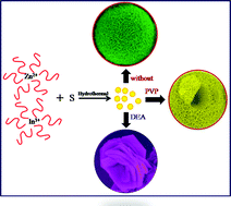
In the present investigation, a surfactant-assisted hydrothermal route has been employed to design self-assembled rose and hollow marigold-like ZnIn2S4 flowers. In the absence of the surfactant, uniform (∼3–5 μm) marigold-like flowers are observed. The self-alignment of the transparent petals (∼3–5 nm thick with a length of ∼25–100 nm) leads to the formation of hollow marigold-like flowers, for which a plausible growth mechanism has also been proposed. Moreover, DEA assisted ZnIn2S4 demonstrates a rose flower-like via self assembly of hexagonal nanoplates. Structural and optical characterization shows the existence of hexagonal structures with a band gap in the range of ∼2.4–2.6 eV. Considering the ideal band gap in the visible region, we have used such unique nanostructured self assemblies of ZnIn2S4 as photocatalysts and demonstrated visible light-driven photocatalytic production of clean hydrogen by toxic hydrogen sulphide, which is abundantly available as a waste gas from oil refineries (15–20%). We believe that continuous efforts in this direction may open up new insights into the design of controllable nanostructures and their potential applications in advanced fields.
Recently, a new class of multi-component sulphides has also been reported to show a high photocatalytic efficiency.8–11 Among these, ZnIn2S4 is a n-type semiconductor with a bandgap of 2.4 eV, which possesses photo- and chemical stablility.12 It exhibits a layered structure in the AB2X4 (A = Cu, Ag, Zn, Cd, etc.; B = Al, Ga, In; C = S, Se, Te) family, which has attracted considerable attention due to its outstanding electrical and optical properties.13 There are several reports of ZnIn2S4 in the literature with different morphologies using effective methods/strategies.13–18 Furthermore, Shen et al. employed nanostructured ZnIn2S4 for hydrogen evolution, and the photocatalytic activity of the ZnIn2S4 was greatly affected by the crystal plane spacing along the c-axis.17 Considering the importance of the band and crystal structure of ZnIn2S4, hierarchically hollow microspheres and faceted growth of nanosheets are usually relevant to the self-assembly process, and are promising in photocatalytic hydrogen evolution. Also, the issue of highly toxic hydrogen sulphide and necessity of producing hydrogen from it by photocatalysis has been described in our previous report.11,19,20 Hence, the fabrication and designing of different morphologies of ZnIn2S4 is highly desirable, and their photocatalytic hydrogen generation from hydrogen sulphide will be significant.
Herein, we have reported nanostructured hollow marigold and rose flower-like ZnIn2S4 using polyvinyl pyrrolidone (PVP) and diethyl amine (DEA) as capping agents. The effect of the concentration of PVP and DEA on the morphology has been demonstrated. The possible growth mechanism has also been discussed in detail. Finally, the effect of different morphologies on the photocatalytic hydrogen generation is investigated.
![]() | ||
Fig. 1 X-ray diffraction patterns of the ZnIn2S4 synthesized without using surfactant (S1) and with 300 ppm PVP (S2) and 0.015 mol DEA (S3). |
In order to investigate the influence of the size and shape on the optical properties, UV-visible diffuse reflectance spectra (DRS) were recorded. Fig. 2 depicts diffuse reflectance spectra of all samples (S1–S3), which show steep absorption edges in the visible region, indicating the formation of a single phase compound. The steep shape of the visible edge, and the strong absorption in the visible region, also reveal that the absorption band of ZnIn2S4 is because of the transition from the valence band to the conduction band. The samples without a capping agent (S1) and with PVP (S2) show an absorption edge at ∼515 nm with a corresponding band gap of 2.4 eV. Meanwhile, the DEA assisted ZnIn2S4 sample (S3) shows an absorption edge at ∼475 nm (2.6 eV) which is due to the reduced nanoparticle size.
Fig. 3a shows the FESEM image of pristine ZnIn2S4 (S1), indicating the formation of uniform marigold flowers originating from well organized petals. The size of the flower is observed to be in the range of 3–5 μm with the thickness of the petal being ∼3–5 nm. The observed size of the ZnIn2S4 flower clearly indicates a high aspect ratio. A more detailed structural analysis of the present marigold-like ZnIn2S4 flower was carried out using a TEM. Fig. 3b shows the TEM micrograph of the petals of the marigold flower. The size of the ZnIn2S4 petal is found to be ∼50–200 nm, which is observed to be thin and transparent. The selected area electron diffraction (SAED) pattern taken from the surface of the ZnIn2S4 petal (Fig. 3e), demonstrates the single crystalline nature of the ZnIn2S4 (Fig. 3c). The lattice fringes in the HRTEM image confirm the high crystallinity of the petal, as shown in Fig. 3d. The HRTEM image is taken on the edge of the same petal, and the interplanar spacing (d) is observed to be ∼3.35 Å which is consistent with the (101) plane of the hexagonal ZnIn2S4. A fast Fourier transform (FFT) pattern taken from the same area is shown in Fig. 3f. The FFT result indicates a single crystalline nature with a well defined hexagonal pattern. This is agreement with the result shown by the XRD pattern.
FESEM studies of the PVP assisted ZnIn2S4 samples with different PVP concentrations were also carried out to see the morphological effect. Surprisingly, the variation in concentration plays an important role in the formation of the hollow marigold flower structures. In the systematic study, at lower PVP concentration (100 ppm), premature distorted flowers are observed (Fig. SI-3a and b†). As the concentration of PVP is increased up to 200 ppm, hollow spheres of size ∼4–6 μm with a ∼2.5 μm cavity are obtained (Fig. SI-3c and d†). However, the puffiness of the flower is observed to be much less, due to the compactness of the petal. When the concentration of PVP is further increased up to 300 ppm (S2), interestingly, puffy flowers with hollow cavities are obtained (Fig. 4). The petals of the flower are fully grown and well separated from each other. These highly porous flowers with a cavity represent the hollow nature of the nanoparticles. Fig. 4b–d reveal that the hierarchical nanostructured hollow marigold flowers are assembled by numerous orderly packed nanosheets (petals). Furthermore, a nitrogen adsorption isotherm was carried out for surface area analysis study (Table SI-1 and Fig. SI-4†). BET surface areas of ∼40 and 55 m2 g−1 are found for samples S1 and S2, respectively. The hollow marigold flowers (S2) show a higher surface area due to their cavity, in comparison to the marigold flowers only (S1).
In order to examine the hollow cavity and crystalline nature of the 300 ppm PVP assisted ZnIn2S4 sample, TEM analysis was carried out. The TEM images in Fig. 5a and b confirm the hollow structure, which is in good agreement with the SEM results. A magnified TEM image (Fig. 5b) reveals that hollow microspheres, with hierarchical nanostructures assembled with nanosheets, were obtained. Significantly, very thin hexagonal petals of ZnIn2S4 with length ∼1 μm are found around the cavity. Fig. 5c shows a high resolution image taken from the edge part, and the magnified HRTEM image (inset Fig. 5c) depicts the lattice fringes of ∼3.29 Å associated with the (101) plane of the hexagonal ZnIn2S4. The corresponding electron diffraction pattern (Fig. 5d) depicts not only the formation of a single crystalline ZnIn2S4 nanostructure but also gives some indication that the blur diffraction ring pattern may be due to a slightly low crystallinity. Furthermore, detailed observations reveal that these electron diffraction spots form a hexagonal geometry in the SAED pattern, which is due to the hexagonal ZnIn2S4.
![]() | ||
Fig. 5 (a–c) TEM images of the samples synthesized with 300 ppm PVP (S2) as a capping agent and (d) the electron diffraction pattern. |
Fig. 6a and b depict the FESEM images of the sample synthesized using diethyl amine (DEA) as a capping agent (S3). This clearly shows the rose flower-like morphology of the ZnIn2S4 with a size of 0.5–1 μm. The individual flower-like structures consist of aggregated hexagonal nanosheets (petals) of thickness ∼50 nm. These petals, with a random array, are connected to each other to build a rose flower-like architecture.
![]() | ||
Fig. 6 FESEM images of the sample synthesized in the presence of (a) 0.01 and (b) 0.015 mol DEA as a capping agent. |
Fig. 7a and b depict the TEM images of the same sample (S3). The presence of a hexagonal sheet with sharp edges is clearly observed. The HR-TEM image shows visible lattice fringes with an inter-planar distance of ∼4.11 Å (Fig. 7c), which represents the (006) plane, indicating the [001] direction, i.e. c-axis oriented growth of the ZnIn2S4. The electron diffraction (ED) pattern (Fig. 7d) indicates that the nanosheet had a low crystallinity, which may be due to smaller crystallites.
![]() | ||
Fig. 7 (a and b) TEM images of the ZnIn2S4 sample synthesized using diethyl amine as a capping agent (S3), (c) HR-TEM image of nanosheets and (d) an electron diffraction pattern. |
The size, shape and growth kinetics of ZnIn2S4 can be explained on the basis of the capping agents and their concentration under hydrothermal conditions, which are given in the following section.
Zn(NO3)2 + In(NO3)3 + H2O → Zn2+ + In3+ | (R1) |
Zn2+ + In3+ + CA → [Zn(CA)]2+ + [In(CA)]3+ | (R2) |
NH2CSNH2 → NH2CN + H2S | (R3) |
H2S + H2O ↔ H3O+ + HS− | (R4) |
HS− + H2O ↔ H3O+ + S2− | (R5) |
The released Zn2+, In3+ and S2− ions combine to form ZnIn2S4 nuclei, as expressed by (R6):
Zn2+ + In3+ + S2− → ZnIn2S4 | (R6) |
Zn2+, In3+ and S2− ions are released gradually (R2–R5), which controls the nucleation rate of ZnIn2S4 and hence the numbers of ZnIn2S4 nuclei formed in the initial stage are limited. Accordingly, there are still large amounts of unreacted Zn2+, In3+ and S2− ions in the reaction solution, in the form of [Zn(CA)]2+, [In(CA)]3+ and NH2CSNH2, respectively. Depending on the concentration of the capping agent and the reaction time, these unreacted forms release ions to develop a particular morphology, which is explained in the “growth mechanism” section.
Initially, Zn–PVP and In–PVP complexes are formed through coordinative bonding with the carbonyl oxygen of the PVP. At elevated temperatures, thiourea starts to release H2S as a sulphur source. This H2S slowly reacts with Zn2+ and In3+ ions by replacing the PVP ligand, and forms ZnIn2S4 nuclei. Here, the ZnIn2S4 nuclei are capped with PVP at the surface, and hence restricted growth of the flower-like morphology is observed. In this case, the precursors are capped with PVP and the nanoparticles then aggregate into bigger particles to decrease their surface energy. At supersaturation conditions, the crystallites on/near the outermost surface of the aggregates will continue to grow at a slow rate.24 This continuous growth of aggregates confers a spherical self assembly, having a core with smaller nanoparticles and an exterior layer with bigger nanoparticles (Scheme 1a). As the concentration of PVP increases up to 300 ppm, the excess of PVP on the ZnIn2S4 nuclei in the core is diffused at 150 °C to the exterior layer (Fig. 4). At this stage, as per the Ostwald ripening phenomenon, there is an aggregation of the primary nuclei, followed by an outward mass transfer through the growth of larger crystallites from those of smaller size, and the migration of crystallites from the inner core to the outer shell. In this process the inner core is completely consumed and a hollow cavity is created due to diffusion. During this process, the excess PVP present in the core is relinquished at 150 °C, to release the pressure created by the PVP. Hence, some of the mass of the ZnIn2S4 diffuses from the core. Due to this reason, a hole or cavity is created on the flower. This phenomenon creates the hollowness in the flower-like assembly which is clearly seen in the FESEM and TEM images (Fig. 4 and 5a and b). The flower-like assembly shows a porous structure due to the steric effect of the PVP molecules. Hence, we find there are many voids in the interior of the spherical aggregates.23
In an extension of this work, we also used diethyl amine (DEA) as a capping agent. At lower concentrations of DEA (0.01 mol), three dimensional growth is observed and hence, aggregated spherical nanoparticles align and show premature nanoplates. As the boiling point of DEA is higher than water, the overall vapour pressure is less than water alone. Hence, growth is restricted to form a random flower-like structure. As the concentration of DEA increases up to 0.015 mol, two dimensional growth of the nanoparticles is accelerated. Hence, at prolonged reaction time, due to the Ostwald ripening phenomenon a hexagonal plate-like structure is conferred. At equilibrium conditions, these hexagonal plates are self organized and the assembly of rose flower-like structure is conferred. Interestingly, at higher concentrations of DEA due to the lower vapor pressure, there is no strain created on the plate, and hence the curving ability is suppressed.18 Therefore, instead of the marigold structure, the rose flower-like structure is obtained.
![]() | ||
Fig. 8 XPS patterns of the as-synthesized ZnIn2S4 at 150 °C for 30 h using 300 ppm PVP as a capping agent (S2) (a), survey spectra and high-resolution spectra for (b) Zn2p (c) In3d, (d) S2p. |
Footnote |
† Electronic supplementary information (ESI) available: Fig. SI-1 and SI-2: XRD study of different concentrations of PVP and DEA capping agents, Fig. SI-3: FESEM images of the samples synthesized using (a and b) 100 and (c and d) 200 ppm PVP as a capping agent. Fig. SI-4: BET surface area and pore width distribution, and Fig. SI-5: error bars of the hydrogen evolution. See DOI: 10.1039/c3ra45767b |
This journal is © The Royal Society of Chemistry 2014 |